




No CrossRef data available.
Article contents
Intravascular ultrasonic assessment of post-operative remodelling of aortic coarctation repaired by use of a subclavian flap
Published online by Cambridge University Press: 18 April 2005
Abstract
Background: We have used intravascular ultrasound in an attempt to clarify the extent of vascular remodelling of the aortic arch after the repair of aortic coarctation by use of a subclavian flap. Methods: We investigated 13 patients with coarctation of the aorta, ranging in age from 1.4 to 43.0 months, with a mean of 20.8 months, who underwent aortoplasty by incorporation of a subclavian flap. The mean postoperative period was 19.6 months, with a range from 0.03 to 41.2 months. The luminal morphology of the aortic arch was evaluated by intravascular ultrasound at the time of post-operative catheterization. Results: We observed 3 cases longitudinally. Over the period of observation, we found three types of morphology of the aorta at the site of incorporation of the subclavian flap, namely a snowman shape with two inflection points, a pisiform shape with one inflection point, and a round shape without any points of inflection. There was a correlation between the cross-sectional shapes at the site of the subclavian flap in the postoperative period (p < 0.01). In each case, we measured the cross-sectional area at the site of subclavian flap, at the descending aorta, and at the distal aortic arch. The cross-sectional area, and the increment of the cross-sectional area at the site of subclavian flap, was larger. Conclusion: The shape of the lumen subsequent to repair of aortic coarctation changes progressively from a snowman, to a pisiform, and finally to a round shape. Greater growth of the subclavian flap compared to the native wall of the aorta was observed for at least the first 4 years after repair. This finding may improve our understanding of the remodelling process of the arterial trunks after surgical repair.
Information
- Type
- Original Article
- Information
- Copyright
- © 2003 Cambridge University Press
Intravascular ultrasound is a useful method for evaluating the morphological characteristics of normal and diseased arteries. The coronary arteries can be seen well by intravascular ultrasound. Thus far, however, the method has not been extensively analyzed for evaluating the morphology and pathology of the great vessels. To the best of our knowledge, there has been no study in which intravascular ultrasound has been used to evaluate the repair of coarctation by incorporation of a subclavian flap, in which the subclavian artery is turned down and used to enlarge the narrow portion at the site of the aortic coarctation (Fig. 1). Both this procedure, and end-to-end repair, is effective for repair of coarctation in young infants, with the two approaches having a similar risk of recurrence.1, 2 The aim of this study was to use intravascular ultrasound to assess the remodelling process of the aorta after repair using a subclavian flap.

Figure 1. Use of the subclavian flap for repair of aortic coarctation. After ligating the left subclavian artery, the aorta and subclavian artery are opened. The subclavian artery is divided distally and turned down to create the flap. ASC Ao: ascending aorta; DESC Ao: descending aorta; BC A: brachiocephalic artery; LCC A: left common carotid artery; LSC A: left subclavian artery.
Materials and methods
Subjects
We studied 13 patients in whom aortic coarctation had been repaired by turning down a subclavian flap (Fig. 1) at Nagano Children's Hospital during the period from January 1996 through October 1999. Of the patients, 6 were boys and 7 girls, with mean age of 20.8, and a range from 1.4 to 43.0 months, at the time of the ultrasonic study. They underwent repair of coarctation at mean age of 37.4 days, with a range from 3 to 120 days. The mean postoperative period was 19.6 months, with a range from 0.03 to 41.2 months. We studied one patient only a day after the operation, the patient having a problem with low cardiac output syndrome due to residual coarctation. This patient, therefore, was catheterized while considering re-operation.
In 3 of the 13 cases, we achieved longitudinal examinations, with the total number of examinations using intravascular ultrasound being 18. In 10 of the 13 cases, there were associated complex cardiac abnormalities. The remaining 3 patients had no intracardiac abnormalities. The initial repair was successful in all but one, being conducted without any pressure gradient or any morphological narrowing. Re-coarctation developed in the outstanding patient, who had successful balloon dilation at the time of catheterization that included the intravascular ultrasonic examination.
Intravascular ultrasound
The luminal morphology of the aortic arch obtained by intravascular ultrasound was evaluated at the time of postoperative catheterization. The mean postoperative period was 19.6 months, with a range from 0.03 to 41.2 months. The diagnostic apparatus used was the Hewlett-Packard M2400A, with a 3.5 French Sonicath (Boston-Scientific: 20/30 MHz, Boston, USA). The fluoroscopic view was displayed in the same cathode-ray tube screen as the intravascular ultrasonic image, permitting recognition of the position of the tip of the ultrasonic catheter. After catheterization, we measured cross-sectional area and the circumference of the vessel lumen by retrospective review of the videotape, measuring these two parameters at the level of the descending aorta, at the subclavian flap, and at the distal arch.
Methods
We compared the luminal cross-sectional configuration at the site of the postoperative aortic isthmus. We measured the cross-sectional area, diameter, and circumference from an intravascular ultrasonic image obtained at end-systole (Fig. 2). We chose the precise site of the subclavian flap by comparing the fluoroscopic view displayed by intravascular ultrasound with aortography. The luminal shape was observed while pulling back the catheter, changing from a circle to a distorted form, and finally returning to a circle. We deemed the site of maximal distortion to be the site of insertion of the subclavian flap.

Figure 2. We measured cross-sectional area and circumference at three different levels, the descending aorta 1 to 2 vertebral bodies above the diaphragm, the subclavian flap, and the distal arch. DAoA: distal aortic arch; SCF: subclavian flap (isthmus); DAo descending aorta.
The architecture of the aortic wall was analyzed to establish whether there was a difference between the native aortic wall and the subclavian flap, and whether the expected mural architecture was observed. These were analyzed carefully from the ultrasonic images as seen on the screen of the cathode-ray tube.
Results
We were able to divide the luminal cross-sectional configurations into “snowman”, “pisiform”, and “round” shapes, with 5 patients showing a snowman configuration, 7 a pisiform shape, and 6 having a round appearance. There were two points of inflection with the snowman shape, one inflection with a pisiform, and no inflections when the shape was round (Fig. 3). There was a significant correlation between the luminal cross sectional configuration and the period subsequent to surgical repair (p < 0.01). The snowman shapes were observed after a period of 3.7 ± 4.5 months, the pisiform shape after 18.6 ± 8.1 months, and the round shape at 33.9 ± 5.9 months after the surgery (Fig. 4). There was no correlation between the cross-sectional shapes and the preoperative morphology of the aortic arch, including the diameter of the subclavian artery and of aortic isthmus before the surgical procedure as measured by angiography. Neither was any correlation found between blood pressure and the postoperative shape of the aortic arch.

Figure 3. Examples of postoperative intravascular ultrasonic findings showing A: the snowman shape, B: the pisiform shape, and C: the round shape. The small arrows show the points of inflection. The white arrow in the lower left panel shows the point of inflection looking brighter.

Figure 4. Comparison of the post-operative period among the three groups.
The morphological changes could be followed longitudinally in three patients, with progression from a snowman through a pisiform to a round shape being seen in one case, from a snowman to a pisiform shape in a second patient, and from a pisiform to a round shape in the other patient.
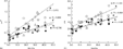
We charted the relationships between the postoperative period in months and the cross-sectional area measured in centimeters squared at the site of distal aortic arch, the subclavian flap, and the descending aorta (Fig. 5a), and also between luminal circumference and the postoperative period (Fig. 5b). Both area and circumference increased during the postoperative period, with the cross-sectional area and the circumferential length of the descending aorta and distal aortic arch increasing in parallel fashion. The relationship between cross-sectional area and postoperative period was expressed as y = 0.015x + 0.26 in the descending aorta, y = 0.013x + 0.26 in the distal arch, and y = 0.031x + 0.34 at the subclavian flap. The relationship between circumferential length and postoperative period was expressed as y = 0.039 + 1.83 in descending aorta, y = 0.036x + 1.89 in distal arch, and y = 0.056x + 2.29 at the subclavian flap. The regression lines for the descending aorta and distal arch mimicked normal aortic growth. In contrast, the steeper slope at the site of the subclavian flap indicates increased growth of the postoperative aortic isthmus. Aortic angiography done at the same time as the intravascular ultrasonic examination showed a dilated segment at the site of the subclavian flap (Fig. 6).

Figure 5. Correlations between (a) the cross-sectional area and the time after repair and (b) the cross-sectional circumference and time after repair, with measurements made at end-systole. ○: at the subclavian flap segment; [utri ]: at descending aorta; [bull ]: at distal arch.

Figure 6. Aortography from a patient after repair using the subclavian flap shows dilation of the isthmic portion (arrow) at the site of the flap.
When assessing the architecture of the aortic wall at the subclavian flap, we found that the points of inflection appeared brighter than the remaining wall in those with snowman and pisiform configurations. No significant differences were found, however, between the original aortic wall and the wall of the subclavian artery except at the points of inflection. Because of this, in those with round configurations it was hard to decide whether any given site was the wall of the native aorta or the subclavian arterial wall. A trilaminar structure was observed at the site of the subclavian flap in 14 (78%) of the ultrasonic studies. In those with pisiform arches, careful and simultaneous comparison of the fluoroscopic and ultrasonic images revealed that the point of inflection on the side of the greater curvature of aortic arch merged earlier than that on the other side in 5 of 7 cases.
Discussion
It is widely accepted that intravascular ultrasound is a useful method for the morphological evaluation of vessels, particularly the characteristics of their walls.3, 4 Intravascular ultrasound is also an important method for quantitative analysis of the vessel lumen, and can provide dynamic images.5, 6 Although some have investigated aortic coarctation using intravascular ultrasound,7, 8 as far as we are aware ours is the first study to examine the remodelling process of the aortic arch subsequent to repair using a subclavian flap. We observed changes in luminal configuration in terms of diameter, cross-sectional area, and circumferential length in patients corrected during infancy.9 We observed significant morphological changes at the site of insertion of the subclavian flap, from a distorted configuration to one of symmetry, and finally to a round shape over time.
It has been noted that, because of the artifactual distortion of intravascular ultrasonic images, circular arteries are seen as noncircular vessels.10 We avoided this artifact by placing the intravascular ultrasound catheter as parallel to the wall of the descending aorta as accurately as was possible.
As the configuration of the repaired segment changed from a snowman to a round shape, the point of inflection at the outer curvature disappeared earlier than did the inflection on the other side. As shown in Figure 5, the increment in the cross-sectional area of the subclavian flap was greater than the expected normal growth as observed in the descending aorta and distal arch, the subclavian flap itself therefore growing more rapidly than normal. The luminal cross-sectional area at this site increased, either by a process of smoothing, or in the process of the merging at the point of inflection. We found a significant correlation between the luminal configuration at the site of the subclavian flap and the period subsequent to operative repair. As time goes by, under the influence of blood pressure, the subclavian flap almost became equivalent to the morphology of the normal aortic wall. In children with pulmonary hypertension, it has been shown that the pulmonary arterial wall is thicker than that of control children, with the ultrasonic images demonstrating a trilaminar appearance for the wall.11, 12 The blood pressure at the aortic isthmus rises after incorporation of the subclavian flap, which may cause endothelial proliferation. As a result, a trilaminar appearance frequently becomes visible over long-term post-operative period. Following long-term post-operative follow-up, the circular intimal layer can no longer be distinguished from a normal aorta, possibly due to uniform growth of intimal cells.
By measuring both circumference and cross-sectional area, we showed that the development of the distal arch was no different from that of the descending aorta. The growth of the subclavian flap itself, in contrast, was more rapid than that of the descending aorta and distal arch, while there was no difference between the luminal cross-sectional area at the site of the subclavian, the distal arch, or the descending aorta shortly after the operation. This suggests that the postoperative aortic wall grows not only on the native aortic side, but also on the side of the subclavian flap.13 Kino et al.14 reported late aneurysmal formation in one patient repaired using subclavian flap aortoplasty. This may be the consequence of an abnormal excessive extension of the subclavian arterial flap, albeit that this complication is rare when the subclavian flap is used for repair.
Thus, we have demonstrated significant remodelling of aorta, including changes in luminal shape and development, after repair of aortic coarctation using the subclavian flap. We have shown that intravascular ultrasound provides a unique technique for evaluating the morphology and the characteristics of the aortic wall, and may help still further to improve the understanding of aortic remodelling. We accept, nonetheless, that the number of our patients examined in a longitudinal fashion is small, and we have made no comparison with alternative methods of repair such as the end-to-end anastomosis.
Acknowledgement
We thank Atsushi Komiyama, Professor of Department of Pediatrics, Shinshu University School of Medicine for invaluable advice.
References
Pandian NG, Weintraub A, Kreis A, Schwartz SL, Konstam MA, Salem DN. Intracardiac, intravascular, two-dimensional, high-frequency ultrasound imaging of pulmonary artery and its branches in humans and animals. Circulation 1990; 81: 2007–2012.Google Scholar
Ge J, Erbel R, Gorge G, et al. Intravascular ultrasound imaging of arterial wall architecture. Echocardiography 1992; 9: 475–483.Google Scholar
Zehr KJ, Gillinov AM, Redmond JM, et al. Repair of coarctation of the aorta in neonates and infants: a thirty-year experience. Ann Thorac Surg 1995; 59: 33–41.Google Scholar
Cobanoglu A, Thyagarajan GK, Dobbs JL. Surgery for coarctation of the aorta in infants younger than 3 months: end-to-end repair versus subclavian flap angioplasty: is either operation better? Eur J Cardiothorac Surg 1998; 14: 19–26.Google Scholar
Chandran KB, Vonesh MJ, Roy A, et al. Computation of vascular flow dynamics from intravascular ultrasound images. Med Eng Phys 1996; 18: 295–304.Google Scholar
Hardt SE, Just A, Bekeredjian R, Kubler W, Kirchheim HR, Kuecherer HF. Aortic pressure-diameter relationship assessed by intravascular ultrasound: experimental validation in dogs. Am J Physiol. 1999; 276 (3 Pt 2): H1078–1085.Google Scholar
Tong AD, Rothman A, Atkinson RL, Shiota T, Ricou F, Sahn DJ. Intravascular ultrasound imaging of coarctation of the aorta: animal and human studies. Am J Card Imaging 1995; 9: 250–256.Google Scholar
Higashidate Y, Tsuda T, Takamuro M, Tomita H. Intravascular ultrasound imaging shortly after balloon angioplasty of native aortic coarctation in two neonates. Pediatr Cardiol 1999; 20: 377–379.Google Scholar
Kornet L, Jansen JR, Gussenhoven EJ, Versprille A. Determination of the mean cross-sectional area of the thoracic aorta using a double indicator dilution technique. Pflugers Arch 1996; 432: 1069–1073.Google Scholar
Delachartre P, Cachard C, Finet G, Gerfault FL, Vray D. Modeling geometric artefacts in intravascular ultrasound imaging. Ultrasound Med Biol 1999; 25: 567–575.Google Scholar
Ivy DD, Neish SR, Knudson OA, et al. Intravascular ultrasonic characteristics and vasoreactivity of the pulmonary vasculature in children with pulmonary hypertension. Am J Cardiol 1998; 81: 740–748.Google Scholar
Nakamoto A, Yoshitake J, Hase T, et al. Intravascular ultrasound imaging of the pulmonary arteries in primary pulmonary hypertension. Respirology 2000; 5: 71–78.Google Scholar
Mellgren G, Friberg LG, Bjorkerud S. Can we predict the long-term function of the subclavian flap angioplasty? J Thorac Cardiovasc Surg 1992; 104: 932–937.Google Scholar
Kino K, Sano S, Sugawara E, Kohmoto T, Kamada M. Late aneurysm after subclavian flap aortoplasty for coarctation of the aorta. Ann Thorac Surg 1996; 61: 1262–1264.Google Scholar

We measured cross-sectional area and circumference at three different levels, the descending aorta 1 to 2 vertebral bodies above the diaphragm, the subclavian flap, and the distal arch. DAoA: distal aortic arch; SCF: subclavian flap (isthmus); DAo descending aorta.

Examples of postoperative intravascular ultrasonic findings showing A: the snowman shape, B: the pisiform shape, and C: the round shape. The small arrows show the points of inflection. The white arrow in the lower left panel shows the point of inflection looking brighter.

Comparison of the post-operative period among the three groups.
Correlations between (a) the cross-sectional area and the time after repair and (b) the cross-sectional circumference and time after repair, with measurements made at end-systole. ○: at the subclavian flap segment; [utri ]: at descending aorta; [bull ]: at distal arch.

Aortography from a patient after repair using the subclavian flap shows dilation of the isthmic portion (arrow) at the site of the flap.
 You have
Access