Introduction
Infertility is a global health problem that is defined as the reduced ability or an inability to conceive after 1 or more years of regular, unprotected sexual intercourse (WHO, 2010). Infertility affects up to 15% of couples globally (Agarwal et al., Reference Agarwal, Mulgund, Hamada and Chyatte2015). Low or absence of sperm production, abnormal sperm function and blockages can all cause male infertility, as can infection, injuries, chronic health problems, and environmental and lifestyle factors (Esteves and Agarwal, Reference Esteves and Agarwal2011). Semen analysis is an essential and systematic method to test male infertility.
Azoospermia is defined as a complete absence of spermatozoa in the ejaculate. This condition is found in 1% of the general male population and in 10–15% of male infertility cases (Elzanaty, Reference Elzanaty2013; Popal and Nagy, Reference Popal and Nagy2013). Azoospermia is divided into two categories: obstructive (OA) and non-obstructive (NOA) azoospermia. OA is defined as the absence of sperm cells in a semen sample due to blockage of the male reproductive tract, whereas NOA results from a failure of spermatogenesis due to impaired sperm production (Wosnitzer et al., Reference Wosnitzer, Goldstein and Hardy2014). NOA is responsible for 60% of azoospermia cases and is the most severe form of male infertility (Hamada et al., Reference Hamada, Esteves and Agarwal2013).
Spermatogenesis is an androgen-dependent event and testosterone is the major androgen source in the testis (Walker, Reference Walker2011). In the testosterone metabolic pathway, the enzyme 5-alpha reductase converts testosterone to dihydrotestosterone (DHT) in the testicular and peripheral tissues. Compared with testosterone, DHT is a much more potent androgen, with a higher binding affinity for androgen receptors and a longer half-life, therefore DHT has a stronger androgen effect on male productive tissues (Zhao et al., Reference Zhao, Wu, Xu, Niu, Cui, Zhang, Wang and Wang2012; Ogino et al., Reference Ogino, Miyagawa and Iguchi2016). In addition, DHT is important for the development of prostate and male external genitalia. Although testosterone is the primary androgen source in spermatogenesis, mammalian studies have shown DHT to have moderator androgen activity in human seminiferous tubules, play a role in the sperm maturation process and sustain normal spermatogenesis (Payne et al., Reference Payne, Kawano and Jaffe1973; Robaire and Viger, Reference Robaire and Viger1995; Hines et al., Reference Hines, Tho, Behzadian and McDonough1999).
The enzyme 5-alpha reductase is known to have three different subtypes: types 1, 2 and 3. Type 2 5-alpha reductase (SRD5A2) is commonly found in the prostate, epididymis, seminal vesicles, prostate, liver and other genital tissues (Eunice et al., Reference Eunice, Philibert, Kulshreshtha, Audran, Paris, Khurana, Pulikkanath, Kucheria, Sultan and Ammini2008). SRD5A2 is located on chromosome 2p23, composed of five exons, and encodes a 28.4 kD protein made up of 254 amino acids (Azzouni et al., Reference Azzouni, Godoy, Li and Mohler2012). It has been postulated that polymorphisms in genes encoding 5-alpha reductase may be associated with impaired male fertility (Suzuki et al., Reference Suzuki, Sasagawa, Itoh, Ashida and Ogata2002; Peters et al., Reference Peters, Saare, Kaart, Haller-Kikkatalo, Lend, Punab, Metspalu and Salumets2010; Zhao et al., Reference Zhao, Wu, Xu, Niu, Cui, Zhang, Wang and Wang2012). To date, there are no data on the association between polymorphisms in the SRD5A2 gene and NOA in Turkish men. The aim of this study was to analyse the genotype frequency of one single nucleotide polymorphism (SNP-rs523349) in Turkish NOA patients and fertile controls.
Materials and methods
Study population
This study was performed between June 2013 and February 2015 at the Bahçeci Umut IVF Center, Istanbul, Turkey. In total, 75 NOA patients and 43 fertile control individuals were included in the study. The study was approved by the Yeditepe University Ethics Committee (decision number 313 on 2 April 2013). Patients were evaluated in accordance with WHO recommendations (WHO, 2010). A diagnosis of azoospermia was based on two sequential semen analyses in which spermatozoa sperm cells were not observed after centrifugation. For the diagnosis of NOA, a detailed andrological examination was performed; the patient’s clinical history was reported, a physical examination was performed, and hormone testing was conducted as an endocrinological evaluation. Patients with a normal karyotype and no Y chromosome microdeletions were included, in the study. Patients that were diagnosed with NOA underwent surgical sperm retrieval by microsurgical testicular sperm extraction (micro-TESE). In the absence of spermatozoa in the tissue samples, the samples were sent to a pathology laboratory for histological examination. NOA patients were further divided into two subgroups according to the success of sperm retrieval via micro-TESE. In total, 43 fertile volunteers who had offspring naturally were selected as the control group. This group underwent a physical examination and hormone testing. The clinical data of all patients and control subjects were gathered from medical records.
DNA extraction and genetic analysis
After obtaining written consent from the patients, 5 ml of peripheral blood was collected for genomic DNA extraction. To perform genomic DNA isolation, an iPrep purification instrument (Invitrogen, Life Technologies; Thermo Fisher Scientific Inc., Waltham, MA, USA) and an iPrep PureLink gDNA blood isolation kit (Invitrogen, Life Technologies; Thermo Fisher Scientific Inc.) were used in accordance with the manufacturer’s protocol. The quality and quantity of DNA were measured using a NanoDrop 2000 spectrophotometer (Thermo Fisher Scientific Inc., Waltham, MA, USA) and using a 1.8 optical density range for genotyping. Final sample concentrations were diluted to approximately 100 ng/µl. The DNA samples were kept at 4°C until genotyping assessment.
SRD5A2 rs523349 polymorphism genotyping was performed using a 7500 Fast Real-Time PCR instrument (Applied Biosystems, Foster City, CA, USA) and the TaqMan Genotyping assay and TaqMan Genotyping Master Mix (TaqMan Reagents, Applied Biosystems, Foster City, CA, USA). The reaction mixture and conditions were applied as suggested by the manufacturer as follows: 10 min at 95°C for the holding stage, followed by 40 cycles of 10 s at 95°C for denaturation and 60 s at 60°C for annealing/extension. Allelic discrimination of the samples was carried out by collecting and interpreting the fluorescence signals of the hybridization probes using the software from the 7500 Fast Real-Time PCR System.
Statistical analysis
Statistical analyses were carried out using the SPSS 24.0 statistical package (IBM Corp. Released 2016). A P-value of ≤0.05 was considered to indicate statistical significance for all statistical tests.
First, the Kolmogorov–Smirnov test was used to determine whether the continuous quantitative parameters measured in the study (age, follicle-stimulating hormone (FSH), luteinizing hormone (LH), total testosterone level and testis volume) were normally distributed or not. Kolmogorov–Smirnov tests determined that none of the continuous quantitative parameters was normally distributed. In all tables, the median and corresponding minimum and maximum values were reported for each specific quantitative measurement. An independent median test was used to show the differences between the NOA and control groups for the median age, FSH, LH, total testosterone level and testis volume.
For the NOA group, further detailed comparisons were performed using the independent median test to identify differences in median age, FSH, LH, total testosterone level and testis volume between NOA patients in whom TESE surgery was successful versus those that were unsuccessful.
Similarly, Pearson’s chi-squared test was used to assess the differences in the proportion of categorical variables by comparing the CC, CG and GG genotype groups and the C and G allelic frequency between the NOA and control groups and between the NOA subgroups.
Results
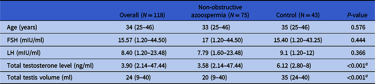
Clinical data of all NOA patients and controls are given in Table 1. The analysis included 75 patients with NOA and 43 control subjects. No statistically significant differences were identified between the NOA and control groups regarding their age (P = 0.576), FSH (P = 0.444) and LH levels (P = 0.366). However, statistically significant differences between the two groups were obtained for the median values of total testosterone level and total testis volume (P < 0.001).
Clinical data of the study population

Values are reported as median (minimum–maximum).
Independent sample median test is used to test the median values between non-obstructive azoospermia and control groups for each characteristic.
a Statistically significant P-value at α = 5% level.
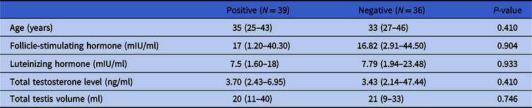
Table 2 provides the detailed clinical data information of the NOA patient subgroups. Of the NOA patients, 36 did not have successful micro-TESE sperm retrieval (sperm negative), while 39 had successful sperm retrieval (sperm positive). There was no significant difference between the micro-TESE-positive and micro-TESE-negative groups in terms of age (P = 0.410), FSH (P = 0.904), LH (P = 0.933), total testosterone level (P = 0.410) and total testis volume (P = 0.746).
Clinical data of non-obstructive azoospermia subgroups
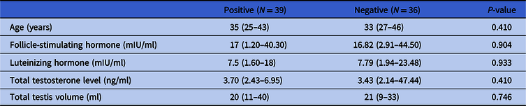
Values are reported as median (minimum–maximum).
Independent samples median test is used to test the median values between testicular sperm extraction (TESE)-positive and TESE-negative groups for each characteristic.
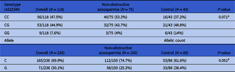
Genotype and allele frequencies for the SRD5A2 rs523349 polymorphism in the NOA and control groups are given in Table 3. There was no significant difference in the frequency of the CC, CG and GG genotype groups. The frequencies of CC, CG, and GG were 37.2, 48.8 and 14% in the control group, whereas in the NOA patient group these were 53.3, 42.7 and 4%, respectively. Allelic frequency comparisons suggested that there were no significant differences in the C and G allele types between the NOA and control groups. The frequencies of the C and G alleles were 61.6% and 38.4% in the control group, whereas these were 74.7% and 25.3% in the NOA group, respectively.
Genotype and allele frequencies between non-obstructive azoospermia and control groups

Proportions are given as n (%).
a Chi-squared test is used to test the proportions between non-obstructive azoospermia and Control groups for genotype rs523349 categories.
b Chi-squared test is used to test the proportions between non-obstructive azoospermia and Control groups for C and G Allele categories.
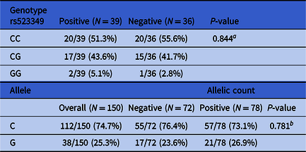
Table 4 details the genotype and allele frequencies for the SRD5A2 gene rs523349 polymorphism in the micro-TESE-positive and negative subgroups. There were no significant differences observed between these subgroups among CC, CG and GG genotypes. The frequencies of CC, CG, and GG were 51.3, 43.6 and 5.1% in the micro-TESE-positive subgroup, whereas these were 55.6, 41.7 and 2.8% in the micro-TESE-negative subgroup, respectively. Additionally, the frequencies of the C and G allele types in the NOA subgroups are reported in the same table. No statistically significant differences between the micro-TESE-positive and micro-TESE-negative subgroups were seen for the C and G allele types. The frequencies of the C and G alleles were 76.4 and 23.6% in the micro-TESE-negative subgroup, respectively. The frequencies of the C and G alleles were 73.1 and 26.9% in the micro-TESE-positive subgroup, respectively.
Genotype and allele frequencies between non-obstructive azoospermia subgroups

Proportions are given as n (%).
a Chi-squared test is used to test the proportions between TESE-Positive and TESE-Negative groups for genotype rs523349 categories.
b Chi-squared test is used to test the proportions between TESE-positive and TESE-negative groups for C and G Allele categories.
Discussion
The present study examined the possible association of the SRD5A2 gene polymorphism (rs523349) with NOA in Turkish patients. We found no genetic link between the SRD5A2 gene rs523349 and NOA in Turkish patients. Several studies have evaluated the relationship between the SRD5A2 gene and male infertility. Suzuki and his team investigated mutations in the SRD5A2 gene in Japanese patients with isolated cryptorchidism and idiopathic azoospermia. The authors screened the SRD5A2 gene within exon 1 using direct sequencing and in exons 2–5 using single-stranded conformational polymorphism analysis. There was no detected mutation within the five exons of the SRD5A2 gene in their study group (Suzuki et al., Reference Suzuki, Sasagawa, Itoh, Ashida and Ogata2002). The polymorphism ‘rs523349’ has been studied in Estonian men and in the Han-Chinese population. Peters and his group analyzed genotype frequencies of five common single nucleotide polymorphisms (SNPs 1–5: rs632148, rs523349, rs2300701, rs2268797 and rs12470143) in the SRD5A2 gene in 132 azoospermic or oligozoospermic and 211 normozoospermic Estonian men. Similar to our results, they did not find any association between the investigated genotypes and male infertility (Peters et al., Reference Peters, Saare, Kaart, Haller-Kikkatalo, Lend, Punab, Metspalu and Salumets2010). Another study investigated the association of SRD5A2 gene polymorphisms (rs4952197, rs2268797, rs13395648, rs523349 and rs632148) and semen quality in the Han-Chinese population. The results suggested that genetic variations in the SRD5A2 gene (rs13395648 and rs642138) may be associated with semen quality, but there was no association between rs523349 and semen quality (Zhao et al., Reference Zhao, Wu, Xu, Niu, Cui, Zhang, Wang and Wang2012), which is akin to our study results.
In the current study, the serum FSH and LH levels were not statistically different between the NOA group and the control group. However, there were significant differences in the total testosterone level and total testis volume between NOA patients and controls. Pezzella and colleagues found no statistically significant differences in serum testosterone levels between patients with normal sperm parameters (in accordance with WHO values) and NOA patients. However, they had findings similar to ours for the mean value of bilateral testicular volume at ultrasonography (Pezzella et al., Reference Pezzella, Barbonetti, D’Andrea, Necozione, Micillo, Di Gregorio, Francavilla and Francavilla2014). Furthermore, the same study group did not find a statistically significant difference in serum FSH or serum testosterone between the TESE-positive and TESE-negative NOA subgroups, which is similar to our findings. Nevertheless, in their results, ultrasonography testicular volume was found to be statistically significant in the TESE-positive group after TESE (Pezzella et al., Reference Pezzella, Barbonetti, D’Andrea, Necozione, Micillo, Di Gregorio, Francavilla and Francavilla2014). In accordance with the study from Franco and colleagues (Franco et al., Reference Franco, Scarselli, Casciani, De Nunzio, Dente, Leonardo, Greco, Greco, Minasi and Greco2016), they did not find any significant discrepancy in the clinical data in terms of age, FSH, LH, or inhibin B levels between positive sperm retrieval and negative sperm retrieval groups.
In conclusion, this study evaluated the relevance of SRD5A2 gene rs523349 polymorphism regarding NOA in Turkish patients. To the best of our knowledge, this study is the first to be conducted in NOA Turkish patients. Our study found no association between the single nucleotide polymorphism (SNP-rs523349) and NOA in Turkish men. As genetic polymorphisms often show ethnic differences, further functional studies are needed to evaluate the genotype and phenotype correlations in larger cohorts of various ethnicities.
Financial support
This research received no specific grant from any funding agency, commercial or not-for-profit sectors.
Conflicts of interest
The authors have no conflicts of interest.
Ethical standards
Ethical approval was granted by the ethics committee in Yeditepe University with decision number 313 on 2 April 2013.