Introduction
Kazachostylops occidentalis Nesov, Reference Nesov1987b is based on a dentary and a maxilla fragments from the upper Paleocene Zhylga (=Dzhylga) locality in the Saryagash District of the Turkistan Region, Kazakhstan (Nesov, Reference Nesov1987b). This is the only Asiatic arctostylopid mammal known outside China and Mongolia. The species epithet ‘occidentalis’ refers to the fact that this taxon is the westernmost occurrence of the Arctostylopida in Asia. Kazachostylops lived on the western margin of the Asiatic landmass in the Paleocene on the shore of the Tethys Ocean, and its remains are found in a nearshore locality together with abundant fossils of marine animals. The non-mammalian vertebrate assemblage from the Zhylga locality includes diverse chondrichthyan and osteichthyan fishes, terrestrial and marine turtles, choristoderes, aquatic snakes, lizards, and various birds (Nesov and Udovichenko, Reference Nesov and Udovichenko1984, Reference Nesov and Udovichenko1986; Nesov, Reference Nesov1986, Reference Nesov1987a, Reference Nesov, Bogdanova and Oshurkova1988a, Reference Nesov1988b, Reference Nesov and Campbell1992a, Reference Nesov1992b, Reference Nesov1995; Nesov and Chkhikvadze, Reference Nesov and Chkhikvadze1987; Averianov et al., Reference Averianov, Panteleev, Potapova and Nesov1991, Reference Averianov, Nesov and Udovichenko1993; Averianov, Reference Averianov2001, Reference Averianov2005). The mammals from the Zhylga locality, besides Kazachostylops, are nyctitheriids Voltaia minuta Nesov, Reference Nesov1987b and Jarveia minuscula Nesov, Reference Nesov1987b, stem rodent Kazygurtia clivosa Nesov, Reference Nesov1987b, ischyromyid Asiaparamys shevyrevae Nesov, Reference Nesov1987b, and enigmatic mammal Asiabradypus incompositus Nesov, Reference Nesov1987b (Nesov, Reference Nesov1987b; Averianov, Reference Averianov1995; Averianov and Martin, Reference Averianov and Martin2001; Lopatin, Reference Lopatin2006).
The specimens of Kazachostylops were considered ‘relatively good’ (Cifelli et al., Reference Cifelli, Schaff and McKenna1989, p. 23). Actually, the holotype of K. occidentalis is one of the most complete dentaries known from Arctostylopida. Nevertheless, Kazachostylops was omitted from the detailed comparison and discussion presented by Cifelli et al. (Reference Cifelli, Schaff and McKenna1989) because the authors had no access to the specimens. Additionally, some authors mistakenly considered Kazachostylops as missing the upper dentition and thus taxa based on upper dentition only often lack comparison with it (Huang et al., Reference Huang, Zheng and Ding2001; Wang et al., Reference Wang, Meng, Ni and Beard2008).
The original description of Kazachostylops by Nesov (Reference Nesov1987b) is extremely brief, and his diagnosis of this taxon is based exclusively on lower dentition characters. Here I provide a more detailed description and better illustrations of Kazachostylops, and discuss the phylogenetic position of this taxon within the Arctostylopida.
Dental nomenclature proposed by Cifelli et al. (Reference Cifelli, Schaff and McKenna1989), with some subsequent modifications (Kondrashov and Lucas, Reference Kondrashov, Lucas, Lucas, Zeigler and Kondrashov2004; Wang et al., Reference Wang, Meng, Ni and Beard2008), is used. Because the molars have a lophodont pattern, I use the suffix “lophid” instead of “cristid” for the lower molar crests. The dental terminology adopted here is shown in Figure 1.
Interpretative drawings and dental nomenclature for Kazachostylops occidentalis (CCMGE 12/12455, right upper P3-4, M1-2, and CCMGE 10/12455, right lower p3-4, m1-3). The mesial end is to the right. Light gray represents enamel surface, dark gray = the exposed dentine, black = the exposed enamel.

Materials
Repositories and institutional abbreviations
AMNH, American Museum of Natural History, New York, USA; CCMGE, Chernyshev's Central Museum of Geological Exploration, Saint Petersburg, Russia; IVPP, Institute of Vertebrate Paleontology and Paleanthropology, Chinese Academy of Sciences, Beijing, China.
Systematic paleontology
Arctostylopida Cifelli, Schaff, and McKenna, Reference Cifelli, Schaff and McKenna1989
Arctostylopidae Schlosser, Reference Schlosser, Broili and Schlosser1923
Kazachostylops Nesov, Reference Nesov1987b
Type species
Kazachostylops occidentalis Nesov, Reference Nesov1987b.
Other species
Type species only.
Revised diagnosis
Differs from Asiostylops Zheng, Reference Zheng1979 by presence of the fossette on the posterior upper premolars and molars, single-rooted p2, relatively longer talonid on p4, and entolophid connecting the ectolophid. Differs from Allostylops Zheng, Reference Zheng1979 by having single-rooted P2, but lacking pseudohypocone. Differs from Bothriostylops Zheng and Huang, Reference Zheng and Huang1986 by m1 shorter than m2, lack of entolophid on m1, m3 hypoconulid as wide as the rest of the talonid, and lack of mesiolabial cingulid on lower molars. Differs from Wanostylops Huang and Zheng, Reference Huang and Zheng1997 by a higher crown, lack of metaconule, and presence of the fossette on the upper molars. Differs from Enantiostylops new genus (Appendix 1) by upper molar parastyles convex, M1–3 lingual cingulum absent, m1–3 mesiolabial cristid present and m1 entoconid absent. Differs from Stenostylops Huang et al., Reference Huang, Zheng and Ding2001 by P4 and upper molars less elongated, M1 protocone centrally placed, and M1 postcingulum less developed and shorter than precingulum. Differs from Anatolostylops Zhai, Reference Zhai1978 by the anterior root of zygoma placed posterior to M2, lack of pseudohypocone, lingual cingula not confluent lingually, relatively longer lower molars trigonid, transverse entolophid on m3, and lack of lingual and labial cingulids on lower molars. Differs from Arctostylops Matthew, Reference Matthew1915 by P3 longer than wide, lack of lingual cingulum on upper cheek teeth, lack of pseudohypocone on upper molars, lack of lingual rib on lower canine, lack of mesiolabial cingulid on p4, by the ectolophid on lower molars lingually attaching the trigonid, and by lack of entolophid on m1. Differs from Migrostylops Tong and Wang, Reference Tong and Wang2006 by single-rooted P2, lack of pseudohypocone, longer trigonid of lower molars, ectolophid attached lingually, entolophid absent on m1, and m3 distinctly longer than m2. Differs from Palaeostylops Matthew and Granger, Reference Matthew and Granger1925 by more symmetrical P3, by lack of lingual cingulum on upper cheek teeth, by lack of pseudohypocone on upper molars, by the ectolophid on lower molars lingually attaching the trigonid, and by lack of entolophid on m1.
Kazachostylops occidentalis Nesov, Reference Nesov1987b
Figures 1–4
Reference Nesov1987b Kazachostylops occidentalis; Nesov, p. 212, pl. 2, figs. 5, 6.
Kazachostylops occidentalis, CCMGE 12/12455, the right maxillary fragment with P3–4 and M1–2 in labial (1), occlusal, (2) stereopair, and lingual (3) views. Zhylga, Kazakhstan; upper Paleocene. Scale bar = 5 mm.

Kazachostylops occidentalis, CCMGE 10/12455, holotype, the left dentary with p3–4 and m1–3 in labial (1), and lingual (2) views. Zhylga, Kazakhstan; upper Paleocene. Scale bar = 10 mm.

Kazachostylops occidentalis, CCMGE 10/12455, holotype, the lower teeth p3–4 and m1–3 in lingual (1), occlusal, (2) stereopair, and labial (3) views. Zhylga, Kazakhstan; upper Paleocene. Scale bar = 10 mm.

Holotype
CCMGE 10/12455, right dentary with c, p3–4, m1–3 and alveoli for i1–3 and p1–2.
Occurrence
Zhylga, sand quarry 8 km NNE of Zhylga railway station in Saryagash District of Turkistan Region, Kazakhstan; light-yellow sands of unnamed formation, upper Paleocene.
Description
The maxilla preserves almost complete facial palatal processes (Fig. 2). The width of the bony plate formed by maxilla is approximately equal to the width of M2, the largest upper molar. The straight suture with the collateral maxilla is preserved for most of its length. The suture is rather thick dorsoventrally, with the maximum thickness at the level of P4–M1. Anteriorly, the palatal process of the maxilla is very thin, and its sutural margin with the collateral maxilla is broken. The alveolar border is set at an angle with the intermaxillary suture, and thus the maxilla tapers anteriorly in ventral view. The alveolar margin is straight. Anterior to P3 there is a large alveolus, likely for the single-rooted P2. Its length is slightly less than the length of the P3 alveolus. More anteriorly, there is a preserved distal margin of the P1 alveolus. This alveolus extends dorsally towards the dorsal margin of the facial process of the maxilla. The dorsal margin of the facial process is very thin and does not bear a sutural facet. It was likely covered by the jugal laterally. The infraorbital canal opens anteriorly above P4. Its width is less than half of the M1 width. The anterior root of the zygomatic process is not preserved; likely it was opposite the missing M3.
The dentary is almost completely preserved (Fig. 3); the tips of the coronoid and angular processes, the anterior tip of the mandibular body, and the condylar process are missing. The dorsal margin is slightly concave posterior to the canine and slightly convex or straight at the canine and anteriorly. The ventral margin of the mandibular body is convex and converges anteriorly towards the straighter dorsal margin. The mandibular body is relatively deep, with the maximum depth at m3 where its height is more than three times the height of m3.
There is a large anterior mental foramen under p1 at the midheight of the mandibular body. A larger posterior mandibular foramen was present under p4, but this part of the dentary is largely damaged. Also, there are two distinct vascular foramina at the alveolar borders of i2 and i3.
The mandibular symphysis is obliquely oriented in medial view. Its posterior end extends to the level between p1 and p2. The posteroventral end of the mandibular symphysis is a prominent tubercle that connects with the distinct ridge extended along the ventral margin of the dentary and converging medially towards this tubercle.
There are oval-shaped alveoli for the three incisors (long axis labiolingual). The alveoli for i2 and i3 are similar in size, while the alveolus for i1 is twice smaller. The p1 and p2 were relatively large, single-rooted teeth, judging from their alveoli. The p2 was somewhat larger than p1.
The ventral margin of the mandibular ramus is deeply convex. The coronoid process is high and vertical, with a convex anterior margin. The masseteric fossa is very shallow, lacking a distinct ventral crest. It is bordered anteriorly by a low, blunt masseteric crest. In anterior view, the anterior margin of the coronoid process is wide mediolaterally at the base, where it is more than three times wider than m3. Dorsally, the anteromedial margin of the coronoid process converges to the straight anterolateral margin, and the dorsal third of the coronoid process is very thin. On the medial side of the mandibular ramus, there is a shallow pterygoid fossa at the base of the angular process. The preserved posterior margin of the mandibular ramus is very thin, while in other mammals there is usually a thickening at the base of the condylar process and variably developed condyloid crest, which is totally absent in Kazachostylops. Another astonishing character of this taxon is the lack of mandibular foramen in the preserved portion of the mandibular ramus.
There are two premolars (P3–4) and two molars (M1–2) preserved from the upper dentition (Fig. 2). These premolars and molars are three-rooted, while P2, known from the alveolus only, is single-rooted, although it is only slightly smaller than the double-rooted P3. The P3 is submolariform, considerably longer than wide (L/W = 1.62). The crown is dominated by the straight ectoloph and large protocone that is located at the distal half of the tooth. The highest point of the ectoloph is placed in the distal half of the crown, but mesial to the protocone apex. The area of exposed dentine occupies almost all the crown occlusal surface. A small space lingual to the ectoloph and mesial to the protocone is occupied by unworn enamel. On the protocone, there is an oval-shaped depression (fossette), which is bordered mesially and distally by two shallow ridges extending from the ectoloph eminence and protocone, respectively. The mesiolabial root is the largest, and the distolabial root is the smallest among the three roots of P3.
The P4 is molariform, with a straight ectoloph and large central protocone (Fig. 2). The crown is longer than wide (L/W = 1.30). There are two folds on the ectoloph, the remnants of the paracone and metacone. The distal fold is higher and located at the level of protocone apex. The mesiolabial side of the crown is concave to accommodate the distal part of P3. Dentine is exposed on most of the crown surface except the oval-shaped area labial to the protocone apex. There is a slight transverse ridge distal to this area of the enamel surface. There is no precingulum. The postcingulum is short and weak. The lingual root is the largest, and the distolabial root is the smallest of the three roots of P4.
The M1 is of triangular shape, almost as wide as long (L/W = 1.08; Fig. 2). In contrast with the premolars, the ectoloph is subdivided by a shallow vertical groove on the labial crown side into the parastyle and more distal part of the ectoloph. The parastyle occupies about one-third of the ectoloph length. Two weak folds exist on the ectoloph, one mesial to the labial notch and the other and smaller one at the distal third of the ectoloph. The ectoloph, in contrast with P4, is not straight, but slightly convex labially. Most of the crown occlusal surface is occupied by the area of exposed dentine, except a depressed area (fossette) preserving enamel surface labial to the protocone apex. The mesial side of the crown is concave for the distal part of p4. There are prominent pre- and postcingulum. The precingulum is weaker, but extends more lingually compared with the postcingulum. The lingual root is much larger than the labial roots.
The M2 is similar in proportions to M1 (L/W = 1.02; Fig. 2), but has a more prominent parastyle, which is directed mesiolabially (mesially in M1). The labial notch separating the parastyle from the rest of the ectoloph is deeper compared with M1. The parastyle occupies about one-fourth of the ectoloph length. The mesial side of the crown is concave, as in P4 and M1. The distal part of the ectoloph is labially convex, as in M1. The two folds of the ectoloph, on the parastyle and near the distal end of the crown, are widely separated. The crown is less worn compared with M1, reflecting the normal erupting sequence of molars in placental mammals. The areas of exposed dentine are along the ectoloph, along the postcingulum, on the precingulum, and labial to the protocone. Within the latter area, there is a deep, small fossette preserving the enamel surface. The postcingulum is stronger than precingulum and its lingual part curves mesially towards the base of protocone. The lingual root is much larger than on M1.
The lower dentition is represented by the canine, p3–4, and m1–3 (Figs. 3, 4). The canine is small and incisiform. The crown is longitudinally elongated. Wear has eliminated much of the labial side of the crown.
The p3–4 and m1–3 are double rooted teeth, while p1–2 are single-rooted. The crown of p3 is blade-like, with three cusps aligned longitudinally and connected with the ridges, forming the continuous shearing line (Fig. 4). The main cusp (protoconid) is centrally placed. The mesial paraconid is half the size of the protoconid, and the metaconid is lower than the paraconid. These cusps are adjacent to the protoconid and connected with the ridges extending mesially (paraconid) and distally (metaconid). The distal metaconid crest is connected with a single talonid cusp, the hypoconid. The paraconid and metaconid are similar in height and slightly lower than the protoconid. The labial part of the crown is longer than the lingual part. On the labial side, there is a concavity opposite the metaconid. On the lingual crown side, there are vertical ridges separating the side cusps from the protoconid. The groove separating the paraconid and protoconid is higher vertically and deeper; both roots are nearly equal in size.
The p4 is molariform, with the trigonid and talonid forming crescentic labially convex crests (the talonid crest is more convex; Fig. 4). The trigonid crest is formed by aligned columnar paraconid, protoconid, metaconid, and the mesial crest connected with the paraconid. The protoconid is only slightly higher than the paraconid and metaconid, which are of similar height. The lingual groove separating the paraconid and protoconid is more prominent compared with the lingual groove separating the protoconid and metaconid. On the talonid, the individual cusps are not discernable. A wide labial groove separates the trigonid and talonid. Both roots are similar in size.
The molars increase in length from m1 to m3, and m2 is distinctly wider than m1 and m3 (Table 1; Fig. 4). The molars have unreduced trigonid with protolophid and paralophid of equal length. The ectolophid contacts the metaconid lingually resulting in a deep ectoflexid. The metaconid is the highest trigonid cusp, and the paraconid is the lowest. The paraconid rib (ectocingulid of Cifelli et al., Reference Cifelli, Schaff and McKenna1989) is prominent, pillar-like on all molars. The paralohid is concave mesially. The labial margin of the protoconid is concave on m1 (with a short mesiolabial cristid = paracristid of Cifelli et al., Reference Cifelli, Schaff and McKenna1989), straight on m2, and convex on m3. The trigonid notch increases in size and depth from m1 to m3. Most of the talonid is occupied by the crescentic ectolophid, which is convex labially. The hypoflexid is deepest on m2 and shallowest on m1. The entolophid is present on m2 and m3, but not on m1. If entolophid was present on m1, it was a low ridge quickly eliminated by wear. The entolophid is transverse on m3 and directed slightly distolabially on m2. On m3 the hypoconulid is higher than the metaconid. The roots are more widely separated on m1 compared with other molars. The mesial root is somewhat smaller than the distal root.
Dental measurements in Kazachostylops occidentalis (CCMGE 12/12455, upper dentition, and CCMGE 10/12455, lower dentition). Measurements: L, crown length; W, crown width.

Material
CCMGE 12/12455, right maxilla with P3–4, M1–2, and alveoli for P1–2.
Measurements
See Table 1.
Remarks
The specimens CCMGE 10/12455 and CCMGE 12/12455 were found in close proximity, but in subsequent years, in 1984 and 1985. Both specimens have similar stage of wear and likely belonging to a single individual. The mammals in Zhylga locality are extremely rare and represented by few individuals.
Principal component analysis
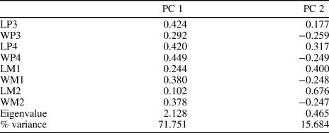
For the principal component analysis (PCA), I used published measurements of arctostylopid specimens compatible in preservation with the upper and lower jaws of Kazachostylops (i.e., having P3–4, M1–2 and p3–4, m1–2; see Supplementary materials 1 and 2 for measurements). The samples used in the analyses include 16 upper dentition specimens representing eight taxa and nine lower dentition specimens representing five taxa. The PCA was performed using the software PAST version 3.01 (Hammer et al., Reference Hammer, Harper and Ryan2001). The principal components (PC) 1 and 2 of the upper dentition analysis explain 87.44% of the variance (Table 2; Fig. 5). The measurements LP3, LP4, WP4, WM1, and WM2 contribute roughly equally to PC1 (Table 2). The greatest contribution to PC2 is from the measurement LM2 (Table 2). In the PC1–2 scatterplot, Kazachostylops is clearly different from all other arctostylopids included in the sample (Fig. 5). It is almost the only taxon with positive values of both PC1 and PC2. The closest taxa to Kazachostylops on this scatterplot are Palaeostylops macrodon Matthew, Granger, and Simpson, Reference Matthew, Granger and Simpson1929 and Migrostylops roboreus Tong and Wang, Reference Tong and Wang2006.
Principal component analysis of arctostylopid specimens based on measurements of P3–4, M1–2 (see SM 1). Abbreviations of taxa: M, Migrostylops; P, Palaeostylops.

Axis loadings and eigenvalue data for PCA of the upper dentition (P3–4, M1–2) of selected arctostylopid taxa.

In the PCA of lower dentition measurements, PCA 1 and 2 explain 97.21% of the variance (Table 3; Fig. 6). The length of m3 has the greatest contribution to PC1 (Table 3). PC2 is most heavily loaded by the length of m2 and m3 (Table 3). On the PC1–2 scatterplot, Kazachostylops is clearly distant from all other arctostylopid taxa included in the sample (Fig. 6). Only Bothriostylops occurs with Kazachostylops in the quarter of negative values of PC1 and positive values of PC2.
Principal component analysis of arctostylopid specimens based on measurements of p3–4, m1–3 (see SM 2). Abbreviation of the taxon: P, Palaeostylops.

Axis loadings for PCA of the lower dentition (p3–4, m1–3) of selected arctostylopid taxa.

The PCA confirms generic separation of Kazachostylops from the other arctostylopid taxa included in the analysis.
Review of arctostylopid taxa
Allostylops periconotus Zheng, Reference Zheng1979
The species is known from the rostral part of the skull (IVPP 5043) from the upper Paleocene Wangwu member of the Chijiang Formation in Jiangxi Province, China (Zheng, Reference Zheng1979).
Anatolostylops dubius Zhai, Reference Zhai1978
The species is based on a single maxillary fragment with M2–3 from the lower Eocene Shisanjianfang Formation in Turpan Basin, Xinjiang, China (Zhai, Reference Zhai1978). The original diagnosis includes such characters as a high crown, long and straight ectoloph, and presence of the fossette (see also Cifelli et al., Reference Cifelli, Schaff and McKenna1989). However, this taxon is unique in having a deep distal groove on M3, between the worn occlusal surface and postcingulum, not present in any other arctostylopid (this groove might also be present on M2, but eliminated by wear; personal observation of the cast of the holotype IVPP 4357).
Anatolostylops zhaii Wang et al., Reference Wang, Meng, Ni and Beard2008
This species is based on jaw fragments and isolated teeth from the lower Eocene (Bumbanian) upper part of the Nomogen Formation in Erlian Basin, Inner Mongolia, China (Wang et al., Reference Wang, Meng, Ni and Beard2008). It was distinguished from the type species, A. dubius, by slightly lower crowns, a complete lingual cingulum, a stronger parastyle fold, a less-elongated ectoloph, and a proportionally smaller M3 relative to M2 (Wang et al., Reference Wang, Meng, Ni and Beard2008). However, the complete lingual cingulum is present also in A. dubius, and other diagnostic characters are vaguely defined. None of the known M3s of A. zhaii has the distal margin intact, so the presence of the unique character of A. dubius—distal groove on M3—cannot be demonstrated for A. zhaii. The upper cheek teeth referred to this species show some intraspecific variation in the development of parastylar fold, development of crenulations on lingual cingulum, and overall size (Wang et al., Reference Wang, Meng, Ni and Beard2008). Undescribed Anatolostylops sp. aff. A. zhaii was cited for an unnamed late Paleocene stratigraphic unit in the Gurbantunggut Desert, Junggar Basin, Xinjiang, China (Ni et al., Reference Ni, Li, Stidham, Li, Lu and Meng2016).
Arctostylops steini Matthew, Reference Matthew1915
The species is known from skull and jaw fragments from several localities in Wyoming, USA: the late Paleocene (late Tiffanian) Silver Coulee beds of the Polecat Bench Formation and the latest Paleocene (Clarkforkian) Willwood Formation in Clark Fork Basin, and unnamed Clarkforkian beds at Togwotee Pass (Matthew, Reference Matthew1915; Jepsen and Woodburne, Reference Jepsen and Woodburne1969; Gingerich and Rose, Reference Gingerich and Rose1977; McKenna, Reference McKenna and Jacobs1980; Rose, Reference Rose1981; Cifelli et al., Reference Cifelli, Schaff and McKenna1989).
Asiostylops spanios Zheng, Reference Zheng1979
The species is known from a rather complete skull and dentary (IVPP V 5042) from the upper Paleocene Lannikong Member of Chijiang Formation in Jiangxi Province, China (Zheng, Reference Zheng1979).
Bothriostylops medius Huang and Zheng, Reference Huang and Zheng2003
The species is known from the upper Paleocene upper part of the Nongshan Formation at Datang, Nanxiong Basin, Guangdong Province, China (Huang and Zheng, Reference Huang and Zheng2003). The species is based on upper and lower jaw fragments from the same individual (IVPP V 13504).
Bothriostylops notios Zheng and Huang, Reference Zheng and Huang1986
It is based on the dentary fragment with p4, m1-3 (IVPP V 7642) from the upper Paleocene Wangwu Member of Chijiang Formation north of Zhulin Hill, Jiangxi Province, China (Zheng and Huang, Reference Zheng and Huang1986). As was already noted (Cifelli et al., Reference Cifelli, Schaff and McKenna1989, p. 22) such characters listed in the diagnosis of Bothriostylops as deep median labial groove and a convex labial wall on lower molars actually do not differentiate this genus from other arctostylopids. Mesiolabial cingulids on lower molars might be diagnostic, but they are poorly recognizable on the published illustrations. The most diagnostic character of this taxon is lobe-like hypoconulid on m3, which is narrower than the rest of the talonid.
Migrostylops roboreus and M. rosella Tong and Wang, Reference Tong and Wang2006
Known from the fragmentary skull, maxillary, and dentary fragments from the lower Eocene Wutu Formation at Wutu coal mine, Shandong Province, China (Tong and Wang, Reference Tong and Wang2006).
Palaeostylops iturus Matthew and Granger, Reference Matthew and Granger1925 and P. macrodon
Both species are usually found together and represented by abundant jaw fragments (the former species is usually more numerous in specimen number). They were recorded from the upper Paleocene Gashato Formation at Bayn Dzak in Mongolia, from the upper Paleocene Zhigden and Naran members of Naran Bulak Formation at Naran Bulak, Tsagan Khushu, and Agui-Dats-Bulak in Nemegt Basin, Mongolia, from the upper Paleocene Nomogen Formation at Nomogen, Bayan Ulan, and Subeng (P. iturus only), Erlian Basin, Inner Mongolia, China (Gradziński et al., Reference Gradziński, Kazmierczak and Lefeld1968; Chow and Qi, Reference Chow and Qi1978; Badamgarav and Reshetov, Reference Badamgarav and Reshetov1985; Meng et al., Reference Meng, Zhai and Wyss1998; Missiaen and Smith, Reference Missiaen and Smith2008; Missiaen et al., Reference Missiaen, Escarguel, Hartenberger and Smith2012). Palaeostylops iturus was also identified in the lower Eocene Shuangtasi Formation in Xuancheng Basin, Anhui Province, China (Huang and Zheng, Reference Huang and Zheng1997).
Palaestylops sp
The third species of Palaeostylops may be represented by AMNH 109521 from the upper Paleocene Nomogen Formation at Bayan Ulan, Erlian Basin, Inner Mongolia, China (Meng et al., Reference Meng, Zhai and Wyss1998). Badamgarav and Reshetov (Reference Badamgarav and Reshetov1985) listed Palaeostylops sp. for the lower Eocene Bumban Member of Naran-Bulak Formation at Tsagan-Khushu, Nemegt Basin, Mongolia.
Sinostylops promissus Tang and Yan, Reference Tang and Yan1976
It is based on a single fragmentary dentary from the upper Paleocene Upper Member of Doumu Formation at Yangxiaowu, Qianshan Basin, Anhui Province, China (Tang and Yan, Reference Tang and Yan1976). Initially, the dentition of the holotype (IVPP 4263) was interpreted as i3, c, p1–4, m1–2 (Tang and Yan, Reference Tang and Yan1976). Cifelli et al. (Reference Cifelli, Schaff and McKenna1989) considered the third from the last tooth on this specimen to be a deciduous tooth because it is remarkably low-crowned compared with preceding and succeeding teeth and the whole dental formula for the specimen was described as i3, c, p1–3, dp4, m1–2. My investigation of the cast of IVPP 4263 revealed that the tooth in question (p4 or dp4) does not differ in height from the adjacent teeth, but is markedly different from the high-crowned last tooth. Moreover, this tooth is not molariform and cannot be p4; it has linearly arranged cusps, as it is typical for the p3 of arctostylopids. Thus, the best interpretation of dental formula in this specimen is i2–3, c, p1–4, m1. The p4 is damaged, and the lingual side of all teeth is destroyed. The specimen IVVP 4263 does not show any diagnostic characters, and ‘Sinostylops promissus’ should be considered a nomen dubium. It is not included in the phylogenetic analysis conducted herein.
Sinostylops progressus Tang and Yan, Reference Tang and Yan1976
The species is based on four dentary and one maxillary fragments (IVPP V 4264) from the lower Eocene Shuangtasi Formation in Xuancheng Basin, Anhui Province, China (Tang and Yan, Reference Tang and Yan1976). Zheng and Huang (Reference Zheng and Huang1986) placed this species within the genus Bothriostylops because of a convex external wall of molars, deep median external groove on molars, rather developed mesiolabial cingulid, and high metaconid. However, mesiolabial cingulids, poorly developed in B. notios (type species of Bothriostylops), are not present in S. progressus. The median external groove (ectoflexid) on molars in B. notios is not much deeper than in other arctostylopids, but in S. progressus it is deep, very narrow, and cleft-like. This condition is not observed in other arctostylopids. Moreover, S. progressus lacks the most diagnostic character of Bothriostylops—the m3 hypoconulid forming a distinct lobe, which is narrower than the rest of m3 talonid. Sinostylops progressus cannot be placed within the genus Bothriostylops. My study of the cast of IVPP 4264.6 revealed that S. progressus possesses a unique modification of the upper molars not seen in any other arctostylopid. The parastylar lobe on M1–2 is not the external fold, as in other arctostylopids, but the internal invagination, forming the groove ribbed by 1–2 ridges. The adjacent metastylar groove on M1 supplements the parastylar groove on M2. This morphology is so unusual that the parastylar groove on M3 of IVPP 4264.6 was mistakenly considered as a damaged area (Cifelli et al., Reference Cifelli, Schaff and McKenna1989, fig. 8F), while all the molars on this specimen are fully preserved. Sinostylops progressus is reassigned here to a new genus, the description of which is provided in Appendix 1.
Huang and Zheng (Reference Huang and Zheng1997) referred to Bothriostylops progressus two dentary and one maxillary fragments from the lower Eocene Shuangtasi Formation in Xuancheng Basin, Anhui Province, China. The lower dentition of these specimens is poorly preserved and the M1 lacks parastylar and metastylar grooves characteristic for S. progressus.
Huang and Chen (Reference Huang and Chen1997) referred to Bothriostylops progressus two dentary fragments of the same individual (IVPP V 12039) from the upper Paleocene Guichi fauna in Tongling Basin, Anhui Province, China.
Two dentary fragments (IVPP V 12665) from the upper Paleocene Tujinshan Formation at Xinzhuang, Anhui Province, China, were also referred to as Bothriostylops progressus (Huang, Reference Huang2003). The dentition of these specimens is poorly preserved.
Stenostylops xiangensis Huang, Zheng, and Ding, Reference Huang, Zheng and Ding2001
The species is represented by an upper dentition (IVPP V 12434) from the lower Eocene Yanmayou Formation at Yanquan, Changtao Basin, Hunan Province, China (Cifelli et al., Reference Cifelli, Schaff and McKenna1989; Huang et al., Reference Huang, Zheng and Ding2001).
Wanostylops youngi Huang and Zheng, Reference Huang and Zheng1997
The species is based on maxillary fragment with M1-2, isolated M3, and dp4 from the lower Eocene Shuangtasi Formation in Xuancheng Basin, Anhui Province, China (Huang and Zheng, Reference Huang and Zheng1997).
Wanostylops promissus Huang, Reference Huang2003
It is based on the maxillary fragment (IVPP V 12666) from the upper Paleocene Tujinshan Formation at Xinzhuang, Anhui Province, China (Huang, Reference Huang2003).
Phylogenetic analysis
For the phylogenetic analysis, I used an original data matrix consisting of 17 taxa and 26 characters (see Appendix 2 and 3 for the description of characters and data matrix). The anagalid Astigale nanxiongensis Zhang and Tong, Reference Zhang and Tong1981 from the early Paleocene of South China was used as an outgroup following the interpretation that this taxon is most similar to the primitive arctostylopid morphotype (Missiaen et al., Reference Missiaen, Escarguel, Hartenberger and Smith2012). Most of the ingroup taxa were scored based on the specimens and casts held in the AMNH collection. For obtaining the tree statistics, consensus tree, and distribution of characters, I used WinClada 1.00.08 (Nixon, Reference Nixon1999). The character-taxon matrix was analyzed using PRAP, parsimony ratchet analysis using PAUP (Müller, Reference Müller2007), and PAUP* 4.0b10 (Swofford, Reference Swofford2002). This analysis produced 323 most parsimonious trees (see Table 4 for the tree statistics). The strict consensus tree of these trees has no resolution, and all ingroup taxa were collapsed to the polytomy. This result is not surprising, taking into account the poor knowledge of the majority of arctostylopid taxa. To increase the tree resolution and recover better phylogenetic signal, I performed a second analysis, including a successive weighting (Farris, Reference Farris1969). Using PAUP, the characters were reweighted by the maximum value of rescaled consistency indices (RC), and a heuristic search with 10,000 random sequence addition replicates, and TBR (tree bisection and reconnection) branch swapping was performed. Eleven and 13 characters got a weight of less than one during first and second reweighting, respectively. Tree statistics stabilized after three successive runs of the reweight analysis (Table 4). The strict consensus of 647 trees obtained in the last analysis is illustrated in Figure 7.
The strict consensus of 647 trees produced by PAUP reweight analysis. Black circles denote unambiguous synapomorphies. Figures at circles are character numbers above and character states below.

Statistics for equal weight (PRAP and PAUP) and reweight (PAUP) analyses.

Phylogeny of Arctostylopidae
Zheng (Reference Zheng1979) divided Arctostylopidae into Asiostylopinae and Arctostylopinae, but did not indicate explicitly which genera belong to each subfamily. From the discussion in that paper, it is evident that he referred Allostylops, Palaeostylops, and Arctostylops to the Arctostylopinae, which is characterized, in particular, by more hypsodont cheek teeth, molariform premolars, presence of pseudohypocone, and entolophid connected to the ectolophid.
Nesov (Reference Nesov1987b) recognized three subfamilies within the Arctostylopidae: Arctostylopinae (Arctostylops and Palaeostylops), Kazachostylopinae (Kazachostylops), and Sinostylopinae (Sinostylops, Allostylops, Asiostylops, and Anatolostylops). He distinguished Kazachostylopinae from Arctostylopinae by long, well-developed paralophids and longer, lower premolars and molars, and from Sinostylopinae by long and transverse entolophid and high paralophid.
Cifelli et al. (Reference Cifelli, Schaff and McKenna1989, fig. 11) provided the first cladogram of the Arctostylopidae based on manually arranged phylogenetic analysis of the distribution of 20 characters among nine taxa. They recognized two lineages. One lineage contains Arctostylops, Palaeostylops (including Gashatostylops), Anatolostylops, and the then undescribed Stenostylops. This lineage is supported by entolophid well developed, ectolophid (=cristid obliqua) attaches labially to rear of trigonid, paraconid rib (=ectocingulid) strong and pillarlike, p4 paraconid shifted labially, and mesiolabial cristid (=paracristid) lost. The other lineage, which includes Sinostylops and Bothriostylops, is supported by ectolophid attaches lingually to rear of trigonid. Asiostylops is the sister taxon to the clade uniting the two lineages. This cladogram was repeated by Cifelli and Schaff (Reference Cifelli, Schaff, Janis, Scott and Jacobs1998, fig. 21.3).
Tong and Wang (Reference Tong and Wang2006) recognized three subfamilies within the Arctostylopidae: Asiostylopinae (Asiostylops), Bothriostylopinae (Bothriostylops, Kazachostylops, Sinostylops, Allostylops, and possibly Wanostylops), and Arctostylopinae (Arctostylops, Migrostylops, Palaeostylops, Anatolostylops, and questionably Stenostylops). Their Bothriostylopinae is a junior subjective synonym of Kazachostylopinae and Sinostylopinae (Nesov, Reference Nesov1987b).
Wang et al. (Reference Wang, Meng, Ni and Beard2008) allied Anatolostylops and Stenostylops based on such upper molar characters as straight and flat ectoloph, enclosed fossette, and M2 distinctly longer than wide. According to these authors, the lower molars of Anatolostylops, Sinostylops, Bothriostylops, and Kazachostylops are similar in having a wide and deep ectoflexid, the ectolophid reaching the metaconid, and an unreduced m3 with a narrow talonid. Wang et al. (Reference Wang, Meng, Ni and Beard2008) considered all these taxa belonging to one group. Wang et al. (Reference Wang, Meng, Ni and Beard2008) consider Arctostylops, Palaeostylops, and Migrostylops representing a different lineage characterized by uniquely specialized dentition, including an extremely reduced trigonid, ectolophid reaching the protoconid, M2 wider than long, and a well-developed pseudohypocone.
The phylogenetic hypothesis presented here does not support dividing the Arctostylopidae in two or more lineages (Fig. 7). Instead, it shows one lineage with several stages of diversification. Asiostylops is the first branching taxon. Such characters as one-rooted P2 (3(0)), M1–3 metaconule present (9(1)), and p4 metaconid lingual to protoconid 14(0) are recovered as autapomorphies for Asiostylops. The more-derived arctostylopids are united into the clade supported by three synapomorphies: P3 longer than wide (4(0)), M1–3 lingual cingulum present (11(1)), and p3 metaconid present (13(1)). This radiation includes the polytomy formed by Allostylops, two species of Bothriostylops, two species of Wanostylops, and the clade of more-derived arctostylopids. Four synapomorphies support the latter clade: high molars crown (1(1)), M1–3 metaconule absent (9(1)), m1–3 entoconid connected with ectolophid by entolophid (20(1)), and m2 wider than m1 and m3 (24(1)). This clade contains polytomy of Enantiostylops, Kazachostylops, Stenostylops, and Arctostylopinae, which is defined here as a node-based taxon including Arctostylops steini, Palaeostylops iturus, their most recent common ancestor, and all descendants. The Arctostylopinae includes Arctostylops, Anatolostylops, Migrostylops, and Palaeostylops, and has three synapomorphies: M1–3 pseudohypocone present (10(1)), m1–3 trigonid mesiodistally reduced (16(1)), and m1–3 ectolophid attached labial on trigonid, ectoflexid absent (19(2)). All five arctostylopid genera that contain two species (Bothriostylops, Wanostylops, Anatolostylops, Migrostylops, and Palaeostylops) are not monophyletic in the resulting cladogram, but they are closely positioned. More specimens and characters are needed to test the monophyly of these genera. According to the performed phylogenetic analysis, Kazachostylops is a sister taxon to the Arctostylopinae.
Acknowledgments
I thank J. Meng and F. Mao for help in the study of the AMNH collection of Paleogene mammals. I thank J. Meng and an anonymous reviewer for reading the paper and providing useful comments. I received support from the Zoological Institute, Russian Academy of Sciences (project АААА-А19-119032590102-7) and Russian Foundation for Basic Research (project 19-04-00057). The work is performed according to the Russian Government Program of Competitive Growth of Kazan Federal University.
Accessibility of supplemental data
Data are available from the Dryad Digital Repository: https://doi.org/10.5061/dryad.k0p2ngf44
Appendix 1. Description of a new genus for ‘Sinostylops’ progressus
Arctostylopidae Schlosser, Reference Schlosser, Broili and Schlosser1923
Enantiostylops new genus
ZooBank: urn:lsid:zoobank.org:act:B5EDE6E5-2AA5-4AFA-8944-59A2A6C154D1
Etymology
From Ancient Greek enantios (ἐνάντιος) ‘opposite,’ allusion on concavity instead of convexity on the upper molar parastylar lobe, and ‘stylops,’ a common suffix for the arctostylopid genera.
Type species
Sinostylops progressus Tang and Yan, Reference Tang and Yan1976 from lower Eocene Shuangtasi Formation in Xuancheng Basin, Anhui Province, China.
Other species
Only type species.
Diagnosis
Differs from all arctostylopid genera by having internal invagination on M1–3 parastyle and narrow, cleft-like ectoflexid on m1–3. Differs from Asiostylops Zheng, Reference Zheng1979 by high crown molars, by M1–3 lingual cingulum present, p3 metaconid present, p4 metaconid distal to protoconid, p4 talonid curved, m1–3 paraconid rib prominent, m1–3 entoconid connected with ectolophid by entolophid, m2 wider than m1 and m3, and m3 hypoconulid as wide as the rest of talonid. Differs from Allostylops Zheng, Reference Zheng1979 by high crown molars and M1–3 pseudohypocone absent. Differs from Bothriostylops Zheng and Huang, Reference Zheng and Huang1986 by high crown molars, M1–3 metaconule absent, M1–3 lingual cingulum present, m1–3 entoconid connected with ectolophid by entolophid, and m2 wider than m1 and m2. Differs from Wanostylops Huang and Zheng, Reference Huang and Zheng1997 by high crown molars and M1–3 metaconule absent. Differs from Kazachostylops Nesov, Reference Nesov1987b by M1–3 lingual cingulum present, m1–3 mesiolabial cristid absent, and m1 entoconid present. Differs from Stenostylops Huang et al., Reference Huang, Zheng and Ding2001 by M1–3 lingual cingulum present. Differs from Anatolostylops Zhai, Reference Zhai1978 by M1–3 pseudohypocone absent, m1–3 labial and lingual cingulids absent, and m2 wider than m1 and m3. Differs from Arctostylops Matthew, Reference Matthew1915 by M1–3 pseudohypocone absent, m1–3 trigonid length about one-third of the crown length, m1–3 ectolophid attached lingual on trigonid, with long ectoflexid, m1–3 labial and lingual cingulids absent, and m3 longer than m2. Differs from Migrostylops Tong and Wang, Reference Tong and Wang2006 by M1–3 pseudohypocone absent, m1–3 trigonid length about one-third of the crown length, m1–3 mesiolabial cristid absent, m1–3 ectolophid attached lingual on trigonid, with long ectoflexid, and m2 wider than m1 and m2. Differs from Palaeostylops Matthew and Granger, Reference Matthew and Granger1925 by M1–3 pseudohypocone absent, m1–3 trigonid length about one-third of the crown length, m1–3 mesiolabial cristid absent, m1–3 ectolophid attached lingual on trigonid, with long ectoflexid, and m3 longer than m2.
Appendix 2. Character list
1. Molars, crown: low (0), or high (1).
2. P1, number of roots: one (0), or two.
3. P2, number of roots: one (0), two (1), or three (2).
4. P3, proportions: longer than wide (0), or wider than long (1).
5. P3–4, parastylar fold: absent (0), or present (1).
6. P4, M1–3, fossette: absent (0), or present (1).
7. M1–3, parastyle: small (0), large (1), or internal invagination (2).
8. M1–3, parastylar fold: absent (0), or present (1).
9. M1–3, metaconule: present (0), or absent (1).
10. M1–3, pseudohypocone: absent (0), or present (1).
11. M1–3, lingual cingulum: absent (0), or present (1).
12. p2, number of roots: one (0), or two (1).
13. p3 metaconid: absent (0), or present (1).
14. p4 metaconid position: lingual to protoconid (0), or distal to protoconid (1).
15. p4 talonid: straight (0), or curved (1).
16. m1–3, trigonid length: trigonid length about one-third of crown length (0), or trigonid mesiodistally reduced (1).
17. m1–3, mesiolabial cristid: absent (0), or present (1).
18. m1–3, paraconid rib: absent (0), or prominent (1).
19. m1–3, ectolophid attachment on trigonid: at the middle of trigonid (0), lingual, with long ectoflexid (1), or labial, ectoflexid absent (2).
20. m1–3, entoconid: separate from ectolophid (0), or connected with ectolophid by entolophid (1).
21. m1–3, labial cingulid: absent (0), or present (1).
22. m1–3, lingual cingulid: absent (0), or present (1).
23. m1 entoconid: present (0), or absent (1).
24. m2 width: as wide as m1 or m3 (0), or wider than m1 and m3 (1).
25. m3 length to m2: longer (0), shorter (1).
26. m3 hypoconulid: forming a distinct lobe, narrower than the rest of talonid (0), or as wide as the rest of talonid (1).
Appendix 3. Data matrix.