Introduction
The use of radiotherapy is mostly appreciated for head and neck (H&N) cancer,Reference Jemal, Siegel and Xu 1 but it also possesses many challenges. Mostly the H&N tumor is in close proximity to critical organs therefore saving these organs from intolerable radiation exposure is a challenge. The accuracy of H&N radiotherapy has been improved with the introduction of intensity-modulated radiotherapy (IMRT).Reference Nutting, James and Kevin 2 IMRT is usually used for H&N cancer but its main drawbacks are the delivery of treatment in a longer time and involvement of a larger number of monitor units (MUs),Reference Purdy 3 , Reference Hall 4 to overcome these limitations volumetric-modulated arc therapy (VMAT) has been introduced. VMAT is the new emerging technology whose supremacy is evidenced for various cancer sites including prostate, H&N, lungs and breast and so on.Reference Zhang, Happersett and Hunt 5 – Reference Studenski, Bar-Ad and Siglin 9 VMAT is an arc-based IMRT having the capability of modulating gantry rotation speed in addition to variable dose rate and changing treatment aperture shapes.Reference Zulkafal, Khan and Ahmad 10 It is the advanced form of IMRT that can deliver the whole treatment within a few arcs with a reduced treatment time.Reference Otto 11 Short treatment time has the benefit of improving patient comfort, reduced intra-fraction motion and enhances overall treatment outcomes. VMAT has the superiority compared to other arc-based techniques in that it can be delivered with a conventional linear accelerator. VMAT optimisation techniques, in that it is the cause of its eminence,Reference Otto 11 that optimise the dose rate, gantry speed and multi leaf collimator (MLC) positions in a single arc until the desired dose distribution is achieved. The optimisation algorithm starts with a limited number of inputs and gradually increases to a large number to give the planned dose profile.Reference Ling, Zhang and Archambault 12 , Reference Verbakel, Cuijpers and Hoffmans 13 Another potential advantage of VMAT is the involvement of a lower number of MUs which significantly lessens the integral dose to the patient thus decreasing the risk of secondary cancer.Reference Ruben, Davis and Evans 14 VMAT is a complex treatment modality and its clinical implementation for H&N cancer requires very accurate acceptance testing, commissioning of delivery and treatment planning system (TPS) and a very comprehensive quality assurance program. Patient-specific quality assurance is a core element of quality assurance procedure, which is emphasised by most of the professional institutions American Association of Physicists in Medicine (AAPM), American College of Radiology (ACR), American Society for Radiation Oncology (ASTRO).Reference Ezzell, Galvin and Low 15 , Reference Hartford, Seung and Larson 16 Patient-specific quality assurance (QA) is a pretreatment validation, which is performed to check the accuracy (feasibility) of the planned treatment. Pretreatment validation is often marked with better radiotherapy results that ensure the delivery of the planned dose to the patient.Reference Rehman, Iqbal and Tailor 17 , Reference Ur Rehman, Isa and Ahmad 18 Various reports and articles of AAPM are published to provide a direction to perform the patient-specific QA. This article has been presented to experimentally verify the dosimetric performance of clinical VMAT through one-dimensional and two-dimensional dosimeters in an Imaging and Radiation Oncology Core Houston (IROC-H) anthropomorphic H&N phantom. The IROC-H phantom is a well known and standard phantom, used for credentialing of institutions.
Materials and Methods
IROC H&N phantom
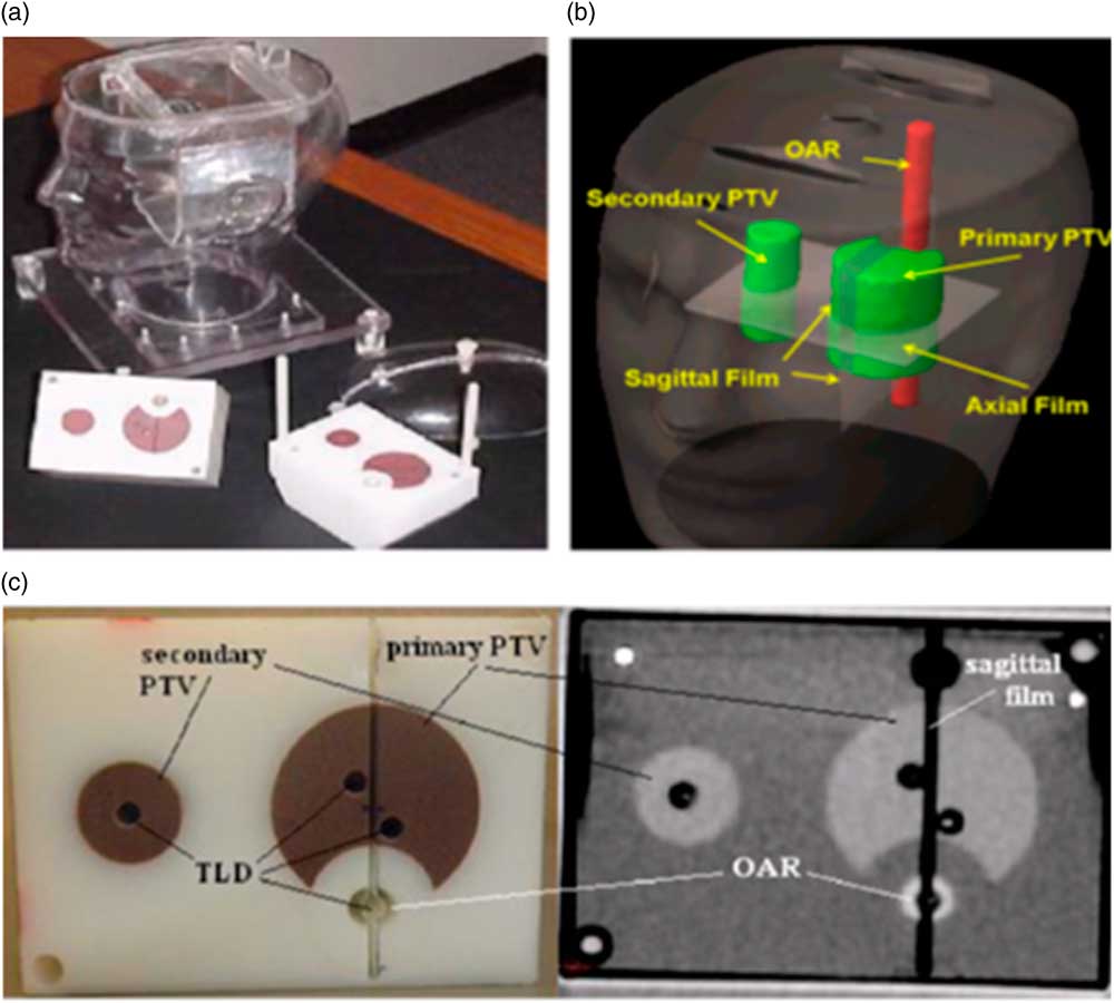
The IROC anthropomorphic H&N phantom, made up of an outer plastic shell with watertight polystyrene insert in it. The plastic shell is filled with water to substitute human tissue and the insert was designed to resemble the oropharynx, related lymph nodes and spinal cord. The oropharynx was decided to be our primary planning target volume (PTV) and secondary planning target volume (STV) was the lymph nodes and spinal cord was determined to be organ at risk (OAR). The plastic insert with its target structures and OAR are imageable being constructed of a material having different density than surrounding water and plastic shell. The insert had slits and holes to hold radio-chromic films and thermoluminescent dosimeters (TLDs).
Accommodation of TLDs and films in phantom
Capsulated TLDs were used to measure delivered point doses and radio-chromic external beam radiotherapy 2 (EBT2) films [GafChromicVR EBT2, International Specialty Products (ISP), NJ] were used for the measurement of planar dose distribution. A total of eight TLDs were placed in insets, four in primary PTV region, two in STV and two in OAR regions. Sheets of EBT2 film were put at the central axial planes of STV and OAR and one EBT2 film piece was placed at the primary PTV in the sagittal plane. The IROC H&N phantom with the insert, TLDs and film are shown in Figure 1.
IROC-H head and neck IMRT phantom (a) and inserts: PTV, STV, OAR, three films (b) and eight TLD capsules. The location of four TLD capsules in the upper part of the insert (c) and another set of four TLD capsules was in the corresponding location in the lower part of the insert; it also shows one axial CT slice of the inserts. Abbreviations: CT, computed tomography; IROC-H, Imaging and Radiation Oncology Core Houston; OAR, organ at risk; PTV, planning target volume; STV, secondary target volume; TLD, thermoluminescent dosimeters.

Treatment planning and delivery
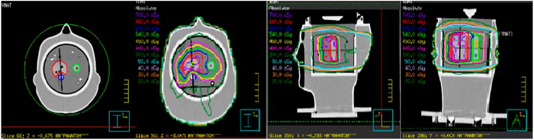
The computed tomography (CT) slice of 1.5-mm thickness of IROC H&N phantom was obtained by Philips CT scanner (Philips Healthcare, Andover, MA). The CT images were imported through Digital Imaging and Communications in Medicine (DICOM) to a pinnacle TPS for segmentation and planning. The contouring of all the structures and TLDs were done manually on TPS and then the procedure of treatment planning started. A dose criteria of 6.6 Gy and 5.4 Gy were prescribed for 95% volume of primary PTV and STV, and maximum dose limit of 4.5 Gy was set for OAR. Four arcs of 182o–178o, 180o–184o, 182o–178o, 180o–184o were planned for VMAT delivery and the plan was optimised by adaptive convolve algorithm until the set goals were achieved. The VMAT plan geometry is shown in Figure 2, where dose distribution for different planes has also been shown. The doses to be delivered were calculated using C.C convolution algorithm. The phantom was laid on the couch of a Varian True beam (Varian Medical Systems, Palo Alto, CA), linear accelerator, to deliver the planned treatment, the delivery of photons was repeated three times for the plan reproducibility.
VMAT plan geometry, dose distribution in the axial, sagittal and coronal view of the phantom containing film from left to right. Abbreviation: VMAT, volumetric-modulated arc therapy.

Dose Measurements and Analysis
Three types of test were performed to see the agreement between the planned and delivered doses to the phantom, given as TLD percent dose difference, isodose line profile and Gamma analysis. TLD-measured doses were compared with the mean-computed dose of TLD calculated from TPS contours, and percentage difference was evaluated between the two. For film analysis, the doses from three tiny pinholes drilled in each film were compared with the planned dose. Pixel resolution of 0.3 mm was set for film study. The film dose was normalised by taking average of corresponding superior and inferior TLD doses. The planar dose distribution was measured from two cross profiles constructed on each film representing a sagittal or axial plans.
Gamma index analysis was performed to find the agreement between the computed dose from TPS and recorded doses from film. The 3% dose difference and 3-mm distance-to-agreement (DTA) criteria were set to analyse the difference between planned and measured doses. Whereas, criteria of 7%/4 mm were used in IROC for the purpose of quality assurance of IMRT and VMAT. Isodose line profiles were also constructed to visualize the agreement between computed and planned dose. All the analysis and graph preparation was performed by Matlab R2008b (The MathWorks, Inc., Natick, MA) software.
Results
TLD result
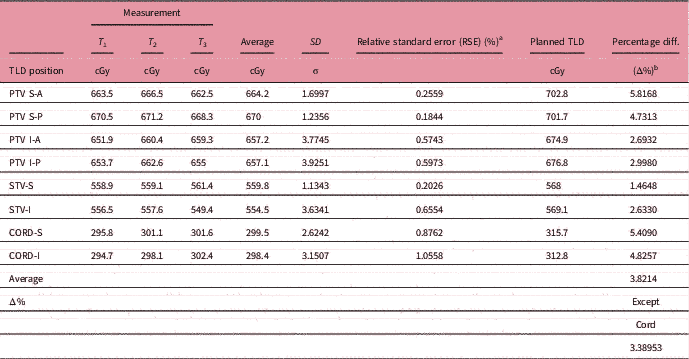
The comparison between the computed and measured point doses for all the eight TLDs is shown in Table 1. In Table 1, the standard deviation (σ) of the three deliveries is calculated which was less than 4% and relative standard error (RSE) was also evaluated that gave the values within 2%. The percentage dose difference evaluation of planned and delivered TLD dose was found to be within 5% in all the eight locations where TLDs were inserted. The average percentage difference of 3.8% was obtained and this gradient reduced to 3.4% when OAR percentage difference was excluded.
The comparison between thermoluminescent dosimeters (TLD) measurements and the dose calculations from treatment planning system (Pinnacle@3)

Note: aRelative standard error=σ/(mean of 3 irradiations)×100.
b(calculated TLD dose-measured TLD dose)/(measured TLD dose)×100.
Abbreviations: PTV, planning target volume; STV, secondary target volume.
Film result
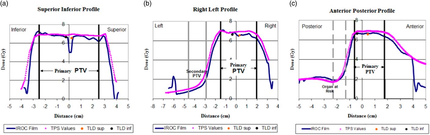
The comparison between pinnacle TPS and EBT2 film in the form of line profiles only for one irradiation, and similar results were observed for the other two deliveries. The line profile comparison of the delivered and planned doses exhibit a good agreement and the slight discrepances which were seen they were also within the tolerance limit of 4 mm, provided by IROC as shown in Figure 3. Maximum disagreements were present at the edges of the line profiles and film doses were bit more rough than the planned TPS dose profile.
Isodose line profile comparison of EBT2 film and pinnacle TPS in inferior–superior, left–right, posterior–anterior for first delivery. Abbreviation: TPS, treatment planning system.

Gamma analysis
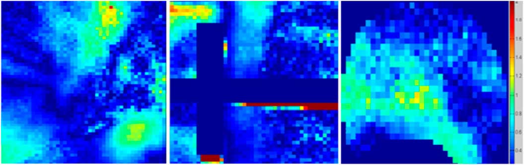
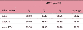
To further authenticate the delivered doses gamma analysis was performed which gives the dose difference and DTA test in a single analysis. Figure 4 shows the gamma analysis performed for one treatment delivery which is representative of all the three irradiations. It was observed that the test passed the criteria of 3%/3 mm used by IROC for the purpose of credentialing of the institutions. The passing percentage for gamma analysis of the three irradiation is given in Table 2.
Gamma map analysis of EBT2 film and TPS in the axial, sagittal and PTV planes for 3%/3 mm criteria. Abbreviations: TPS, treatment planning system; PTV, planning target volume.

Percentage of points passing gamma analysis of 3%/3 mm criteria for all the three deliveries

Abbreviations: PTV, planning target volume; VMAT, volumetric-modulated arc therapy.
Discussion
In this research study, VMAT treatment planning for H&N cancer has been verified using IROC anthropomorphic phantom with one-dimensional and two-dimensional dosimeters. The agreement between the planned and measured doses has been studied through evaluating percent dose difference, constructing isodose line profile and analysing the 2D gamma maps. The IROC passing criteria of 3% dose difference and a DTA of 3 mm is used within PTV regions. The OAR region has been excluded from analysis being very sensitive to error. TLD analysis has given a dose difference of 3.3% without spinal cord and 3.8% with spinal cord, and a DTA of 1.5 mm has been observed between planned and measured isodose line profiles. The gamma analysis has passed the criteria of 3%/3 mm with average passing pixels of more than 98%. The disagreement in all cases are within the acceptable limit, therefore VMAT can be delivered to patient with better target coverage and better organ sparing. Delivering planned VMAT dose to H&N cancer site is a challenging task, except the phantom setup error, limitation of dosimetric tools, contouring challenges, VMAT planning and delivery itself is a complex procedure. The Planning VMAT delivery taking into account the MLCs penumbra and leakage radiation, its tongue and groove effect, linac head scattering are very complicated. Calculating dose for such an uncertain system in addition to considering patient heterogeneities makes VMAT process inaccurate.Reference LoSasso, Chui and Ling 19 In VMAT, many parameters have to be modulated, and significant dosimetric errors have been evidenced due to continuous variation of gantry speed and MLC shape.Reference Song, Zhang and Burman 20 Another problem is a heavy load of treatment machine head which cannot be changed always, therefore sometimes larger standard deviations are observed in treatment deliveries, as it can be seen in this experiment in Table 1 as well. MLC acceleration during irradiation also reduces VMAT accuracy and the greater speed of MLC is mostly accompanied with increasing errors in delivering the planned doses.Reference Park, Wu and Kim 21 Film and TLD’s energy dependence, fading factor, inaccurate film processing and TLD readout methods also reduce the accuracy of measurement of radiation doses.Reference Lynch, Kozelka and Ranade 22 , Reference Hasabelrasoul 23 Our VMAT QA has successfully attained the IROC established criteria despite these all sources of errors, therefore VMAT can be delivered safely to an H&N cancer patient.
Conclusion
VMAT is an advanced modality of delivering external beam radiotherapy with the potential advantage of short treatment time. H&N cancer radiotherapy which is quite challenging can be benefited by VMAT. The dosimetric performance of these complex procedures can be verified using anthropomorphic H&N phantom with TLDs and films.
Acknowledgements
The authors are thankful to the Higher Education Commission (HEC), Pakistan for providing a scholarship for Mr. Jalil ur Rehman.