Device closure is now the standard of care for patients with ostium secundum atrial septal defect with adequate margins. Although infrequent, few patients require surgical intervention to treat late complications associated with the procedure. Reference Abaci, Unlu, Alsancak, Kaya and Sezenoz1 We report a rare case of a young female patient who presented with systemic desaturation and exertional dyspnoea 5 years after device closure of atrial septal defect requiring surgical extraction of the septal occluder.
Case report
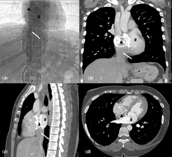
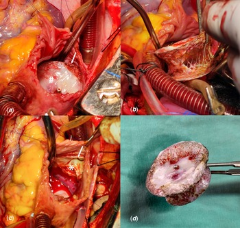
A 36-year female patient presented with exertional dyspnoea and palpitation 5 years after device closure (36 mm Cocoon Septal Occluder, Sanare, Philippines) of ostium secundum atrial septal defect. She was found to have exercise-induced desaturation from 92% at room air to 68% during peak exercise. Transthoracic echocardiography showed an in situ device with residual peri-device shunt (right to left) and turbulence in the inferior vena cava flow. During catheter study to estimate shunt fraction, guide wire could not be passed into the right atrium via the inferior vena cava, however, the contrast was seen streaming into the left atrium (Fig 1A). Room air arterial partial pressure of oxygen was 64 mmHg. Contrast CT scan showed malposition of the device obstructing the inferior vena cava inflow into the right atrium (Fig 1B-1D). The patient was planned for device retrieval and atrial septal defect closure. Intraoperative transesophageal echocardiography was suggestive of malposition of the device with partial obstruction of inferior vena cava and right to left across the occluder (Supplementary Figure 1). Surgery was performed on cardiopulmonary bypass with bicaval cannulation under moderate hypothermia and the heart was arrested with Del Nido cardioplegia. Left atrium vent was placed via the right upper pulmonary vein. Intraoperative assessment showed the right atrial disk of the device adherent to the anterior margin of the inferior vena cava, directing the inferior vena cava blood into the left atrium. Dense adhesions of the device with inferior vena cava margin and fossa ovalis were released using electrocautery and the device was retrieved (Fig 2). The atrial septal defect was closed with an untreated autologous pericardial patch. The patient was weaned off cardiopulmonary bypass and had an uneventful postoperative course.
Preoperative images A: Catheter study showing contrast injected into the IVC is streaming into LA around the region of the ASD device. B-D: CT scan showing the position of the device in relation to the IVC. Triangle – ASD device, hash (#) – left atrium (LA), Asterix (*) – right atrium (RA) and arrow – inferior vena cava (IVC).

Intraoperative images. A: ASD device seen in-situ, with the anteroinferior edge of the device seen adherent to the anterior margin of the IVC opening. B: ASD device being extracted. C: anterior and posterior margins of the IVC after device retrieval. D: Explanted 36 mm ASD device (Cocoon Septal Occluder, Sanare, Philippines) hash (#) – ASD device, Asterix (*) – posterior margin of IVC opening and arrow – anterior margin of IVC opening.

Comment
Introduced by King and Mills in 1976, device closure has evolved to be the preferred treatment for atrial septal defect. Reference King, Thompson, Steiner and Mills2 The peri-procedural major complication rate is 1%, with the need for surgical retrieval of the device seen in 0.7% of patients, the majority of whom had device embolisation. Medium-term follow-up demonstrated that 0.1% of patients required surgical intervention to retrieve the device. Reference Abaci, Unlu, Alsancak, Kaya and Sezenoz1
Device embolisation and malposition warrants surgical device retrival. Reference Berdat, Chatterjee, Pfammatter, Windecker, Meier and Carrel3,Reference Soo, Healy, Walsh and Wood4 In our case, device malposition led to the streaming of inferior vena cava blood into the left atrium. Consequently, there was an effective right to left shunt across the atrial septum and systemic desaturation. The unusual presentation posed a diagnostic challenge, and the exact nature of the problem could not be entirely appreciated on transthoracic echocardiography and catheter study. A combination of CT and transesophageal echocardiography demonstrated anterior malposition of the device impeding the inferior vena cava flow and directing it into the left atrium.
Our case highlights the rare prospect of late systemic desaturation after device closure of atrial septal defect due to device malposition. It emphasises the fact that long-term follow-up and surveillance are required for all patients undergoing transcatheter device closure to detect late device-related complications.
Supplementary material
To view supplementary material for this article, please visit https://doi.org/10.1017/S1047951122003833
Financial support
This research received no specific grant from any funding agency, commercial, or not-for-profit sectors.
Conflicts of interest
None.
Ethical standards
Maintained.