Introduction
In cereal seeds (caryopses), stored starch provides a primary source of carbon and energy for seed germination and seedling growth. The degradation of starch in the endosperm is a major metabolic process following germination, which supplies soluble sugars to growing tissues. Several excellent reviews (e.g. Peng and Harberd, Reference Peng and Harberd2002) have been published on the regulation of starch digestion in the endosperm by gibberellins, which induce the activity of α-amylase in the aleurone layers. The hexoses produced by starch degradation can be taken up by scutellum cells of the embryo. It has been suggested that hexoses are re-synthesized into sucrose in the scutellum, based on changes observed in sucrose content and 14C-labelled sugar tracing (Edelman et al., Reference Edelman, Shibko and Keys1959; Nomura et al., Reference Nomura, Kono and Akazawa1969). Sucrose in the scutellum is loaded into vascular bundles, transported and then unloaded to growing tissues through apoplastic or symplastic pathways (Aoki et al., Reference Aoki, Scofield, Wang, Offler, Patrick and Furbank2006). Sucrose synthesis in the embryo was considered to be a response to growth environment, for example, anaerobic conditions (Guglielminetti et al., Reference Guglielminetti, Loreti, Perata and Alpi1999).
On the other hand, starch is re-synthesized in some tissues of rice, wheat and maize seeds during and following germination. This deposition of starch almost always occurs in tissues that do not have high accumulations of starch during seed maturity (Halmer, Reference Halmer1985; Feng et al., Reference Feng, Dong, Gao, Hu and Wang2005). Starch that is re-synthesized in germinating and germinated seeds is termed as transitory starch because it disappears soon after re-synthesis (Halmer, Reference Halmer1985). However, the mechanisms that regulate starch synthesis in germinating and germinated seeds remain unclear. The objective of this study was to investigate the regulation of starch re-synthesis in rice embryos during germination and seedling growth. To elucidate the regulatory mechanisms of transitory starch accumulation in germinating and germinated rice embryos, the effects of sink strength of growing tissues on transitory starch accumulation in the embryo were investigated. The expression of genes and enzymes responsible for starch biosynthesis and metabolism in the embryo under different sink demand conditions was also analysed.
Materials and methods
Seed germination and plant growth
Rice (Oryza sativa L. cv. Tainung 67) seeds were sterilized with 1% (v/v) sodium hypochlorite containing approximately 0.1% (v/v) Tween 20 for 15 min and washed with distilled water three times. Seeds were then germinated at 37°C in the dark for 3 d and moved to the phytotron for growth at 30/25°C (12/12 h) under natural light.
Embryo and seedling treatments
Experiment 1
To investigate the effects of exogenous sugar on starch accumulation in the embryo, rice seeds were imbibed in water at 37°C for 3 d in the dark and the embryos were dissected and treated with water, 100 mM glucose or 100 mM mannitol for 3 d at 30/25°C before starch accumulation in embryonic tissues was analysed.
Experiment 2
To investigate the effects of shoots on starch metabolism in other tissues, shoots were removed from the seedlings 6 d after imbibition (DAI). The shoot-less seedlings were maintained continuously under the same conditions. Seedlings were collected 14 and 22 DAI and cut into halves for analysis of starch distribution.
Experiment 3
To investigate the effects of reduced photosynthetic efficiency on starch accumulation in embryonic tissues, seedlings were cultured under the phytotron conditions until 17 DAI and then moved to dark conditions. The seedlings were harvested 19, 21 and 23 DAI and cut into halves for starch analysis by iodine staining.
Localization of starch
For localization of starch, seedlings or isolated embryos were cut into halves and soaked in an iodine–potassium iodine solution (0.05% KI, 0.005% I2, 0.01 N HCl) for 1 min, washed with water and analysed under a stereoscopic microscope.
Quantification of carbohydrates
For carbohydrate determination, ten embryos were ground to powder in liquid nitrogen, and then extracted with 1 ml of 80% (v/v) ethanol at 80°C for 5 min before centrifugation at 3000 g. The pellet was analysed for starch content according to the procedure described by Smith and Zeeman (Reference Smith and Zeeman2006). The supernatant was analysed for glucose, fructose and sucrose following the method described by Spackman and Cobb (Reference Spackman and Cobb2002).
Enzyme assays
Proteins were prepared using the method described by Nakamura et al. (Reference Nakamura, Yuki, Park and Ohya1989) with modification. Briefly, 50 embryos were ground to powder in liquid nitrogen with a mortar and pestle. The powder was mixed with 0.5 ml of extraction buffer [100 mM tricine-NaOH, pH 8.0, 2 mM EDTA, 8 mM MgCl2, 12.5% (v/v) glycerol, 5% (w/v) polyvinylpolypyrrolidone-40 and 50 mM 2-mercaptoethanol]. After centrifugation at 10,000 g for 5 min at 4°C, the pellet including the starch granules was assayed for granule-bound starch synthase (GBSS) activity. Soluble proteins in the supernatant were assayed for ADP-glucose pyrophosphorylase (AGPase), soluble starch synthase (SSS) and starch branching enzyme (SBE) activities.
AGPase activity was assayed with the method described by Nakamura et al. (Reference Nakamura, Yuki, Park and Ohya1989). Briefly, a crude extract of soluble proteins (50 μl) was mixed with 150 μl of reaction buffer [133 mM HEPES–NaOH, pH 7.4, 6 mM MgCl2, 1.4 mM ADP-glucose, 4 mM sodium pyrophosphate and 5 mM dithiothreitol (DTT)] and incubated at 30°C for 60 min. Then the mixture was boiled to inactivate enzymes. After centrifugation, 150 μl of supernatant was mixed with 40 μl of 4 mM NADP+, to which 0.4 U phosphoglucomutase and 0.35 U glucose-6-phosphate dehydrogenase were added before the absorbance at OD340 was measured. Data are presented as the rate of glucose-1-phosphate (G1P) production.
The activity of SSS was assayed using the method described by Nishi et al. (Reference Nishi, Nakamura, Tanaka and Satoh2001). Briefly, a crude extract of proteins (50 μl) was first incubated with 50 μl of reaction solution 1 [50 mM HEPES-NaOH, pH 7.4, 1.6 mM ADP-glucose, 0.23% (w/v) glycogen and 16.7 mM DTT] at 30°C for 60 min. Samples were then boiled to inactivate enzymes. The mixture was supplemented with 25 μl of reaction solution 2 (50 mM HEPES-NaOH, pH 7.4, 10 mM phosphocreatine, 200 mM KCl and 2 U creatine phosphokinase). After incubation at 30°C for 30 min, the sample was boiled. Then, samples (90 μl) were mixed with 135 μl of reaction solution 3 (50 mM HEPES-NaOH, pH 7.4, 20 mM MgCl2, 10 mM glucose, 2 mM NADP+ and 0.4 U glucose-6-phosphate dehydrogenase), and the absorbance at OD340 was determined and used as the blank. The increase in the absorbance at OD340 was measured after hexokinase was added.
The assay for GBSS activity was similar to that for SSS, except that GBSS, which contains starch granules, was used for the reaction primer in solution 1 instead of the glycogen primer. The SBE activity was measured with the iodine-stain method described by Guan and Preiss (Reference Guan and Preiss1993). Extraction of α-amylase from rice embryos and activity assays were carried out according to the instructions provided in the CERALPHA kit (Megazyme International Ireland Ltd, Wicklow, Ireland).
For all enzyme activity assays, three independent experiments were performed and the data are presented as mean ± SE.
Quantitative real-time reverse transcription-polymerase chain reaction
RNA extracted from the embryos was treated with DNase to remove contaminating genomic DNA. Then, DNA-free total RNA (200 ng) was used as a template for quantitative real-time reverse transcription-polymerase chain reaction (RT-PCR) analyses with the Brilliant SYBR Green QRT-PCR Master Mix (Stratagene, La Jolla, California, USA). PCR reactions were performed with a Multiplex 3000P Real-Time PCR System (Stratagene). The gene-specific RT-PCR primers for GBSSI were 5′-GCGAAGAACTGGGAGAATG-3′ and 5′-CACTACAACAAACAAACCACTG-3′; the primers for GBSSII were 5′-AACTGCATGGCTCAAGACCT-3′ and 5′-ACAGGCAAATGCATGCCATC-3′; the primers for SBEI were 5′-GCCCTAACTCATTCAAAGTCC-3′ and 5′-TCTCCCCACTTGTTGCTTC-3′; the primers for SBEIII were 5′-TGAGAACACCAAGAGGCAG-3′ and 5′-CGCAAAGCACAAAAGCAAG-3′; and the primers for SBEIV were 5′-TTGAGGAGGAGGAAGAGATT-3′ and 5′-TGGAGCCTGCACAGCAAAC-3′. To quantify the relative expression levels of these genes, the C T value of the target gene was normalized to the C T value of the ubiquitin (ubi) gene. The PCR primers for the ubi gene were 5′-CGCAAGTACAACCAGGACAA-3′ and 5′-TGGTTGCTGTGACCACACTT-3′. For all real-time RT-PCR analyses, three independent experiments were carried out, and the data are presented as the mean ± SE.
Results and discussion
Changes in starch content and distribution in rice seeds during germination and seedling growth
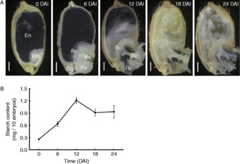
To investigate the metabolism and distribution of carbohydrate reserves among different tissues of rice seeds during and following germination, starch was visualized with iodine staining. Starch stored in the endosperm was initially degraded near the scutellum. Starch degradation gradually spread to the entire endosperm during seed germination and seedling growth, and continued until it completely disappeared from the endosperm around 18 DAI (Fig. 1A). Starch re-synthesis was observed in embryonic tissues during seed germination and seedling establishment (Fig. 1A and B). Starch content in the scutellum increased by 6 DAI (Fig. 1A) and decreased during the next few days. Later, starch was re-synthesized in cells surrounding the vascular tissues in the embryonic axis (Fig. 1A; 12–24 DAI). The quantitative analysis showed that starch accumulation in the whole embryo peaked around 12 DAI (Fig. 1B), which was consistent with the changes observed with iodine staining (Fig. 1A).
Changes in starch accumulation in germinating and germinated rice seeds. (A) Rice seeds were collected from 0 to 24 DAI at 6-d intervals, the growing shoots and roots were dissected and discarded, and starch was visualized by iodine staining (purple/blue colour; see online for a colour version of this figure). En, endosperm; Em, embryo; Sc, scutellum; Sv, shoot vascular bundle; Rv, root vascular bundle. Scale bars indicate 1 mm. (B) Starch content in the embryo of germinating and germinated rice seed. The embryos were dissected from seeds at the indicated times, the shoots and roots were dissected and discarded, and starch content of the rest of embryo was determined.

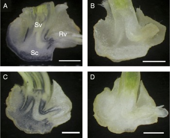
Several studies have shown that dry embryos contain soluble sugars, particularly sucrose (Guglielminetti et al., Reference Guglielminetti, Loreti, Perata and Alpi1999; Matsukura et al., Reference Matsukura, Saitoh, Hirose, Ohsugi, Perata and Yamaguchi2000). We investigated whether the carbon source for starch synthesis in the embryo was already stored as soluble sugars in the embryo or was imported from the endosperm following the degradation. The endosperm was removed from seeds 3 DAI and the isolated embryo was continuously cultured in water for 3 d. In contrast to the embryo in intact seeds (Fig. 2A), there was no detectable starch in the embryo that was separated from the endosperm and germinated in water (Fig. 2B). However, starch accumulation was recovered when 3-DAI isolated embryos were cultured in glucose solution (Fig. 2C). Starch accumulation was not observed in 3-DAI embryos incubated in mannitol solution (Fig. 2D), suggesting that starch accumulation in the embryo induced by exogenous glucose was not due to its osmotic effect. These results suggested that the carbon source for starch synthesis in the embryo was provided primarily from the endosperm.
Starch accumulation in isolated embryos induced by exogenous glucose. Starch distribution was determined with iodine staining in (A) the embryos of 6-DAI seed and (B–D) embryos isolated from 3-DAI seeds and incubated for three more days in (B) water, (C) 100 mM glucose or (D) 100 mM mannitol. Sc, scutellum; Sv, shoot vascular bundle; Rv, root vascular bundle. Scale bars indicate 1 mm. (See online for a colour version of this figure.)

Effects of the demand of growing embryonic tissues on the regulation of transitory starch accumulation
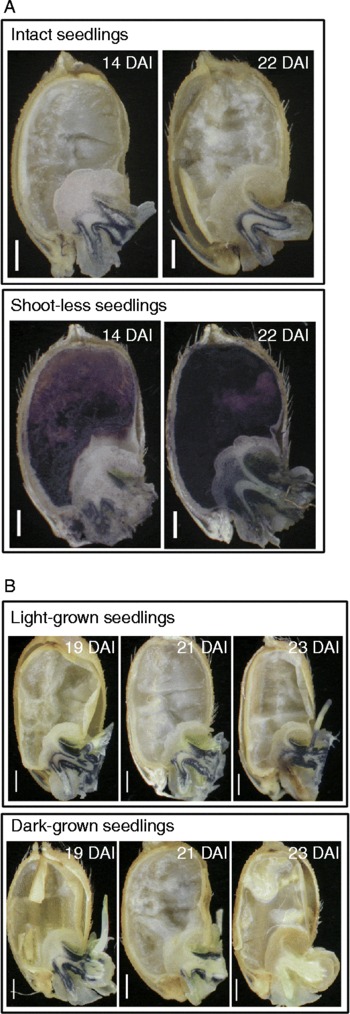
During seed germination and early seedling growth, the endosperm functions as a carbon source and the growing shoot functions as a sink tissue. The pattern of starch allocation shown in Fig. 1 suggested that at least part of the embryo played the role of a transitory carbon storage tissue during early germination and seedling establishment, which in turn became a carbon source tissue later. However, those experiments did not provide information about whether sink–source interaction influenced starch metabolism in the endosperm and embryo during germination and seedling growth. To study this, the shoots of 6-DAI seedlings were removed to reduce sink strength. Then changes in starch content of the source tissues (endosperm and embryo) were evaluated 14 and 22 DAI. Starch reserved in the endosperm of intact seedlings was completely hydrolysed by 14 DAI (Fig. 3A, upper panel). In contrast, starch remained undigested in the endosperm of seedlings that lacked shoots 14 DAI (Fig. 3A, lower panel). Furthermore, starch accumulation in the embryo was more enhanced in the shoot-less seedlings than in intact seedlings (Fig. 3A).
Effect of sink strength of growing tissues on starch accumulation in the embryo. (A) Changes in starch distribution in intact and shoot-less seedlings. Upper panel: starch accumulation in seed tissues of intact seedlings. Lower panel: starch accumulation in shoot-less seedlings. Shoots were removed 6 DAI and the rest of the seed was incubated further. Seedlings were harvested 14 and 22 DAI and cut into halves for iodine staining (purple/blue colour; see online for a colour version of this figure). Scale bars indicate 1 mm. (B) Effect of dark treatment on starch accumulation in the embryo. Upper panel: changes of starch content in seedlings incubated under natural light 19, 21 and 23 DAI. The starch distribution was determined with iodine staining (purple/blue colour; see online for a colour version of this figure). Lower panel: starch distribution in dark-grown seedlings. The seedlings were transferred to dark conditions 17 DAI, grown and collected after 2 d (19 DAI), 4 d (21 DAI) and 6 d (23 DAI). Scale bars indicate 1 mm.

In other experiments, three-leaf-stage seedlings (17 DAI) were transferred to dark conditions to reduce the photosynthetic efficiency, and changes in starch accumulation in the embryo were assessed every 2 d. Starch content of the embryo of dark-treated seedlings was lower than that in the embryo of light-grown seedlings (Fig. 3B). Spyropoulos and Lambiris (Reference Spyropoulos and Lambiris1980) have reported the effects of environmental stresses on carbohydrate metabolism in germinating legume seeds. Other studies have demonstrated that a buffer comprised of transitory carbohydrate reserves played a role in supporting young growing sink tissue. For example, the leaf sheaths of the upper leaves in rice plants accumulated abundant starch before panicle heading. Starch accumulated in the leaf sheaths was rapidly degraded after heading and provided sugars, which were transported to developing grains as a carbon source (Yoshida, Reference Yoshida1972; Yang et al., Reference Yang, Zhang, Wang, Xu and Zhu2004; Chen and Wang, Reference Chen and Wang2008).
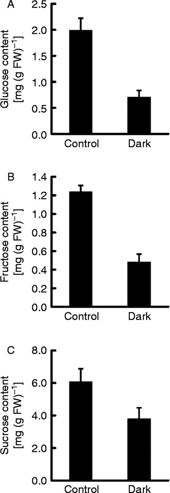
To determine whether a decrease in soluble sugars had contributed to the low starch accumulation in embryonic tissues of dark-grown rice seedlings, soluble carbohydrate content was determined 4 d after dark treatments (21 DAI). The data showed that glucose, fructose and sucrose levels were reduced in the dark-grown embryos (Fig. 4). These results suggested that reserved soluble sugars in the embryo could serve as a carbon source to support shoot growth. Thus, when the environment is unfavourable for photosynthesis, the carbohydrates in the embryo can be transported to growing tissues instead of remaining in the embryo for starch biosynthesis.
Effect of dark treatment on soluble sugar content in embryonic tissues of rice seedlings. Seedlings were transferred to dark conditions 17 DAI, and the levels of (A) glucose, (B) fructose and (C) sucrose in the embryo were determined after 4 d (21 DAI). Control seedlings were grown under natural light conditions and their embryos were collected 21 DAI. The data represent mean values ± SE (n = 3).

Taken together, the results from the shoot-removal and dark-treatment experiments suggested that carbohydrate metabolism in the endosperm and starch biosynthesis in the embryo are regulated by the demands of the sink. Starch re-synthesis in the embryo might be a mechanism to control the osmotic status of embryonic cells.
Metabolic enzyme activities and expression of starch biosynthesis genes in the embryos of dark-grown seedlings
Starch is a glucan-polymer complex that consists of amylose and amylopectin. Amylose is an α-1,4-linked linear glucan polymer. Amylopectin is a highly branched glucan polymer with its branch points linked by α-1,6 glycosidic bonds. In the starch biosynthetic pathway, AGPase is the key enzyme for converting ATP and glucose-1-phosphate into ADP-glucose, a substrate for starch biosynthesis (Tsai and Nelson, Reference Tsai and Nelson1966; Preiss, Reference Preiss and Preiss1988). GBSS and SSS perform glucose chain elongation in the synthesis of amylose and amylopectin, respectively (Tsai, Reference Tsai1973, Reference Tsai1974; Visser et al., Reference Visser, Somhorst, Kuipers, Ruys, Feenstra and Jacobsen1991; Craig et al., Reference Craig, Lloyd, Tomlinson, Barber, Edwards, Wang, Martin, Hedley and Smith1998; Gao et al., Reference Gao, Wanat, Stinard, James and Myers1998). SBE plays a role in catalysing the formation of α-1,6-linkages (Smith et al., Reference Smith, Denyer and Martin1997; Nishi et al., Reference Nishi, Nakamura, Tanaka and Satoh2001; Nakamura, Reference Nakamura2002). In germinated cereal seeds, α-amylase plays an important role in hydrolysing starch (Beck and Ziegler, Reference Beck and Ziegler1989). We determined whether the reduction of starch content in the embryos of dark-grown seedlings was due to a decrease in starch biosynthesis or an increase in starch degradation.
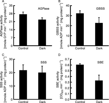
The seedlings were transferred to dark conditions 17 DAI, and the activities of enzymes related to starch biosynthesis and degradation in the embryo were assayed 21 DAI. Seedlings grown under light conditions were compared to those grown under dark conditions. The activities of AGPase and SSS were not different in seedlings grown in dark and light conditions (Fig. 5A and C). However, GBSS and SBE activities were reduced 1.6-fold and 1.5-fold, respectively, in the dark-grown samples compared to those grown under light conditions (Fig. 5B and D). Both GBSS and SBE are important factors for determining starch content and structure (reviewed by Smith et al., Reference Smith, Denyer and Martin1997). Thus, down-regulation of GBSS and SBE activities could be a mechanism for reducing starch accumulation in the embryos of dark-grown seedlings.
Effect of dark treatment on activities of starch biosynthesis enzymes in the embryos of rice seedlings. Seedlings were transferred to dark conditions 17 DAI, and the activities of AGPase, GBSS, SSS and SBE in the embryos were determined after 4 d. Control seedlings were grown under natural light conditions, and their embryos were collected 21 DAI. The data represent mean values ± SE (n = 3).

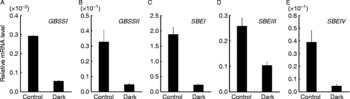
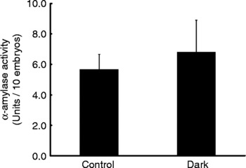
There are two isoforms of GBSS, GBSSI and GBSSII. RT-PCR results indicated that the accumulation of GBSSI and II transcripts were significantly reduced in the embryos of seedlings grown for 4 d in the dark, compared to those grown under normal conditions (Fig. 6A and B). The expression of the three SBE genes, SBEI, III and IV, were also reduced in seedlings grown in the dark compared to the expression of these genes under normal conditions (Fig. 6C, D and E). In addition, we evaluated the activity of α-amylase, the key enzyme for starch degradation. Small differences were observed in α-amylase activity in light- and dark-grown seedlings (Fig. 7). Several studies using different plant species have shown that gene expression and enzyme activities of starch synthesis-related enzymes were regulated by light (Guan and Janes, Reference Guan and Janes1991; Bae and Liu, Reference Bae and Liu1997). Other studies have demonstrated that sugars influenced starch synthesis-related gene expression in various plant tissues (Wang et al., Reference Wang, Yeh and Tsai2001; Dian et al., Reference Dian, Jiang, Chen, Liu and Wu2003; Akihiro et al., Reference Akihiro, Mizuno and Fujimura2005; Baguma et al., Reference Baguma, Sun, Borén, Olsson, Rosenqvist, Mutisya, Rubaihayo and Jansson2008). In the present study, glucose, fructose and sucrose were reduced in embryonic tissues of dark-grown seedlings compared to their levels in seedlings grown under light conditions (Fig. 4). However, it remains to be determined whether the lack of light or low sugar content caused the down-regulation of GBSS and SBE expression in embryonic tissues of the dark-grown seedlings (Fig. 6).
Effect of dark treatment on the expression of starch biosynthesis genes in the embryos of rice seedlings. The seedlings were transferred to dark conditions 17 DAI, and the transcript levels of GBSSI, GBSSII, SBEI, SBEIII and SBEIV in the embryos were determined after 4 d. Control seedlings were grown under natural light, and the embryos were collected 21 DAI. The data represent mean values ± SE (n = 3).

Effect of dark treatment on α-amylase activity in the embryos of rice seedlings. The seedlings were transferred to dark conditions 17 DAI, and α-amylase activity in the embryos was determined after 4 d. Control seedlings were grown under natural light conditions and embryonic tissues were collected from seedlings 21 DAI. The data represent mean values ± SE (n = 3).

In conclusion, the re-synthesis of starch was observed in rice embryos during seed germination and seedling growth. Transitory starch accumulation and metabolism in rice embryos were closely related to the sink strength of growing tissues, such as seedling leaves.
Acknowledgements
We thank Dr Chih-Wen Wu at Kaohsiung District Agricultural Research and Extension Station in Taiwan for providing rice seeds.