The Scope of the Problem: Adolescent Suicide Attempt
More than 36,000 people in the United States die from suicide annually, and suicide is the third leading cause of death in adolescence, with 4% of adolescents aged 10–18 attempting suicide. 1 Suicide attempt thus remains a leading cause of adolescent morbidity and mortality. 1 , Reference Lubell, Kegler, Crosby and Karch 2 A suicide attempt is defined as self-injurious behavior, associated with at least some intent to die as a result of the act.Reference Posner, Oquendo, Gould, Stanley and Davies 3 Despite identification of risk factors and protective factors for suicidal behavior,Reference Bridge, Goldstein and Brent 4 we have limited understanding of the mechanisms underlying risk for a suicide attempt. The prevailing framework guiding this exploration is the stress-diathesis model, wherein certain individuals possess traits that render them vulnerable to suicidal behavior in reaction to stressors which would not induce suicidal behavior in the general population.Reference Van Heeringen 5 In particular, differences in cognitive rigidity, decision making, impulse control, and poor emotional regulation have been implicated. Adolescence is a time of high risk for suicidal behavior, as well as a time that intervention and treatment may have the greatest impact because of structural brain changes and significant psychosocial development during this period.Reference Maalouf and Brent 6 Functional magnetic resonance imaging (fMRI) studies show promise to yield markers of risk for suicidal behavior in adolescents because they can help identify neurobiological underpinnings of pathophysiologic mechanisms that are not observable at the behavioral level, and can also provide targets for future neurobiological interventions. Markers of risk for suicidal behavior are beginning to be elucidated, but as yet have not been applied to the clinical management of adolescents at risk for suicide.
Structural Neuroimaging Studies of Suicide Attempt
Structural neuroimaging studies in adults who have attempted suicide suggest abnormally decreased gray matter volume in cortical regions. One study reported significantly decreased gray matter volume in the fronto-striatal-limbic network in adults who attempted suicide versus healthy controls, and diminished rostral anterior cingulate volume compared with nonsuicidal individuals with major depressive disorder (MDD).Reference Wagner, Koch, Schachtzabel, Schultz, Sauer and Schlösser 7 Adult females who attempted suicide showed decreased gray matter volume in the bilateral orbitofrontal cortex and greater right amygdala volumes than non-attempters.Reference Monkul, Hatch and Nicoletti 8 Furthermore, a postmortem study of suicide victims found diminished right parahippocampal cortex volume relative to healthy controls.Reference Altshuler, Casanova, Goldberg and Kleinman 9 A meta-analysis of 12 studies in adults with a history of suicide attempt revealed that a history of suicidal behavior is interrelated with structural and functional deficits in brain areas that contribute to decision making, defined as “reduced motivational control over the intentional behavioral reaction to salient negative stimuli.”Reference Van Heeringen, Bijttebier, Desmyter, Vervaet and Baeken 10 While studies in nonsuicidal adolescents with MDD report abnormally decreased volumes of the prefrontal cortex, whole brain,Reference Steingard, Renshaw and Hennen 11 hippocampus,Reference MacMaster and Kusumakar 12 and amygdalaReference Rosso, Cintron, Steingard, Renshaw, Young and Yurgelun-Todd 13 , there are few structural neuroimaging studies of adolescent attempters.
We recently showed that adolescents (ages 14–17) with depression and history of suicide attempt had smaller right superior temporal gyrus volumes than healthy controls.Reference Pan, Ramos, Segreti, Phillips and Brent 14 The right superior temporal gyrus is involved in attention to emotion, spatial perception and exploration, and face processing. Given our previous findings of salience neural network abnormalities in adolescent attempters during processing of socially relevant stimuli, angry faces,Reference Pan, Batezati-Alves and Almeida 15 , Reference Pan and Phillips 16 the finding of abnormally decreased right superior temporal gyrus volume may be a structural neural marker of social-emotional information evaluation abnormalities in adolescent attempters.
Structural studies in adolescent suicide attempters have also revealed relationships between white matter intensities and suicidal behavior. A study of 102 adolescent and child (14.6±3.4 years) inpatients with a history of depression with or without a history of suicide attempt revealed that adolescents with a history of an attempt had a greater total number of white matter hyper-intensitiesReference Ehrlich, Noam, Lyoo, Kwon, Clark and Renshaw 17 and more parietal concentration of white matter hyper-intensities.Reference Ehrlich, Noam, Lyoo, Kwon, Clark and Renshaw 18 However, it was impossible to determine if these white matter intensities preceded or followed the attempt.
Functional Neuroimaging Studies of Suicide Attempt
Functional neuroimaging studies of adult suicide attempters indicate neural circuitry abnormalities. One study in adult suicide attemptersReference Oquendo, Placidi and Malone 19 reported lower glucose uptake in the prefrontal cortex and dorsal anterior cingulate gyrus (dACG) in high versus low lethality suicide attempters. Lethality was scored using the Beck Medical Lethality Scale based on the degree of medical damage caused by the attempter’s most lethal attempt; high-lethality attempters required hospitalization for treatment of sequelae related to the suicide attempt.Reference Beck, Beck and Kovacs 20 With regard to emotion processing, vulnerability to suicidal behavior has been associated with differences in response to negative emotion. Specifically, adult males with a history of suicide attempt showed greater activity in the right lateral orbitofrontal cortex and decreased activity in the right superior frontal gyrus to 100% intensity angry versus neutral faces relative to healthy and depressed non-attempter controls. In addition, cognitive inflexibility (inability to change strategy) is well documented in studies of adult patients with a history of suicide attempt (especially high lethality).Reference Jollant, Bellivier and Leboyer 21 , Reference Keilp, Sackeim, Brodsky, Oquendo, Malone and Mann 22
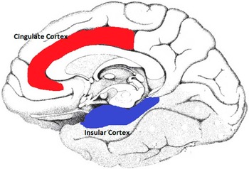
There are few functional neuroimaging studies exploring the neural circuitry underlying adolescent suicidal behavior. This is an important oversight because suicide is one of the leading causes of death in adolescence, and the developing brain may provide a window into risk for suicidal behavior and allow for earlier intervention to prevent suicide. Our preliminary functional neuroimaging studies indicate differences in emotion processing and cognitive control of emotion neural circuitry in adolescents (ages 14–17) with a history of depression and suicide attempt relative to healthy control adolescents.Reference Pan, Hassel, Segreti, Nau, Brent and Phillips 23 Here, we have shown differences particularly in the salience networkReference Ham, Leff, de Boissezon, Joffe and Sharp 24 (Figure 1).
The salience network. Bilateral dACG and insulae are identified nodes in the salience network, which are implicated in sensory processing, modulation of attention to emotion, and fluid reasoning capacity (the ability to solve problems logically in novel situations).Reference Ham, Leff, de Boissezon, Joffe and Sharp 24

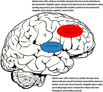
Specifically, we have shown increased attentional control network activity and decreased functional connectivity between the dACG and the insular cortex when viewing angry faces, but not happy faces, in adolescents with a history of depression and suicide attempt compared with healthy controls and adolescents with a history of depression but no history of suicide attempt (Figure 2). The dACG is implicated in attentional and emotional control, including attentional control of emotion. The insular cortex is a neural region associated with interoceptive processing of emotion. This may indicate inefficient recruitment of attentional control neural circuitry when regulating attention to mild intensity angry faces. In contrast, adolescents with a history of depression and suicide attempt showed no abnormalities in levels of performance accuracy or dACG activation in tasks of attentional control compared with healthy controls. These findings may indicate greater attention to negative emotional stimuli in adolescents with a history of suicide attempt and depression than healthy controls, despite normal recruitment of attentional control circuitry during non-emotional attentional control tasks. In summary, we have found that while adolescents with a history of attempt showed normal cognitive performance, they showed the greatest abnormalities when processing angry emotion. Taken together, these data indicate rapid and fixed responses to cognitive problems and impaired emotion processing in adolescents with a history of suicide attempt.
Summary of described functional neuroimaging abnormalities in adolescents with a history of suicide attempt.

Additionally, there are limited neuroimaging studies exploring circuitry in adolescents engaging in nonsuicidal self-injury (NSSI). NSSI includes self-injurious behaviors, such as self-cutting, burning, or biting, that cause direct and deliberate harm to oneself without lethal intent. Prior NSSI is one of the strongest predictors of future suicide attempts.Reference Hamza, Stewart and Willoughby 25 One pilot study examined emotional processing in female adolescents (ages 14–18) with a history of NSSI using fMRI.Reference Plener, Bubalo, Fladung, Ludolph and Lulé 26 The subjects were shown pictures with self-injurious reference. Adolescents with NSSI rated the pictures significantly more arousing than healthy controls without a history of NSSI. On imaging, adolescents with NSSI showed increased activity in the amygdala and in the ACG, along with increased activity in inferior and middle orbitofrontal cortex bilaterally when shown such images.
Future Directions
The structural and functional neuroimaging studies reviewed in this article indicate abnormalities in neural circuitry underlying emotional processing and emotion regulation in adolescent suicidal behavior. Functional neuroimaging to identify biomarkers of risk and elucidate neural circuitry underlying psychopathology is a relatively young field. There is a paucity of studies in functional neuroimaging in adolescent suicidal behavior. The work that has been done indicates differences in adolescents with a history of suicide attempt compared with adult suicide attempters. The application of these techniques to adolescents has focused primarily on diagnostic categories. The identification of potential markers of risk in adolescents who have attempted suicide is a first step toward understanding both which adolescents will progress to suicide attempt and the pathophysiology underlying suicidal behavior. Future studies building on these findings may aid in the clinical identification of those at risk for suicide, create a platform for longitudinal study of suicidal behavior, and allow for future intervention development.
Disclosures
Petra Martin and Thomas Zimmer have nothing to disclose. Lisa Pan has the following disclosures: NIMH/NICHD, K23 MH0828844, Grant; Klingenstein Third Generation Foundation Award, Grant; American Foundation for Suicide Prevention Young Investigator Award, Grant.