Introduction
Fasciola hepatica, a well-known zoonotic trematode, is the causative agent of fascioliasis in both humans and animals, with a worldwide distribution which causes severe complications in the liver and biliary tract in humans (Cwiklinski et al., Reference Cwiklinski, Allen, LaCourse, Williams, Paterson and Hodgkinson2015; Machicado et al., Reference Machicado, Machicado, Maco, Terashima and Marcos2016; Nazar et al., Reference Nazar, Azmoudeh-Ardalan, Mobedi, Dashti, Salahshour and Ahmadinejad2017). According to the World Health Organization, several millions of people are at risk of fascioliasis and 2.4 million people in more than 70 countries are infected with this parasite (Amer et al., Reference Amer, ElKhatam, Zidan, Feng and Xiao2016).The clinical diagnosis of fascioliasis is very difficult because its clinical symptoms are similar to those of other infections and systemic diseases. Parasitic infections such as ascariasis, clonorchiasis, opisthorchiasis, ruptured hydatid cyst and other diseases such as liver abscess, malignancy, sclerosing cholangitis and cholecystitis may have the same symptoms as fascioliasis (Lim et al., Reference Lim, Mairiang and Ahn2008; Koç et al., Reference Koç, Ulusan and Tokmak2009). Currently, conventional diagnostic methods (coprological examination) are known as a gold standard for the diagnosis of fascioliasis (Valero et al., Reference Valero, Periago and Pérez-Crespo2012). Nevertheless, this method is generally inadequate before establishment of the fluke in the bile ducts (acute phase) and egg-laying of the parasite (Ezzat et al., Reference Ezzat, Karboli, Kasnazani and Hamawandi2010). The parasite eggs may not be seen in the stool, even after maturation of the flukes due to sporadic release of eggs, and in cases of mild infections, ectopic cases or even in repeated examinations (Yamaguti, Reference Yamaguti1970; Valero et al., Reference Valero, Perez-Crespo, Periago, Khoubbane and Mas-Coma2009; Mas-Coma et al., Reference Mas-Coma, Bargues and Valero2014). Because Fasciola antibodies are commonly identified about one to two weeks after infections, the use of serological approaches can play a major role in the early detection of the disease and in ectopic migrations, especially when the eggs are not confirmed in the stool examination (Espinoza et al., Reference Espinoza, Timoteo and Herrera-Velit2005; Sarkari & Khabisi, Reference Sarkari and Khabisi2017). Most immunoglobulin G (IgG) enzyme-linked immunosorbent assay (ELISA) commercial kits are designed based on the excretory–secretory antigens or crude extracts that have cross-reactivity with other helminthiasis, including schistosomiasis, paragonimiasis, hydatidosis, strongyloidiasis and toxocariasis (Ikeda et al., Reference Ikeda, Oikawa and Nishiyama1996; Figueroa-Santiago et al., Reference Figueroa-Santiago, Delgado and Espino2011; Adela Valero et al., Reference Adela Valero, Victoria Periago, Pérez-Crespo, Rodríguez, Jesús Perteguer, Gárate, González-Barberá and Mas-Coma2012; Cabán-Hernández et al., Reference Cabán-Hernández, Gaudier, Ruiz-Jiménez and Espino2014; Gottstein et al., Reference Gottstein, Schneeberger, Boubaker, Merkle, Huber, Spiliotis, Müller, Garate and Doherr2014; Shafiei et al., Reference Shafiei, Sarkari and Sadjjadi2015; Mokhtarian et al., Reference Mokhtarian, Akhlaghi, Meamar, Razmjou, Naeini, Gholami, Samei and Falak2016).The presence of common antigenic regions in several trematode proteins can cause cross-reactions and lead to false positive results, which may reduce the specificity of the test. Recent studies have been conducted on several recombinant antigens to identify IgG immunoglobulins by the indirect ELISA method. Fasciola hepatica cathepsin-L1 (CL1), which is located in a variety of cysteine proteases and widely secreted throughout the life cycle of the parasite, is generally utilized for the diagnosis of fascioliasis (O'Neill et al., Reference O'Neill, Parkinson, Strauss, Angles and Dalton1998; Santana et al., Reference Santana, Dalton, Camargo, Parkinson and Ndao2013). Saposin-like protein 2 (SAP-2) is another antigen of F. hepatica that has been proposed as one of the candidate diagnostic proteins for the design of an indirect ELISA kit. It has also been shown that this protein induces a strong antibody response in the acute and chronic phases of the disease (Figueroa-Santiago et al., Reference Figueroa-Santiago, Delgado and Espino2011; Cabán-Hernández & Espino, Reference Cabán-Hernández and Espino2013). Some studies have shown that 16.5-kDa tegument-associated protein (TP16.5) has a high serodiagnostic potential for human fascioliasis (Gaudier et al., Reference Gaudier, Cabán-Hernández, Osuna and Espino2012; Cabán-Hernández et al., Reference Cabán-Hernández, Gaudier, Ruiz-Jiménez and Espino2014). These results reveal that the use of recombinant/purified antigens for the diagnosis of fascioliasis in ELISA kits could lead to commercialization of the test and reduction of the costs, with still some cross-reactions remained. Hence, lack of a standardized and practical method for the preparation of high-quality antigens with the least cross-reactivity has caused different concerns in the serological tests.
Therefore, it seems that the combination of recombinant proteins can reduce the cross-reaction problems. The use of immunoinformatic approaches to predict and identify appropriate antigenic determinant regions in protein antigens can resolve these problems (Assis et al., Reference Assis, Sousa, Pinto, Silva, Vaz, Andrade, Carvalho and De Melo2014). To the best of our knowledge, some studies have applied the immunoinformatic tools to perform proper serodiagnostic tests and to develop vaccines for some flatworms, including flukes (trematodes) and tapeworms (cestodes). Previous studies have employed epitope prediction of CL1 of F. hepatica and evaluated the IgG-ELISA method for serodiagnosis of Fasciola in human and cattle, respectively (Cornelissen et al., Reference Cornelissen, Gaasenbeek, Boersma, Borgsteede and van Milligen1999; Rokni & Gharavi, Reference Rokni and Gharavi2002). Recently, the findings have shown that synthetic peptides containing B- and T-cell epitopes of F. hepatica cathepsin cysteine proteases and amoebapore proteins (SAPs) can be good candidates to be used in serodiagnostic methods and to develop vaccines for fascioliasis (Torres & Espino, Reference Torres and Espino2006; Jaros et al., Reference Jaros, Zygner, Januszkiewicz, Jaros and Wędrychowicz2010; Martínez-Sernández et al., Reference Martínez-Sernández, Muiño and Perteguer2011; Rojas-Caraballo et al., Reference Rojas-Caraballo, López-Abán, Del Villar, Vizcaíno, Vicente, Fernández-Soto, Del Olmo, Patarroyo and Muro2014, Reference Rojas-Caraballo, López-Abán, Moreno-Pérez, Vicente, Fernández-Soto, Del Olmo, Patarroyo and Muro2017). The novel diagnostic peptide candidates proposed as antigens have provided acceptable results for the neglected tropical diseases caused by cestodes (Zimic et al., Reference Zimic, Gutiérrez, Gilman, López, Quiliano, Evangelista, Gonzales, García and Sheen2011; Miles et al., Reference Miles, Navatta, Dematteis and Mourglia-Ettlin2017, Reference Miles, Portela, Cyrklaff, Ancarola, Frischknecht, Durán, Dematteis and Mourglia-Ettlin2019). Further, a research on immunoinformatic approach has shown the development of a universal peptide vaccine against Schistosoma japonicum using epitope prediction (Abdalrahman et al., Reference Abdalrahman, Ahmed and Elnaeem2019).
In the current study, we designed and confirmed a recombinant multi-epitope peptide composed of F. hepatica antigenic epitopes using bioinformatic tools to assess the indirect ELISA technique for serodiagnosis of the disease.
Materials and methods
Retrieving the sequence of amino acids
The full-length amino acid sequence CL1 (Protein Id. AAB41670), SAP-2 (Protein Id. AAF88069.1) and TP16.5 (Protein Id. AAW34056) proteins of F. hepatica were obtained from the National Center for Biotechnology Information (https://www.ncbi.nlm.nih.gov) database.
Prediction of secondary and tertiary structures of proteins
The secondary structures of these proteins were predicted using the Self-Optimized Prediction Method with Alignment (SOPMA) server (https://npsa-prabi.ibcp.fr/cgibin/npsa_automat.pl?page=npsa_sopma.html), which was performed as described previously (Espino & Hillyer, Reference Espino and Hillyer2003; Gaudier et al., Reference Gaudier, Cabán-Hernández, Osuna and Espino2012). Four conformational states (alpha-helices, beta-turns, extended strands and random coils) were predicted by this method. This improved SOPMA method is based on the homology method and primary sequence of the proteins (Levin et al., Reference Levin, Robson and Garnier1986).The signal peptide regions were also predicted by the Signal P 4.1 server (http://www.cbs.dtu.dk/services/SignalP/). The prediction of transmembrane helices of the antigens was carried out by the hidden Markov model (HMM) using the TMHMM Server v. 2.0 (http://www.cbs.dtu.dk/services/ TMHMM).
Furthermore, the tertiary structures (3D structure) of CL1, SAP-2 and TP16.5 were predicted by the Iterative Threading Assembly Refinement (I-TASSER) server (http://zhanglab.ccmb.med.umich.edu/I-TASSER). This online server predicts the protein structure and function, and uses threading by a homology modelling approach and various parameters, including the secondary structure and surface accessibility (Yang & Zhang, Reference Yang and Zhang2015).Validation of the modelled structures was performed by the Ramachandran plots (PROCHECK; https://servicesn.mbi.ucla.edu/PROCHECK).
Design of antigenic multi-epitope protein
The Immune Epitope Database (IEDB) (http://tools.immuneepitope.org/bcell/), LBtope (http://crdd.osdd.net/raghava/lbtope/protein.php) and ABCpred (http://crdd.osdd.net/raghava/abcpred/) online prediction servers were used to determine the linear B-cell epitope prediction of CL1, SAP-2 and TP16.5 antigens of F. hepatica. The methods used for predicting the B-cell epitopes by the IEDB server were based on the physicochemical properties of amino acids, including hydrophilicity (Parker et al., Reference Parker, Guo and Hodges1986), surface accessibility (Emini et al., Reference Emini, Hughes, Perlow and Boger1985), flexibility (Karplus & Schulz, Reference Karplus and Schulz1985), beta-turn (Chou & Fasman, Reference Chou and Fasman2009) and antigenicity (Kolaskar & Tongaonkar, Reference Kolaskar and Tongaonkar1990). The location of these epitopes was predicted based on the sequence properties of the peptides using the amino acid propensity scale and HMM, which are associated with the continuous epitopes. Based on the score of residues, the larger score represented a higher probability to be part of the epitope (yellow graphs). Parker hydrophilicity prediction implies surface amino acids, which are potentially antigenic regions. Emini surface accessibility reveals the probability being found on the surface of polypeptides, antibody binding sites and binding affinity. This index shows the higher possibility of epitope formation. Karplus and Schulz flexibility correlates with protein docking, protein–ligand interactions and certain amino acid residues, which form the solvent-accessible surface area. With greater flexibility, there is higher bending and folding. The Chou and Fasman beta-turn scale, which has a good performance and correctly predicts 70% of the epitopes (Pellequer et al., Reference Pellequer, Westhof and Van Regenmortel1993), correlates with antigenic sites and reflects the stability and globular shape of the proteins. It is a suitable candidate for molecular recognition and, subsequently, interactions between epitopes and their receptors. The Kolaskar and Tongaonkar antigenicity parameter predicts the efficacy of the generation and binding of 3D epitopes to their specific antibodies. The authors have offered a scale that uses a combination of hydrophobicity, flexibility and surface accessibility parameters with about 75% accuracy. Currently, the BepiPred linear epitope method is included in the IEDB program and determines the position of linear B-cell epitopes using a combination of the HMM and propensity scale method. The ABCpred predictive tool uses an artificial neural network information-processing paradigm and yields 66% maximum accuracy (Saha & Raghava, Reference Saha and Raghava2006). LBtope is a web server that has been developed based on the support vector machine (SVM) models, with the overall accuracy of about 81% (Singh et al., Reference Singh, Ansari and Raghava2013). Furthermore, the conformational B-cell epitopes based on protein sequence are predicted by the CBTope in silico tool (http://crdd.osdd.net/raghava/cbtope/) using the SVM model, and can predict these discontinuous B-cell epitopes with 86.59% accuracy using multiple propensities (Ansari & Raghava, Reference Ansari and Raghava2010).The default settings were applied to all the above-mentioned tools. Discontinuous epitopes were selected based on the high probability scale of each amino acid sequence mapped.
Linear B-cell epitopes contain a continuous amino acid sequence fragment of a protein in their primary structure, while conformational B-cell epitopes consist of discontinuous amino acids corresponding to the folded protein structure. The Chimera 1.8 software (Pettersen et al., Reference Pettersen, Goddard, Huang, Couch, Greenblatt, Meng and Ferrin2004) was used to analyse, check and verify the accurate positions of the selected epitopes on the 3D structure of the three proteins.
Final antigenic multi-epitope was designed following an agreement among all prediction programs with respect to the 3D structure of the peptide in the native proteins of the selected epitopes CL1, SAP-2 and TP16.5.
To gain a high-level expression of multi-epitope construct in Escherichia coli (strain K12), codon optimization was used via the Java Codon Adaptation Tool (J-CAT). The length of optimized codon sequence was 549 nucleotides. The Codon Adaptation Index of the optimized nucleotide sequence was improved to 1.0 and the guanine and cytosine (GC) content of our sequence was 53.18%, showing a possibly good expression of multi-epitope peptide in the E. coli host. The ideal GC content ranged from 30% to 70%. Two short oligonucleotides were placed at the two ends of the construct as enzymatic sites (BamHI and XhoI). Finally, the protein sequence translated by J-CAT software was inserted into the pET-22b (+) vector.
Cloning and protein expression
The multi-epitope sequence nucleotide was synthesized into the pUC57 transfer vector (T-vector) (GeneCust, Ellange, Luxembourg). Then, using restriction enzymes (BamHI and XhoI), the gene fragment (multi-epitope sequence nucleotide) was isolated from the pUC57T-vector and was ligated into the pET-22b(+) expression plasmid using 200 U/μl of T4 DNA ligase (Vivantis, Subang Jaya, Malaysia) at a 1:3 molar ratio of vector to insert, respectively. The ligation reaction was incubated with 20 μl of the ligation reaction at 22°C for 2 h. The inserted DNA sequence was confirmed by DNA sequencing (IDT, Singapore, Singapore). The recombinant multi-epitope-pET-22b (+) peptide (rMEP-pET-22b (+)) was transformed into E. coli BL21 (DE3) (Invitrogen, Shanghai, China). In order to optimize the expression of rMEP-pET-22b (+), we tested different IPTG (Isopropyl-β-D-thiogalactopyranoside) concentrations (0.5, 1, 1.5, 2 and 2.5 mm), periods (2, 4, 6 and 8 h after induction) and temperatures (25°C and 37°C). Protein concentration was measured using the Bradford assay (Bradford, Reference Bradford1976).
After induction, bacteria were harvested by centrifugation and cell pellets were suspended in the lysis buffer (50 mm Tris pH 8, 10% glycerol and 0.1% Triton X-100) and incubated for 30 min on ice. Lysis was achieved by sonication and cell debris and insoluble proteins were removed by centrifugation at 9000×g for 20 min at 4°C. The pellet was resuspended in the sample loading buffer (0.1% glycerol, 2% sodium dodecyl sulphate SDS, 0.5 M ß-mercaptoethanol, 0.25% bromophenol blue and 0.5 M Tris pH 6.8) and boiled for 5 min. Then, rMEP-pET-22b(+) was electrophoresed onto a 15% SDS polyacrylamide gel (SDS-PAGE) in addition to the protein marker (SMOBIO, Hsinchu, Taiwan). The expression of recombinant protein was compared with that of the control samples (pET-22b (+) expression vector without insert and E. coli BL21).
Sera
The pooled human serum samples were obtained from the fascioliasis patients who were coprologically confirmed and from the healthy subjects who had no history of fascioliasis in a non-endemic area. Some pooled sera were obtained from non-fascioliasis patients, including hydatidosis, strongyloidiasis, toxocariasis, ascariasis and amebiasis. Sera from schistosomiasis patients were not available because this infection is not endemic and is eradicated in Iran (Alavi & Salmanzadeh, Reference Alavi and Salmanzadeh2016).
Western blot analysis
Western blot analyses were carried out using an anti-His tag antibody to confirm the expression of 6xHis-tag recombinant multi-epitope-pET-22b (+) protein and using fascioliasis human sera samples to study the immunoreactivity of the purified recombinant multi-epitope peptide. First, rMEP-pET-22b (+) peptide, E. coli BL21 (DE3), as a negative control sample, and purified rMEP (purification under native conditions on Ni-NTA resin (Qiagen®, Hombrechtikon, Switzerland)) were separated by the SDS-PAGE method using 15% polyacrylamide gel. After electrophoresis, the proteins were electro-transferred onto nitrocellulose membrane (Millipore, Billerica, USA). The membrane with blotted antigens was fixed under the ultraviolet cross-linker and washed with phosphate-buffered saline (PBS-1X) and phosphate-buffered saline with Tween 20 (PBST-1X) (0.05% Tween 20) buffers, respectively. The paper was blocked with 5% skimmed milk in PBS [weight by volume (w/v)] (MERK, Darmstadt, Germany) for 75 min at 37°C. After three times of washing with PBST and PBS wash buffers, the membrane related to rMEP-pET-22b (+) peptide and negative control sample was incubated with monoclonal anti-polyhistidine peroxidase conjugate (1:1000 in PBS, 0.05% Tween 20 and 1% BSA) (Sigma-Aldrich, Cleveland, USA) for 2 h at 37°C. Following the washing stage of the purified rMEP blotted membrane, it was cut into strips and incubated for 90 min at 37°C with sera (at 1:100 dilution) of fascioliasis patients, healthy subjects and non-fascioliasis patients. The strips were again washed and incubated with goat anti-human IgG horseradish peroxidase conjugate (1:5000, Razi Biotech, Kermanshah, Iran) for 1 h at 37°C. After washing both the paper and the strips, they were exposed to the chromogenic substrate 3, 3′-Diaminobenzidine, H2O2 and 1 M Tris solution for 15 min at 37 °C. The reactions were stopped by distilled water.
Results
Secondary and tertiary structures of proteins
The secondary structure of the three proteins was predicted using the SOPMA server. The results showed alpha-helices (α-helices), beta-turns (β-turns), random coils and extended strands of these proteins. The frequencies of random coils, α-helices, extended strands and β-turns of CL1 protein were 42.02%, 31.90%, 19.33% and 6.75%, respectively, and those of SAP-2 were 16.83%, 67.33%, 8.91% and 6.93%, respectively. The secondary structure predicted for TP16.5 kDa included 52.14% random coil, 29.91% extended strands, 15.38% β-turns and 2.56% α-helices, indicating that these three proteins were likely to have highly antigenic epitopes (table 1, fig. 1).
The secondary structure, transmembrane topology and signal peptide of CL1, SAP-2 and 16.5 kDa proteins.

Prediction of secondary structure of the proteins using the SOPMA server: (a) CL1; (b) SAP-2; (c) 16.5 kDa protein.

The signal peptide regions and transmembrane topology of the proteins were predicted by the Signal P 4.1 server and TMHMM Server v. 2.0. The signal peptide sequences were deleted. The sequence lengths of CL1, SAP-2 and TP16.5 kDa proteins were 326, 101 and 117, respectively. These sequences included all external regions of the three proteins located on the positions 1–326, 1–101 and 1–117, respectively. The results are shown in fig. 1 and table 1.
The prediction of conformational B-cell epitopes is based on the 3D structures of the proteins.
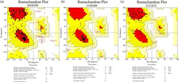
Using the I-TASSER server, the 3D structures of the CL1, SAP-2 and TP16.5 kDa proteins were predicted (fig. 2). Ramachandran plots were used to validate the 3D models of the three proteins. The Ramachandran plot analysis of the modelled CL1 protein revealed that 78.4% of the residues were located in the most favoured region, 19.7% (additionally allowed + generously allowed) in the allowed regions and only 1.9% in the disallowed region. These values were 87.0%, 13.0% and 0.0% for the modelled SAP-2 protein and 66.3%, 33.7% and 0.0% for the modelled TP16.5 kDa protein, respectively (fig. 3). These results provide reliable modelled structures.
3D structure of Fasciola hepatica proteins using the I-TASSER server: (a) CL1; (b) SAP-2; (c) 16.5 kDa protein.

Ramachandran modelling structure for the predicted model using the PROCHECK server: (a) CL1; (b) SAP-2; (c) 16.5 kDa protein.

Predicted antigenic multi-epitope protein
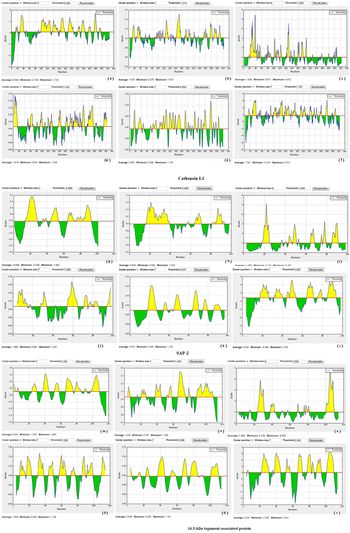
The combination of the predictions using IEDB, BepiPred, LBtope and ABCpred generated 149 linear epitope regions from the CL1, SAP-2 and 16.5 kDa proteins. In total, 13 conformational epitope regions were selected, mostly located in the linear epitope regions (fig. 4, tables 2–4). Using the IEDB server, the highest values calculated for epitopic regions (antigenic regions) with good hydrophilicity, high accessibility, high flexibility, high turn propensity and strong antigenicity were identified as the final epitopic regions. The total values of the linear B-cell epitope regions predicted by these servers were 71, 32 and 46 for CL1, SAP-2 and 16.5 kDa proteins, respectively. The CBTope program was employed to predict the conformational B-cell epitopes of the three proteins. The results of CL1, SAP-2 and TP16.5 kDa proteins were 8, 2 and 3 for the high-scoring conformational epitope regions, respectively (tables 2–4).
Prediction of CL1, SAP-2 and 16.5 kDa proteins and B-cell epitope using the IEDB server: (a, g, m) BepiPred linear epitope; (b, h, n) Chou and Fasman β-turn; (c, i, o) Emini surface accessibility; (d, j, p) Kolaskar and Tongaonkar antigenicity; (e, k, q) Karplus and Schulz flexibility; (f, l, r) Parker hydrophilicity.

Prediction of B-cell epitopes using ABCpred, LBtope, CBTope and IEDB for CL1protein.

Prediction of B-cell epitopes by ABCpred, LBtope, CBTope and IEDB for SAP-2 protein.

Prediction of B-cell epitopes using ABCpred, LBtope, CBTope and IEDB for 16.5 kDa protein.

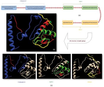
For prediction of antigenic multi-epitope, a consensus of the predicted results by the linear and conformational prediction servers was considered, with the highest scores and shared regions among them as well as maintained 3D structure of the polypeptide. In the selected epitope regions of CL1 protein, the epitopic regions 32–60 and 91–136 were in agreement with four (IEDB, BepiPred, ABCpred and CBTope) and five (IEDB, BepiPred, LBtope, ABCpred and CBTope) tools, respectively. In SAP-2 protein, the regions 27–38 and 64–84 were consensus regions in four (IEDB, BepiPred, ABCpred and LBtope) and five (IEDB, BepiPred, LBtope, ABCpred and CBTope) servers, respectively. In TP16.5 kDa protein, the epitope regions 33–45 and 89–109 were in agreement with all five programs. On the other hand, the results showed that a total of six epitope regions were selected and designed for the three antigens; two epitope regions for CL1 (located at positions 32–60 (region 1) and 91–136 (region 2)), two epitope regions for SAP-2 (located at positions 27–38 (region 1) and 64–84 (region 2)) and two epitope regions for TP16.5 kDa (located at positions 33–45 (region 1) and 89–109 (region 2)) (table 5). The six predicted epitope regions of the three antigens were fused together with flexible protein linkers, including glycine and serine amino acids, to form the final multi-epitope construct containing a total of 183 amino acid residues (fig. 5).
The six epitopes of amino acid sequence for CL1, SAP-2 and 16.5 kDa of Fasciola hepatica proteins.

(a) Design of multi-epitope structure for CL1, SAP-2 and 16.5 kDa proteins of Fasciola hepatica; (b) the 3D structure model prediction of a multi-epitope sequence; (c) recognition of the parts of CL1, SAP-2 and 16.5 kDa epitopes on the 3D structure model of the multi-epitope.

Expression, SDS-PAGE and Western blot analysis
We observed that the optimal polytope protein expression occurred in 8 h at 37°C after induction with 1 mM IPTG. The protein concentration was measured using the Bradford assay, which was obtained to be 8 mg/ml. The results of SDS-PAGE analysis showed the recombinant multi-epitope was successfully expressed with a strong band of about 23 kDa, which was related to the expected molecular weight of the multi-epitope peptide (fig. 6).
SDS-PAGE analysis of the expressed rMEP-pET-22b (+) polytope in Escherichia coli BL21 (DE3). Lane 1: intact E. coli BL21; lanes 2 and 3: induced intact E. coli BL21; lane 4: BL21 with pET-22b (+); lanes 5 and 6: induced BL21 containing pET-22b (+); lane M: protein marker (240 kDa) (SMOBIO, Taiwan); lane 7: BL21 containing rMEP-pET-22b (+); lanes 8–11: induced BL21 including rMEP-pET-22b (+).

Using Western blot analysis, the rMEP-pET-22b (+) and purified rMEP were probed with 1:1000 dilution of His-tag antibody and 1:100 dilution of human sera samples, respectively. The results showed a single strong band of about 23 kDa (figs 7 and 8a), whereas the band was not observed in immunoblots probed with negative control sample (E. coli BL21) and in the healthy subjects or sera of non-fascioliasis patients (figs 7 and 8b).
Immunoblotting of the recombinant MEP-pET-22b (+) of Fasciola hepatica and Escherichia coli BL21 (DE3) as negative control sample using anti-His tag antibody (lanes 1 and lane 2). M, protein marker (SMOBIO, Taiwan).

Immunoblotting of purified recombinant multi-epitope peptide of Fasciola hepatica with sera from fascioliasis and non-fascioliasis patients and healthy controls. (a) Pooled sera from fascioliasis patients (lane 1), protein marker (SMOBIO, Taiwan) (lane 2); (b) pooled sera from healthy controls (lane 1), pooled sera from non-fascioliasis patients (lanes 2–6).

Discussion
The antigens commonly used in commercial diagnostic ELISA kits are the crude extracts or excretory–secretory antigens. However, the use of such complex antigens reduces the specificity of the serological tests due to cross-reactivity with parasites sharing common epitopes (Losada et al., Reference Losada, Chacon, Colmenares, Bermudez, Lorenzo, Pointier, Theron, de Noya and Noya2005; Salimi-Bejestani et al., Reference Salimi-Bejestani, McGarry, Felstead, Ortiz, Akca and Williams2005; Sabry et al., Reference Sabry, Taher, Farag Allah and Mahgoub2014).
Finding information about the prediction of antigenic epitopes using immunoinformatic tools has become an important issue for the early diagnosis of the disease, enhancement of the specificity of diagnostic method and production of vaccines (Ali et al., Reference Ali, Pandey, Khatoon, Narula, Mishra and Prajapati2017; Adhikari et al., Reference Adhikari, Tayebi and Rahman2018; Lata et al., Reference Lata, Kumar, Vaghasia, Sharma, Bhairappanvar, Soni and Das2018). Such methods can improve the quality of epitope characterization and are precise, accurate, effective, economical and simple. Further, these efforts can substantially save time and reduce the experimental costs (Wang et al., Reference Wang, Wang and Cai2016; Rohani et al., Reference Rohani, Hajighasemi and Sefid2018). Besides, the use of multi-parametric and -method analysis greatly increases the accuracy of the epitope prediction (Assis et al., Reference Assis, Sousa, Pinto, Silva, Vaz, Andrade, Carvalho and De Melo2014; Pan et al., Reference Pan, Chen and Lu2017).
In the present study, we designed and confirmed an antigenic recombinant polytope of three protein epitopes (CL1, SAP-2 and TP16.5) of F. hepatica using immunoinformatic tools.
We predicted the secondary structure of the proteins using the SOPMA server. The results showed high scores for the antigenicity, hydrophilicity and accessibility of regions, which are important for epitope formation and identification and ligand binding. Thus, it was deduced that these selected antigens could interact with other biological molecules, antibodies. Hence, the most probable highly antigenic linear and conformational B-cell epitopes were selected, the majority of which were common to all the programs (IEDB, LBtope, ABCpred and CBTope).
There are different studies mapping the B-cell epitopes on the proteins of F. hepatica and identifying these regions using immunodetection methods. Some surveys have demonstrated the presence of dominant B-cell epitopes on the amino acid sequence of FhSAP-2 protein (21–30 aa (P4) and 76–85 aa (P16)), with high immunoreactivity (Torres & Espino, Reference Torres and Espino2006). In comparison with our study, region 1 of SAP-2 (27–38 aa) has been located in the part of amino acid residues P4 and P5 and shares some conserved (containing two conserved cysteine residues) and non-conserved regions. The highly reactive residues 27–30 are entirely located in an extended region, exerting a strong effect on the results of the antibody recognition (Vázquez-Talavera et al., Reference Vázquez-Talavera, Solís, Medina-Escutia, López, Proaño, Correa and Laclette2001). Also, region 2 of SAP-2 (64–84 aa) (including one conserved cysteine residue) shared residues 76–84 with the P16. In other studies on CL1 protein, the putative peptides include the amino acid residues 25–42, 103–122 and 110–129, which are similar to our selected CL1 epitopic regions (region 1 and region 2) with high sensitivity and specificity results and no cross-reaction with other helminth infections (Cornelissen et al., Reference Cornelissen, Gaasenbeek, Boersma, Borgsteede and van Milligen1999, Reference Cornelissen, Gaasenbeek, Borgsteede, Holland, Harmsen and Boersma2001; Intapan et al., Reference Intapan, Tantrawatpan, Maleewong, Wongkham, Wongkham and Nakashima2005). The investigations on the detection of proper marker antigens for TP16.5 kDa and SAP-2 proteins to improve the serodiagnosis of fascioliasis have shown satisfactory results with no cross-reactivity (Espinoza et al., Reference Espinoza, Timoteo and Herrera-Velit2005; Gaudier et al., Reference Gaudier, Cabán-Hernández, Osuna and Espino2012).
The above-mentioned surveys were in agreement with our study due to the employment of appropriate bioinformatic tools to predict the localization of epitopic regions in the three proteins (CL1, SAP-2 and TP16.5) of F. hepatica and, consequently, their localization experimentally, which revealed a high reaction between total IgG antibody of the sera from fascioliasis patients and the specific polytopic antigen (fig. 8a).
Some studies have indicated relative homologies between Fasciola and some parasites because of the presence of conserved repeats and protein homologous in the entire amino acid sequence of some proteins within these parasites. Therefore, it can lead to cross-reactivity in serological assays (Espino & Hillyer, Reference Espino and Hillyer2003; Intapan et al., Reference Intapan, Tantrawatpan, Maleewong, Wongkham, Wongkham and Nakashima2005; Torres & Espino, Reference Torres and Espino2006). Our immunoblot analysis showed no cross-reactivity of multi-epitope antigen with some non-fascioliasis sera (fig. 8b). This may be associated with specific selection of B-cell epitopes considering minimum conserved regions and similarity with the proteins of other helminths.
In the current study, our findings for the design and laboratory confirmation of the highly antigenic combined B-cell epitopes (multi-epitope) demonstrated the presence of potential epitopes, which could lead to an increase in the specificity of epitope peptides in the serological detection methods.
However, further serological studies are needed to examine a larger number of sera from patients with fascioliasis, schistosomiasis and other helminthiasis to validate the diagnostic value of this synthetic peptide for serodiagnostic targets.
Conclusion
Multi-epitope designing by in silico strategies and laboratory analysis of its results is a suitable method due to appropriate and acceptable protein expression. This study predicted the important biological data of F. hepatica to determine the antigenic multi-epitope regions and confirm the antigen specificity in vitro for efficient diagnosis of fascioliasis in humans and domestic herbivores. However, the suggested recombinant multi-epitope peptide is needed to be further researched in order to evaluate the specificity and sensitivity of the indirect ELISA technique with more serum samples.
Financial support
This study was a part of S.A.’s PhD dissertation, which was supported by Shahid Beheshti University of Medical Science, Tehran, Iran (grant number 10676.IR.SBMU.MSP.REC.1396.181).
Conflicts of interest
None.