INTRODUCTION
Patients with mild cognitive impairment (MCI) have subjective and objective (verified in cognitive testing or clinical assessment) cognitive impairment but not dementia (Petersen, Reference Petersen2004; Petersen et al., Reference Petersen, Smith, Waring, Ivnik, Tangalos and Kokmen1999). The rate of conversion from this condition to dementia or Alzheimer’s disease (AD) is between 6 and 25% per year (Petersen et al., Reference Petersen, Stevens, Ganguli, Tangalos, Cummings and DeKosky2001). Subjective cognitive impairment (SCI) may be a pre-MCI stage and the first detectable stage in the SCI-MCI-AD development (Reisberg & Gauthier, Reference Reisberg and Gauthier2008; Reisberg et al., Reference Reisberg, Prichep, Mosconi, John, Glodzik-Sobanska and Boksay2008).
Neuropsychological studies indicate that prodromal AD is characterized by subtle deficits in learning and memory, executive functioning, processing speed, attention, and semantic knowledge (see Bondi et al., Reference Bondi, Jak, Delano-Wood, Jacobson, Delis and Salmon2008, for a review). Usually, reduced episodic memory is the first detectable indicator of AD development, at a very early stage, often involving difficulty in encoding (Twamley, Ropacki, & Bondi, Reference Twamley, Ropacki and Bondi2006). The disruption of the medial temporal lobe memory system leads directly to disproportionate memory impairment, but fronto-striatal change may also underlie mild memory difficulties associated with other neuropsychological deficits (Buckner, Reference Buckner2004). Executive functions such as working memory, attentional control, and inhibition may also be impaired in the incipient stages of the disease, and may contribute to the observed memory deficit (Storandt, Reference Storandt2008). Co-occurrence of memory and executive dysfunction has been reported in MCI (Buckner Reference Buckner2004; Phillips et al., Reference Phillips, Chertkow, Leblanc, Pim and Murtha2004; Shim et al., Reference Shim, Yoon, Shon, Ahn and Yang2008).
Significant neural dysfunction occurs well in advance of the clinical diagnosis of AD and is characterized by brain atrophy and microscopic appearance of neuritic plaques and neurofibrillary tangles. Accumulation of neurofibrillary tangles begins in the medial temporal lobes (MTL) (i.e., entorhinal cortex) and then progresses to the association cortices of the temporal, parietal, and frontal lobes (Braak & Braak, Reference Braak and Braak1991). The hippocampus has a central position in the memory network because of converging inputs and connectivity to medial and lateral frontal regions (Greicius et al., Reference Greicius, Krasnow, Boyett-Anderson, Eliez, Schatzberg, Reiss and Menon2003). Impaired memory and hippocampal atrophy in MCI are commonly recognized as strong indicators of incipient AD (Dubois et al., Reference Dubois, Feldman, Jacova, Dekosky, Barberger-Gateau and Cummings2007; Van Petten, Reference Van Petten2004) and predict conversion from MCI to AD (Apostolova et al., Reference Apostolova, Dutton, Dinov, Hayashi, Toga, Cummings and Thompson2006; Fischl et al., Reference Fischl, Salat, Busa, Albert, Dieterich and Haselgrove2002; Jack et al., Reference Jack, Petersen, Xu, O’Brien, Smith, Ivnik and Boeve1999, Reference Jack, Petersen, Xu, O’Brien and Smith2000; Phillips et al., Reference Phillips, Chertkow, Leblanc, Pim and Murtha2004). Significant correlations between verbal memory and left hippocampus volume (HcV) have been reported in MCI patients (Müller et al., Reference Müller, Greverus, Dellani, Weibrich, Wille and Scheurich2005).
Most imaging studies of MCI have focused on gray matter alterations (Fjell et al., Reference Fjell, Walhovd, Amlien, Bjørnerud, Reinvang and Gjerstad2008a; Whitwell et al. Reference Whitwell, Shiung, Przybelski, Weigand, Knopman and Boeve2008), although cortical-subcortical and cortico-cortical disconnection also may result in cognitive dysfunction due to white matter tract disruption (Stenset et al., Reference Stenset, Grambaite, Reinvang, Hessen, Cappelen and Bjørnerud2007). Diffusion tensor imaging (DTI) provides a method for delineating the anatomy of white matter pathways by measuring the extent and directionality of water diffusion. In normal white matter, water molecules move more freely in the direction parallel to the nerve fiber tracts, while movements are relatively restricted across the tracts, causing white matter diffusion anisotropy. Directional diffusion can be quantified by fractional anisotropy (FA), calculated from the diffusion tensor eigenvalues (Bihan, Mangin, & Poupon, Reference Bihan, Mangin and Poupon2001; Charlton et al., Reference Charlton, Barrick, McIntyre, Shen, O’Sullivan and Howe2006), which describe diffusivity parallel (λ1) or perdendicular (λ2 and λ3) to the axonal tracts [axial (DA) and radial (DR) diffusivity]. It has been suggested that increased DR may represent demyelination, and that decreased DA may be a measure of axonal loss (Concha, Gross, & Wheatley, Reference Concha, Gross and Wheatley2006; Kraus et al., Reference Kraus, Susmaras, Caughlin, Walker, Sweeney and Little2007; Song et al., Reference Song, Sun, Ramsbottom, Chang, Russell and Cross2002, Reference Song, Sun, Ju, Lin, Cross and Neufeld2003).
Recent findings suggest that magnetic resonance imaging (MRI) volume measures (e.g., HcV) and DTI are complementary methods and their combination may improve diagnostic accuracy (Müller et al., Reference Müller, Greverus, Dellani, Weibrich, Wille and Scheurich2005). The hippocampal formation is connected to other structures in the Papez circuit (Papez, Reference Papez1995) (mamillary bodies, anterior thalamic nucleus, and the cingulate gyrus), a large fraction of both afferent and efferent fibers passing through the fornix. Fornix lesions (disrupting Papez’s circuit) have been related to loss of episodic memory (Aggleton & Brown, Reference Aggleton and Brown1999), although the disruption of the frontal lobe connections to the fornix may also explain executive deficits. DTI abnormalities in AD have been found in the corpus callosum (CC), the white matter of the parietal, temporal, and occipital lobes, the posterior cingulum, and the hippocampus (Fellgiebel et al., Reference Fellgiebel, Wille, Müller, Winterer, Scheurich and Vucurevic2004; Head et al., Reference Head, Buckner, Shimony, Williams, Akbudak and Conturo2004). Shim et al. (Reference Shim, Yoon, Shon, Ahn and Yang2008) reported decreased FA values in the genu/splenium of the CC, bilateral frontal, and temporal regions in MCI patients, as compared to controls. Changes in CC may be related to neuropsychological deficits in MCI and AD because of its neocortical connections (Braak et al., Reference Braak, Griffing, Arai, Bohl, Bratzke and Braak1999). Zhang et al. (Reference Zhang, Schuff, Jahng, Bayne, Mori and Schad2007) found reduced FA in the cingulum fibers in MCI and AD, as well as in the splenium of the CC in AD. The authors argued that the entire connection between hippocampus and posterior cingulum may be affected in the early stage of AD, and that FA in the cingulum fibers, in conjunction with HcV, may be early predictors of future cognitive decline and development of AD. Hence, it follows that memory deficit in MCI patients may be explained by effects of both hippocampal and white matter changes. However, it is not known whether changes of memory network white matter diffusivity (e.g., posterior cingulum, the genu/splenium of the CC, and the fornix) may serve as unique predictors of memory function independently of hippocampal atrophy.
While neuropsychological and MR findings are important markers for AD, recent evidence indicates that pathological markers derived from cerebrospinal fluid (CSF) may add significantly to diagnostic sensitivity and specificity (Dubois et al., Reference Dubois, Feldman, Jacova, Dekosky, Barberger-Gateau and Cummings2007; Leon, Desanti, & Zinkowski, Reference Leon, Desanti and Zinkowski2004; Petersen, Reference Petersen2004). CSF total tau protein (T-tau) levels are associated with the intensity of neuronal damage and degeneration, and have been studied as an adjunctive marker for “probable AD,” as a moderate to marked increase is found in AD (Blennow, Reference Blennow2004; Hansson et al., Reference Hansson, Zetteberg, Buchhave, Londos, Blennow and Minthon2006). Experimental and clinico-pathological evidence implicate axonal transport deficiency and morphological alterations in incipient AD (Stokin et al., Reference Stokin, Lillo, Falzone, Brusch, Rockenstein and Mount2005, Reference Stokin, Almenar-Queralt, Gunawardena, Rodrigues, Falzone and Kim2008). Also, the physiological role of tau in stabilizing axonal microtubules and the localization of hyperphosphorylated tau in AD plaques suggest that tau hyperphosphorylation and subsequent dysfunction may contribute to the development of AD (Adalbert, Gilley, & Coleman, Reference Adalbert, Gilley and Coleman2007; Iqbal, Liu, Gong, Alonso, & Grundke-Iqbal, Reference Iqbal, Liu, Gong, Alonso and Grundke-Iqbal2009).
In one recent study with a sample overlapping with the present sample, the differences in HcV between the MCI and the healthy control groups were only found for patients with pathological CSF biomarkers, and tau-pathology (combined score of T-tau and phosphorylated tau) was found to be related to reductions in HcV and memory (Fjell et al., Reference Fjell, Walhovd, Amlien, Bjørnerud, Reinvang and Gjerstad2008a). In another report from the same cohort, Stenset et al. (Reference Stenset, Bjørnerud, Fjell, Walhovd, Hofoss and Due-Tønnessen2009) focused on white matter diffusivity changes and found increased DR in the genu of CC, left forceps major, and left cingulum in SCI/MCI patients with CSF T-tau pathology compared to controls, as well as increased DR in the left cingulum compared to SCI/MCI patients without T-tau pathology. This may suggest altered axon myelination and early axonal damage in patients with T-tau pathology. Studying white matter lesions (WML) in patients with more pronounced cognitive symptoms, Stenset et al. (Reference Stenset, Hofoss, Johnsen, Skinningsrud, Berstad and Negaard2008) found that WML were significantly related to cognition only in patients with normal CSF amyloid-β42 (Aβ42), suggesting that amyloid pathology as measured in CSF may override effects of WML on cognition. In another recent study, MCI patients with normal CSF T-tau and Aβ42 were shown to score higher on tests of episodic memory and speed/attention compared to MCI patients with CSF abnormalities (Nordlund et al., Reference Nordlund, Rolstad, Klang, Lind, Pedersen and Blennow2008). For pre-dementia cases, all these findings suggest that more homogeneous groups may be obtained by using CSF biomarkers to stratify patient groups.
In a previous study (Fjell et al., Reference Fjell, Walhovd, Amlien, Bjørnerud, Reinvang and Gjerstad2008a), we showed that HcV was reduced in an MCI group with pathological tau levels compared to normal controls, but not in the normal tau MCI group. Both groups did, however, show regional cortical thinning compared to controls and some relationships between morphometric and CSF variables and memory were observed. The present study follows up on these results by adding measures of white matter diffusivity, contrasting groups with and without CSF T-tau pathology (Sjögren et al., Reference Sjögren, Vanderstichele, Agren, Zachrisson, Edsbagge and Wikkelsø2001). Memory difficulties are expected to be related to reduction of HcV in the group with pathological T-tau levels, with effects of HcV loss and incipient AD probably overriding any effects of white matter affection (Stenset et al., Reference Stenset, Hofoss, Johnsen, Skinningsrud, Berstad and Negaard2008). With near normal HcV (Fjell et al., Reference Fjell, Walhovd, Amlien, Bjørnerud, Reinvang and Gjerstad2008a), mechanisms for memory affections in the normal tau group may differ from that in the pathological T-tau group with reduced HcV. Thus, we predict that memory performance in the normal T-tau group can be explained by white matter diffusivity in areas included in the memory-network, namely posterior cingulum, genu CC, splenium CC, and fornix.
METHODS
Participants
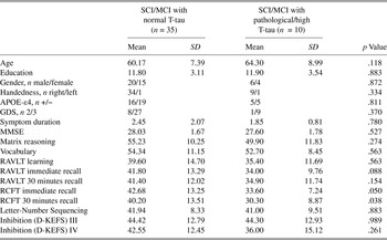
Forty-five patients [mean age 61.1 (43–78) years, SD = 7.9; mean Mini Mental State Examination (MMSE; Folstein, Folstein, & McHugh, Reference Folstein, Folstein and McHugh1975) 27.9 (23–30), SD = 1.7] with cognitive symptoms lasting six months or longer attending a university-based memory clinic between September 2005 and December 2007 were assessed for inclusion. Inclusion criteria were subjective memory impairment, preserved general intellectual function, no or very mild activities of daily living (ADL) problems and a Global Deterioration Scale (GDS) score of 2 (9 patients) or 3 (36 patients) (Auer & Reisberg, Reference Auer and Reisberg1997; Reisberg, Ferris, de Leon, & Crook, Reference Reisberg, Ferris, de Leon and Crook1988) corresponding to subjective and mild cognitive impairment (SCI and MCI), respectively (Gauthier et al., Reference Gauthier, Reisberg, Zaudig, Petersen, Ritchie and Broich2006; Petersen et al., Reference Petersen, Smith, Waring, Ivnik, Tangalos and Kokmen1999; Reisberg & Gauthier, Reference Reisberg and Gauthier2008; Reisberg et al., Reference Reisberg, Prichep, Mosconi, John, Glodzik-Sobanska and Boksay2008; Winblad et al., Reference Winblad, Palmer, Kivipelto, Jelic, Fratiglioni and Wahlund2004), as determined from a clinical interview and screening tests. Screening tests included parameters 13–20 (memory, disorientation, abstract thinking, visuospatial ability, language, sensory aphasia, visual agnosia, and apraxia) from the stepwise comparative status analysis (STEP; Edman, Mahnfeldt, & Wallin, Reference Edman, Mahnfeldt and Wallin2001; Wallin et al., Reference Wallin, Edman, Blennow, Gottfries, Karlsson, Regland and Sjögren1996), word fluency, interference, and numeral-letter items from the I-FLEX (Royall, Mahurin, & Gray, Reference Royall, Mahurin and Gray1992), and items from the Neurobehavioral Cognitive Status Examination (Cognistat; Kiernan, Mueller, Langston, & Van Dyke, Reference Kiernan, Mueller, Langston and Van Dyke1987), as well as MMSE (Folstein et al., Reference Folstein, Folstein and McHugh1975). Subjects classified as GDS 2 scored ≥28 on MMSE, 0 on STEP variables, the total sum was <2 on elements from I-FLEX, and it was a maximum one Clinical Dementia Rating (CDR; Morris, Reference Morris1993) domain where they scored 0,5. Subjects classified as GDS 3 scored ≥26 on MMSE, ≤1 on STEP variables, ≤2 on I-FLEX variables, and there were more than one CDR-domain where they scored 0,5, albeit none where they scored 1. One patient with GDS = 3 had MMSE = 23, but was included as she had normal employment and was self-supported. Criteria for exclusion were established psychiatric disorder, cancer, drug abuse, solvent exposure, or anoxic brain damage. The project was approved by the South-Eastern Norway committee for medical research ethics. All included gave informed consent. The sample was stratified according to normal (n = 35) or pathological (n = 10) CSF T-tau levels following established age-specific cut-off values (Sjögren et al., Reference Sjögren, Vanderstichele, Agren, Zachrisson, Edsbagge and Wikkelsø2001). Table 1 contains descriptive demographic and neuropsychological data.
Demographic and behavioral data comparison between subjective and mild cognitive impairment (SCI/MCI) patients with and without CSF T-tau pathology

Note
APOE = Apolipoprotein E, GDS = Global Deterioration Scale, MMSE = Mini-Mental State Examination, RAVLT = Rey Auditory and Verbal Learning Test, RCFT = Rey Complex Figure Test, D-KEFS = Delis-Kaplan Executive Function System. T-scores, which are depicted for all neuropsychological measures except MMSE, are age corrected test-specific normative data. Data for D-KEFS inhibition test is missing for two patients from the group with normal T-tau, because another version of Stroop inhibition test was used in the initial phase of the project. The comparison of gender, handedness, APOE ε4, and GDS distribution in two patient groups was made with a chi-square test (Pearson chi-square test p value is shown). Mann-Whitney U-test was used to compare the groups for the remaining variables in the table.
Neuropsychological Assessment
All participants received a neuropsychological test battery assessing general mental status (MMSE; Folstein et al., Reference Folstein, Folstein and McHugh1975) and other tests weighted on general cognitive ability, executive, and memory function. Rey Auditory and Verbal Learning Test (RAVLT) was used to measure verbal learning and episodic memory (Schmidt, Reference Schmidt1996). The number of correct responses on all five learning trials was summed to one learning score. The mean of the immediate and 30 minutes recall was termed verbal memory. Rey Complex Figure Test (Meyers & Meyers, Reference Meyers and Meyers1995) was used to investigate visual memory (the mean score of the immediate and 30 minutes recall). D-KEFS (Delis-Kaplan Executive Function System) Color-Word Interference Test, which is based on the Stroop (Reference Stroop1935) procedure (Delis, Kaplan, & Kramer, Reference Delis, Kaplan and Kramer2001), was used as a measure of inhibition, defined as the mean score (time in seconds) of the conditions 3 (Inhibition) and 4 (Inhibition/Switching), regressed on conditions 1 and 2 in order to control for processing speed. The positive values of inhibition scores were then converted into negative values, so that the higher values mean better performance when used in further analyses. The Letter-Number Sequencing task from Wechsler Adult Intelligence Scale (WAIS-III), which requires the patient to order sequentially a series of numbers and letters orally presented in a specified random order, is a measure of auditory working memory (Wechsler, Reference Wechsler1997, Reference Wechsler2003). Vocabulary was measured using the vocabulary subtest of the Wechsler Abbreviated Scale of Intelligence (WASI). Matrix reasoning (WASI/WAIS-III) presents a series of increasingly difficult visual pattern completion and analogy problems and is a measure of nonverbal abstract problem-solving (Wechsler, Reference Wechsler1997, Reference Wechsler1999, Reference Wechsler2003). Nineteen of the subjects were tested with Matrix reasoning from WASI and 26 subjects were tested with Matrix reasoning from WAIS-III. T-scores, based on norms for specific tests, are depicted in Table 1.
MRI/DTI Acquisition and Region Of Interest Analysis
For practical reasons, two different 1.5T MRI scanners were used in this study. Conventional axial fluid-attenuated inversion recovery (FLAIR) and T2-weighted sequences were acquired along with two 3D magnetization prepared, gradient echo (MP-RAGE), T1-weighted sequences. The T1 acquisition protocols were: For Siemens Symphony, acquisition matrix = 128 × 128, pixel size = 1.8 × 1.8 mm2, TE = 131 ms, TR = 4300 ms, maximum b value = 700 s/mm2, section spacing/thickness = 1.5 mm/5 mm. For Siemens Espree, acquisition matrix = 192 × 192, pixel size = 1.2 × 1.2 mm2, TE = 117 ms, TR = 6100 ms, maximum b value = 750 s/mm2, section spacing/thickness = 0.9 mm/3 mm). The DTI scans were acquired with a 2D spin echo planar imaging (EPI) sequence. Diffusion weighting was applied along 12 noncollinear directions with two (Siemens Symphony) and five (Siemens Espree) repetitions per direction. In addition, two and five images, respectively, without diffusion weighting (b = 0) were acquired.
MRI hippocampus segmentations
MRI segmentations were performed using FreeSurfer 4.0.1 (http://surfer.nmr.mgh.harvard.edu/). For HcV calculations, an automated, fully 3D whole-brain segmentation procedure was used (Fischl et al., Reference Fischl, Salat, Busa, Albert, Dieterich and Haselgrove2002), where a probabilistic atlas is used and a Bayesian classification rule is applied to assign a neuroanatomical label to each voxel. The atlas consists of a manually derived training set created by the Center for Morphometric Analysis (http://www.cma.mgh.harvard.edu/) from 40 other subjects across the age range, including individuals with AD. The segmentation uses three pieces of information to disambiguate labels: (1) the prior probability of a given tissue class occurring at a specific atlas location, (2) the likelihood of the image given that tissue class, and (3) the probability of the local spatial configuration of labels given the tissue class. This latter term represents a large number of constraints on the space of allowable segmentations, and prohibits label configurations that never occur in the training set (e.g., hippocampus is never anterior to amygdala). A newly developed atlas normalization procedure was used, which has been shown to increase the robustness and accuracy of the segmentations across scanner platforms (Han & Fischl, Reference Han and Fischl2007). The present segmentation of the hippocampal formation includes dentate gyrus, cornu ammonis (CA) fields, subiculum/ parasubiculum, and the fimbria (Makris et al., Reference Makris, Meyer, Bates, Yeterian, Kennedy and Caviness1999). Estimated intracranial volume (ICV) was used to correct the volumetric data. This was calculated by use of an atlas-based normalization procedure, where the atlas scaling factor is used as a proxy for ICV, shown to correlate .93 with manually derived ICV (Buckner et al., Reference Buckner, Head, Parker, Fotenos, Marcus, Morris and Snyder2004). The standardized residuals, received from the raw scores of HcV after effects of age, ICV, and MRI site were regressed out, were used in the analyses. HcV could not be calculated for four participants, because a too narrow field of view for application of the whole brain segmentation, on which this is based, was mistakenly applied for the T1 sequence for these four.
Diffusion tensor imaging analysis
DTI data were processed using the nICE Basis and Diffusion Module (NordicImagingLab AS, Bergen, Norway). Nine elliptical regions of interest (ROIs) were manually drawn in the FA map (left and right forceps minor/major, genu, and splenium CC, left and right posterior cingulum, and fornix fibers), and ROI sizes ranged from 11.2–34.4 mm2. The placement of ROIs is explained in detail elsewhere (Stenset et al., Reference Stenset, Bjørnerud, Fjell, Walhovd, Hofoss and Due-Tønnessen2009; Zhang et al., Reference Zhang, Schuff, Jahng, Bayne, Mori and Schad2007). White matter segmentations and color-coded eigenvector (cDTI) maps were used for orientation to avoid partial CSF or gray matter volume, and ROIs were placed in the part of the fiber tract with the highest signal intensity. ROI values were extracted from all processed images (FA, ADC, λ1, λ2, λ3) and DR was calculated [(λ2+λ3)/2] for each ROI. ROIs were placed by one rater (VS), and the interrater correlation coefficient (Intraclass correlation coefficient) was calculated to be 0.89. FA, DR, and DA values for the chosen ROIs are provided in Supplementary Table 1. For forceps minor/major and posterior cingulum, mean scores of the left and the right sides were calculated. The scores regressed on age and a MRI scanner was used in the analyses.
Lumbar Puncture and Cerebrospinal Fluid (CSF) Analysis
Lumbar puncture (LP) was obtained for all patients, and CSF T-tau was routinely examined with a commercially available kit (Innogenetics, Belgium) adapted to a Tecan Robotic Microplate 150 Processor (Tecan AG, Switzerland). Age-dependent clinical cut-off values for CSF T-tau (Sjögren et al., Reference Sjögren, Vanderstichele, Agren, Zachrisson, Edsbagge and Wikkelsø2001) were used to define the MCI patients into two groups: SCI/MCIabnormal T-tau (n = 10) if the CSF T-tau level was abnormal (T-tau ≥ 300 ng/L for patients under 50 years, ≥ 450 ng/l for patients from 50 to 69 years, and ≥ 500ng/L for patients 70 years and older), and SCI/MCInormal T-tau (n = 35) if the CSF T-tau level was within normal range.
Statistical Analysis
The Statistical Package for Social Sciences (SPSS 16.0) was used for all statistical analyses. Outlier analyses were conducted for all continuous variables. There were no outliers found among the scores of cognition, HcV, and white matter diffusivity values. When analyzing T-tau scores, one outlier was detected (T-tau = 1399, age = 76.) The outlier was not excluded from further analyses as it is known that T-tau is increasing with age and other pathological conditions were controlled by exclusion criteria. All T-tau scores were further categorized (high/normal) according to established age-specific cut-offs. To compare group demographic characteristics, Mann-Whitney U-test and Chi-Square test were used (see Table 1). The Mann-Whitney U-test was also used to analyze group differences with regard to neuropsychological performance (age, gender, and education level were regressed out and the standardized residuals were used in all analyses, except for the comparative analyses of T-scores, presented in Table 1), white matter diffusivity (age and MRI site were regressed out and the standardized residuals were used in all analyses), and hippocampus volume (age, ICV, and MRI site were regressed out, and the standardized residuals were used in all analyses). Nonparametric methods were used because of small and unequal sample sizes (n = 35 vs. n = 10). To estimate effect sizes, Cohen’s d, defined as the difference between two means divided by the pooled standard deviation for the data, was calculated. As pooled standard deviation, the square root of the average of the squared standard deviations was used (Cohen, Reference Cohen1988). Pearson’s correlation method was used to test for associations between imaging and cognitive variables. To investigate the uniqueness of the FA predictive value to variance in memory function within the group with normal T-tau, hierarchic regression analyses were performed with learning and memory as the dependent variables. The independent variables, HcV, and FA values in the areas, showing significant (p < .05) or significant at trend level (p < .10) correlations with memory, and interactions between HcV and FA, were subsequently entered in the analyses.
RESULTS
For the whole SCI/MCI group, the mean CSF T-tau value was 342 ± 230 ng/L. The average CSF T-tau values of the groups with pathological/high and normal levels of this biomarker were, respectively, 652 ± 296 ng/L and 254 ± 96 ng/L. The subjects with deviating results on T-tau and those with normal results were compared, which is described next.
The two patient groups were quite similar according to their demographics, as seen from Table 1. Age, level of education, gender, frequency of ε4 alleles of the Apolipoprotein E gene (APOE-ε4), symptom duration, MMSE, as well as general cognitive ability (Matrix reasoning and Vocabulary) were not significantly different (p > .10) between the groups. A trend toward significance was seen for RAVLT immediate recall (p = .09), and significant differences were observed for both RCFT immediate recall (p = .05) and RCFT 30-minute recall (p = .04), when age corrected test-specific normative T-scores were used.
Figure 1 provides standardized residuals of neuropsychological measures (raw scores, regressed on age, gender, and education, and based on means and standard deviations for the whole MCI sample) in groups with normal and pathological T-tau levels. Visual memory was significantly better (p = .04) in the normal T-tau group. The Cohen’s d statistics were computed for learning (.24), verbal memory (.47), visual memory (.83), working memory (.16), and inhibition (.33) in order to more accurately evaluate group differences.
Neuropsychological measures for the groups with normal (n = 35) and high (n = 10) CSF T-tau levels. The standardized residuals, after effects of age, gender, and education were regressed out from the raw scores are shown in the figure. *Significant differences between the groups (p < .05). Error bars represent 95% confidence intervals.

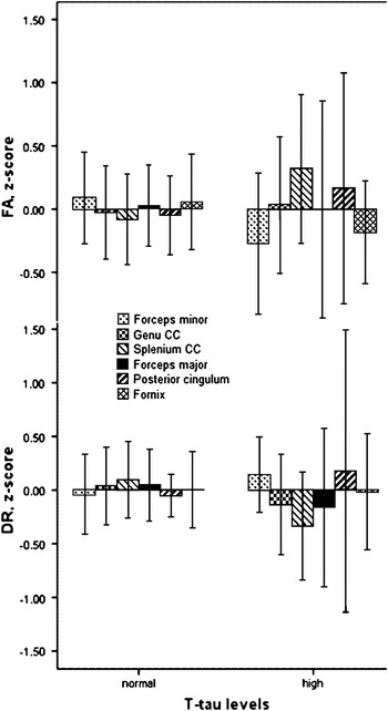
Figure 2 shows FA and DR values (z-scores, corrected for age and MRI scanner). There were no statistically significant differences between the groups with normal and pathological T-tau levels. The Cohen’s d statistics were calculated for FA and DR values, respectively: forceps minor (.38 and .22), forceps major (0 and .21), genu CC (.04 and .19), splenium CC (.44 and .49), posterior cingulum (.19 and .17), and fornix (.28 and .02).
Fractional anisotropy (FA) and radial diffusivity (DR) values (z-scores) for the groups with normal (n = 35) and high (n = 10) CSF T-tau levels. The standardized residuals after age and MRI scanner were regressed from the raw scores are depicted. There were no statistically significant differences between the groups. Error bars represent 95% confidence intervals.

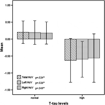
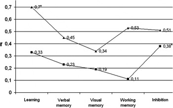
Larger HcV was found in the group with normal T-tau levels than the group with pathological levels (Figure 3). Significant differences were observed for the left (p = .03; Cohen’s d = .93), the right (p = .05; Cohen’s d = .78), and total HcV (p = .02; Cohen’s d = .92). Correlations between total HcV and cognitive performance are depicted in Figure 4. Total HcV was not significantly related to learning or memory, but bigger HcV was significantly associated with better performance on the inhibition task in the normal T-tau group. Despite small sample size (n = 10), better performance on learning was significantly related to larger HcV in the T-tau pathology group.
Hippocampus volume (HcV) for the groups with normal (n = 31) and high (n = 10) CSF T-tau levels. The standardized residuals after age, ICV, and MRI scanner were regressed from the raw scores of HcV are depicted. *Significant differences between the groups (p < .05). Error bars represent 95% confidence intervals.

Correlations between the total hippocampus volume and cognitive performance. ▲ = patients with pathological T-tau levels. ■ = patients with normal T-tau levels. * Significant correlations (p < .05). Higher neuropsychological scores mean better performance.

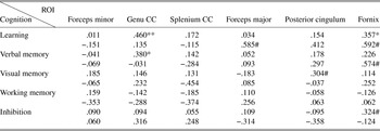
The correlation coefficients between FA ROIs and neuropsychological function in the groups of patients with normal and pathological T-tau levels are presented in Table 2. Both verbal memory (p < .05) and learning (p < .01) correlated positively with FA in genu CC in the normal T-tau group. Learning in this group also correlated significantly with FA in fornix (p < .05), whereas visual memory correlated at trend level (p < .10) with FA in posterior cingulum. None of the ROIs correlated significantly with working memory or inhibition. The corresponding analyses in the T-tau pathology group showed no significant correlations between FA and cognition, but small sample size precludes a firm conclusion.
Correlations between cognitive domains and fractional anisotropy (FA) regions of interest (ROI)

Note
CC = corpus callosum. The results for the group with normal T-tau (n = 35) are presented in gray-shadowed background. The results for the group with pathological T-tau (n = 10) are presented in white background.
*p < .05; **p < .01; #p < .10 (trend).
As the correlation between neuropsychological function and FA was significant (p < .05) in the normal T-tau group, we further analyzed if this relationship was a result of changes in DR or DA. There was a negative correlation between DR and memory function. Learning was significantly (p < .05) related to DR genu CC (r = –.40) and DR fornix (r = –.35), and verbal memory was significantly (p < .05) related to DR genu CC (r = –.33). No relationships were observed between cognitive function and DA.
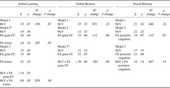
To further determine if FA explained unique variance in learning and memory in the normal T-tau group beyond that which can be explained by HcV, we performed a hierarchic regression analysis entering HcV in step 1, FA values in the areas significantly (or at trend level) correlated with memory were entered in step 2, and interaction terms between HcV and FA were entered in step 3. The results are shown in Table 3.
Hierarchic regression analyses with memory variables as the dependent variables in the patients with normal T-tau levels

Note
HcV = hippocampus volume, FA = fractional anisotropy, CC = corpus callosum.
* Indicates significant model (p < .05). N = 35.
FA genu CC served as a unique predictor of verbal learning, whereas HcV did not. HcV alone explained 11% (p = .07) of variance in learning, and, adding both FA genu CC (p = .04) and FA fornix (p = .18) to the model, the explained variance increased significantly (p = .03) to 31% and resulted in a significant model (F = 4.01, p = .02). The interaction variables did not add significantly (p > .05) to the explained variance in learning.
In the corresponding analysis, with verbal memory as the dependent variable, HcV alone explained only 5% (p = .22) of variance in memory function, and, adding FA genu CC (p = .06) to the model, the explained variance became 16%. This increase in variance over the previous model was significant at a trend level (p = .06). When the interaction term was added, the explained variance became 25%. The increase of variance was significant at a trend level (p = .08), and a significant total model was obtained (F = 3.05, p = .045).
In the analysis with visual memory as the dependent variable, no single variable or combination of HcV and FA resulted in a significant predictive model. HcV explained only 5% of variance in memory (p = .26), and adding FA posterior cingulum to the model, the explained variance became 16%, and the increase was significant at trend level (p = .07). When adding the interaction term, the explained variance increased, not significantly (p = .14), to 23%.
DISCUSSION
This study set out to examine neuropsychological function and its relation to white matter diffusivity in SCI/MCI patients with normal and pathological CSF T-tau levels, and the impact of HcV for these relationships in the normal T-tau group. The results of the study indicate that the size of HcV, as well as memory performance, may differ according to levels of T-tau pathology. Smaller HcV and lower visual memory performance were found to be characteristic for the T-tau pathology group. Verbal learning was significantly related to HcV in this group. In contrast, learning was found to be not significantly related to HcV in the larger group with normal T-tau levels. This group had less hippocampal atrophy, which could explain this weak correlation. Further analyses of the normal T-tau group showed a relationship between learning and directional diffusivity (FA and DR) in fornix, and this fits with knowledge that this fiber tract is a part of the Papez circuit (Papez, Reference Papez1995). FA and DR in the genu of the CC, which is known to connect frontal cortices (Huang et al., Reference Huang, Zhang, Jiang, Wakana, Poetscher and Miller2005), was also related to verbal learning and memory in the normal T-tau group. These relationships were found to be independent of HcV.
Episodic memory encoding and retrieval are known to be associated with prefrontal cortex (PFC) activations. Activations tend to be left lateralized during encoding and right lateralized during retrieval in younger adults, whereas asymmetry reductions have been observed in older adults (the HAROLD model). Optimal memory performance in older adults may be increasingly dependent on coordinated bilateral frontal activation (Cabeza, Anderson, Locantore, & McIntosh, Reference Cabeza, Anderson, Locantore and McIntosh2002), which, in turn, is mediated by callosal transfer. Correlation with memory performance in the normal T-tau group may thus be a function of normal age-related variation. Stenset et al. (Reference Stenset, Bjørnerud, Fjell, Walhovd, Hofoss and Due-Tønnessen2009) have found FA/DR/DA differences in a sample of SCI/MCI patients (overlapping with the present sample) with normal T-tau levels versus those with pathological T-tau levels, while differences between patients with normal T-tau and healthy controls were not statistically significant. Similarly, strikingly small neuropsychological differences were found between MCI patients without CSF T-tau and Aβ42 abnormalities and healthy controls (Nordlund et al., Reference Nordlund, Rolstad, Klang, Lind, Pedersen and Blennow2008).
It has been reported that age-related changes are more prominent in the most anterior portions of the CC (e.g., genu CC; Head, Snyder, Girton, Morris, & Buckner, Reference Head, Snyder, Girton, Morris and Buckner2005; Zhang et al., Reference Zhang, Schuff, Jahng, Bayne, Mori and Schad2007) and reduction of FA with age seem to be partly caused by increased radial diffusion (Fjell et al., Reference Fjell, Westlye, Greve, Fischl, Benner and van der Kouwe2008b). This fits with the results of this study, in which the FA relation to learning and memory is accounted for by DR changes. However, Fjell et al. (Reference Fjell, Walhovd, Amlien, Bjørnerud, Reinvang and Gjerstad2008a) found widespread cortical thinning compared to controls in a group overlapping with the present normal T-tau group, which may account for some of the present findings and suggest that the group with normal T-tau may not represent a group of normal aging. Here, we show that the cortical changes are accompanied by subtle white matter alterations. Further longitudinal studies are needed to determine the sequence of these changes (i.e., whether white matter changes precede cortical thinning, or vice versa). As we can see from Table 1, the group with normal T-tau has relatively low memory results (T score is approximately one SD below normative values for the tests) and almost half (46%) of the subjects from this group have at least one APOE-ε4 allele, known to be a risk factor for AD and generally impaired cognitive function (Wehling, Lundervold, Standnes, Gjerstad, & Reinvang, Reference Wehling, Lundervold, Standnes, Gjerstad and Reinvang2007), which again suggests that patients with normal T-tau may also have incipient ongoing neurodegenerative disease.
Our study has several limitations. Data on white matter diffusivity were not supplemented with data on cortical atrophy (except HcV) for this particular cohort, which together could provide a better understanding of neuropsychological function. The choice of statistical methods was limited. Nonparametric methods in the analyses of group differences were used because of small and unequal sample sizes. Although verbal memory function and white matter diffusivity (FA and DR) did not differ significantly between the groups, some of these differences could have been significant with a larger sample size. For instance, effect sizes for verbal memory and splenium CC were moderate (exceeded .40), which would have been significant with larger samples. The results from all analyses are not corrected for multiple comparisons, and therefore cannot be regarded as final conclusions, but should be replicated by future studies. Despite these limitations, we were able to demonstrate that mechanisms for affection of cognition in patients with and without CSF T-tau pathology may differ. The results allow us to assess the clinical neuropsychological impact of the microstructural white matter changes in SCI/MCI patients with normal T-tau levels, so as to improve our understanding of this particular patient subsample.
Cut-off values for T-tau are based on limited empirical evidence (Sjögren et al., Reference Sjögren, Vanderstichele, Agren, Zachrisson, Edsbagge and Wikkelsø2001) and are not a gold standard criterion for AD, though pathological levels support the diagnosis (Dubois et al., Reference Dubois, Feldman, Jacova, Dekosky, Barberger-Gateau and Cummings2007). Patients with pathological T-tau levels probably have increased risk for AD (Hansson et al., Reference Hansson, Zetteberg, Buchhave, Londos, Blennow and Minthon2006), but the prognosis of SCI and MCI patients with normal T-tau levels is not clear, and probably heterogeneous. Some patients may gradually convert to pathological T-tau levels and reduced HcV. AD patients often have extensive white matter changes, but experimental and clinico-pathological findings suggest that minor axonal changes may precede other aspects of AD pathology (Stokin et al., Reference Stokin, Lillo, Falzone, Brusch, Rockenstein and Mount2005, Reference Stokin, Almenar-Queralt, Gunawardena, Rodrigues, Falzone and Kim2008; Iqbal et al., Reference Iqbal, Liu, Gong, Alonso and Grundke-Iqbal2009). Subtle changes of memory and white matter tracts in patients without T-tau pathology may be related to other factors than AD-related changes, as, for example, chronic ischemic disease or aging, or may also be an early factor in AD development. Follow-up of these patients will help determine the long-term clinical significance of these relationships.
ACKNOWLEDGMENTS
The study is supported by grants from South-Eastern Norway Regional Health Authority (Helse Sør-Øst) and Akershus University Hospital. We thank Lisbeth Johnsen for her work with organizing the study, Kjersti Træland Hanssen, Per Selnes, Sissel Bardosen, and Turid Hegde for their contribution to data collection, Atle Bjørnerud for assistance with DTI data analysis, and Jurate Saltyte-Benth for her assistance with statistical analyses. We are grateful to the participants of this study for donating their time. The authors have no financial relationships that could be interpreted as a conflict of interest.