Introduction
The human immunodeficiency virus (HIV) has arguably caused the biggest change in cancer patterns in Africa, with Kaposi's sarcoma now the most common cancer in males and the third most common in females.Reference Parkin, Sitas, Chirenje, Stein, Abratt and Wabinga1 Human immunodeficiency virus positive patients now have a longer life expectancy due to the widespread use of highly active antiretroviral therapy. However, this may lead to the development of diseases that require a long latency period, such as cancer.Reference Ledergerber, Egger and Telenti2, Reference Hessol, Pipkin, Schwarcz, Cress, Bacchetti and Scheer3 Among HIV-positive patients, the most common cancers in the head and neck region have previously been Kaposi's sarcoma and non-Hodgkin's lymphoma. However, oral squamous cell carcinoma (SCC) is now being diagnosed much more frequently in HIV-positive patients.Reference Frisch, Biggar, Engels and Goedert4
We present 16 relatively young HIV-positive patients diagnosed with advanced stage oral SCC.
Materials and methods
We recruited into the study all HIV-positive patients with orofacial lesions seen at the oral and maxillofacial surgery clinic at the University of Nairobi.
Data were recorded prospectively for analysis and comparison. The patients' demographic data and social habits (e.g. alcohol and tobacco use) were noted. Investigations included incisional biopsy, radiography and neck ultrasonography where indicated. Examination details were recorded for patients with diagnosed oral SCC, including lesion site, size, extent and tumour-node-metastasis (TNM) stage. Broder's classification was used to grade the level of cellular differentiation within the carcinoma.
Patients were treated with surgery and chemoradiation or palliative therapy, as appropriate.
Results
We identified 200 HIV-positive patients with an orofacial malignancy, of whom 16 (8 per cent) had oral SCC. The female:male ratio was approximately 1:1, and the age range was 17 to 43 years (mean age, 31.7 years). Most (68.8 per cent) of the patients denied using tobacco or alcohol, while the remainder (31.3 per cent) used one or both. None reported having a positive family history of cancer.
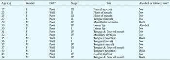
The oral cavity sites affected were the tongue or the floor of the mouth (62.5 per cent), the buccal mucosa (12.5 per cent), the lower lip (12.5 per cent) and the maxillary or mandibular alveolus (12.5 per cent) (Table I).
HIV-Positive patients with oral SCC: data

*Histological differentiation (diff) by Broder's classification. †Tumour-node-metastasis staging. HIV = human immunodeficiency virus; SCC = squamous cell carcinoma; y = years; F = female; M = male
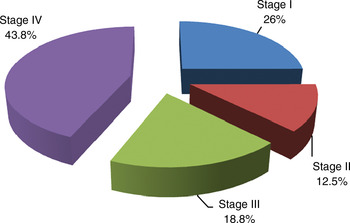
The majority (62.5 per cent) of patients had TNM stage III or IV disease, while the rest (37.5 per cent) had stage I or II disease (Figure 1).
Distribution of tumour-node-metastasis staging in patient population.

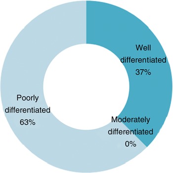
Using Broder's classification, 10 patients had poorly differentiated SCC and six had well differentiated SCC; none had moderately differentiated SCC (Figure 2).
Distribution of histological differentiation in patient population, using Broder's classification.

Discussion
The cancer burden in Africa is likely to rise as a result of increases in HIV-associated malignancies, lifestyle changes due to economic development, and increasing population age.Reference Parkin, Sitas, Chirenje, Stein, Abratt and Wabinga1
Cancers associated with HIV infection tend to present at a more advanced stage, to be diagnosed in younger patients, and to follow a more aggressive clinical course.Reference Haigentz5
The literature associating oral SCC and HIV infection is limited to a few case series, which show a younger age group (median age 40–45 years), more advanced local disease and a higher tumour stage, compared with non-HIV-positive oral SCC patients.Reference Singh, Balwally, Shaha, Rosenfeld, Har-El and Lucente6, Reference Roland, Rothstein and Mittal7 Our results were similar, in that oral SCC presented in younger patients, at a more advanced stage. Epidemiological studies have identified an increased risk of cancers of the oral cavity and lip amongst people infected with HIV; in contrast, there is no strong evidence associating HIV infection with other head and neck cancers.Reference Frisch, Biggar, Engels and Goedert4, Reference Grulich, Wan, Law, Coates and Kaldor8–Reference Grulich, Li, McDonald, Correll, Law and Kaldor11 In the present series, the sites most affected – the tongue and the floor of the mouth – are associated with a poor prognosis.
Other risk factors for SCC include tobacco and alcohol usage, poor nutrition, genetic characteristics, and oncogenic viruses.Reference Shiboski, Shiboski and Silverman12 We suspect that many of our patients did not accurately report their current and previous alcohol and tobacco consumption; therefore, a combination of immunosuppression and other risk factors may have played a key role in the development of oral SCC in these patients.
Similar to our own findings, other authors have reported that SCC of the upper aerodigestive tract may be more aggressive in HIV-infected patients than non-infected individuals.Reference Singh, Sabin, Rofim, Shaha, Har-El and Lucente13 The prognosis of HIV-infected patients with poorly differentiated, TNM stage IV oral SCC is poor. Furthermore, this combination of disease presents a management challenge.
• This study assessed 200 human immunodeficiency virus (HIV) positive patients with oral cavity lesions
• Oral squamous cell carcinoma (SCC) was found in 8 per cent
• Patients' mean presentation age was 31.7 years (range, 17–43), much younger than non-HIV-positive oral SCC patients
Human immunodeficiency virus positive patients treated with highly active antiretroviral therapy have a longer life span, and their HIV infection may predispose them to the eventual development of malignancy. This may influence the overall morbidity and mortality associated with HIV infection.
Findings from the present series may indicate that further research is needed to investigate patterns of oral SCC in HIV-infected patients, compared with non-infected individuals.