Implications
In recent years, the ban on antibiotics contributed to increased prevalence of enteric diseases in the farms, causing epidemiological and economic damage in the poultry industry. In post-antibiotics era, prebiotics have been proposed as a solution to the intestinal problems of poultry. This work demonstrates that in ovo injection of different prebiotics increases number of positive bacteria in chicken feces and improves overall broilers performance. Different routes of prebiotics delivery (in ovo v. in-water v. in ovo+in-water) had comparable impact on performance traits; so a single in ovo injection of prebiotics can replace prolonged in-water supplementation post hatching.
Introduction
Prebiotics are commonly used in poultry production to stimulate growth and development of a healthy microflora in chickens. By definition, prebiotics contain fermentable oligosaccharides that selectively stimulate growth of beneficial bacteria in the guts. In general, they share common properties, including resistance to acidity, hydrolysis and absorption in the gastrointestinal tract; fermentability by the intestinal microflora and expression of the positive effect on the growth and activity of beneficial intestinal microorganisms (Gibson and Fuller, Reference Gibson and Fuller2000; Roberfroid, Reference Roberfroid2007).
There are different ways to deliver prebiotics into avian gastrointestinal tract; but, to achieve desired efficacy, prebiotics must be administered to an animal as early in life as possible. Conventionally, in-feed or in-water supplementation has been used at first hours/days post hatching. However, this approach relies on amount of feed and/or water intake, the quality of water (chlorinated) and other experimental factors (Waldroup et al., Reference Waldroup, Fritts and Fengland2003; Ciesiolka et al., Reference Ciesiolka, Gulewicz, Martinez-Villaluenga, Pilarski, Bednarczyk and Gulewicz2005; Schneitz, Reference Schneitz2005; Biggs et al., Reference Biggs, Parsons and Fahey2007; Midilli et al., Reference Midilli, Alp, Kocabagli, Muglali, Turan, Yilmaz and Cakir2008; Huyghebaert et al., Reference Huyghebaert, Ducatelle and Van Immerseel2011). As a consequence, consumed dose of prebiotics varies in the first hours/days after hatching. Moreover, during early post-hatching period, infection of chicks by detrimental bacteria is also possible. Therefore, in ovo approach for injection of prebiotics directly to the incubating egg has been developed. It allows for a precise delivery of the bioactive substance to all embryos at early stage of development, which unifies the effects of prebiotics across the flock and assures proper development of gut microflora in all chicks. Based on chemical and physical features and dose of the injected substance, different site of injection (i.e. the embryo, the amnion, the allantois, the air cell or the yolk sac) and embryo age (0, 12, 17 or 18 days of incubation) have been used (Casas-Perez and Edens, Reference Casas-Perez and Edens1995; Pilarski et al., Reference Pilarski, Bednarczyk, Lisowski, Rutkowski, Bernacki, Wardenska and Gulewicz2005; Uni et al., Reference Uni, Ferket, Tako and Kedar2005; Cheled-Shoval et al., Reference Cheled-Shoval, Amit-Romach, Barbakov and Uni2011; Ebrahimnezhad et al., Reference Ebrahimnezhad, Salmanzadeh, Aghdamshahryar, Beheshti and Rahimi2011).
In our earlier studies, we have determined that day 12 of incubation is the optimal time for prebiotic injection into the air cell of the incubating egg (Villaluenga et al., Reference Villaluenga, Wardenska, Pilarski, Bednarczyk and Gulewicz2004). At this time, embryo is totally immersed in amniotic fluid. Allantochorion is completely developed and highly vascularized, allowing for transfer of the bioactive solution from air cell to embryonic gastrointestinal tract. This method has been successfully used for prebiotic (Pilarski et al., Reference Pilarski, Bednarczyk, Lisowski, Rutkowski, Bernacki, Wardenska and Gulewicz2005; Bednarczyk et al., Reference Bednarczyk, Urbanowski, Gulewicz, Kasperczyk, Maiorano and Szwaczkowski2011) or synbiotic (Maiorano et al., Reference Maiorano, Sobolewska, Cianciullo, Walasik, Elminowska-Wenda, Slawinska, Tavaniello, Zylinska, Bardowski and Bednarczyk2012; Slawinska et al., Reference Slawinska, Siwek, Zylinska, Bardowski, Brzezinska, Gulewicz, Nowak, Urbanowski, Plowiec and Bednarczyk2014a and Reference Slawinska, Siwek and Bednarczyk2014b; Madej and Bednarczyk, Reference Madej and Bednarczyk2016; Madej et al., Reference Madej, Stefaniak and Bednarczyk2015; Pruszynska-Oszmalek et al., Reference Pruszynska-Oszmalek, Kolodziejski, Stadnicka, Sassek, Chalupka, Kuston, Nogowski, Mackowiak, Maiorano, Jankowski and Bednarczyk2015) in ovo delivery. As a consequence, in ovo delivery of prebiotics not only have improved performance traits, such as the growth rate, feed intake (FI), nutrient digestibility (Bednarczyk et al., Reference Bednarczyk, Urbanowski, Gulewicz, Kasperczyk, Maiorano and Szwaczkowski2011) and meat quality (Maiorano et al., Reference Maiorano, Sobolewska, Cianciullo, Walasik, Elminowska-Wenda, Slawinska, Tavaniello, Zylinska, Bardowski and Bednarczyk2012), but also significantly increased total activity of pancreatic enzymes (amylase, lipase and trypsin) (Pruszynska-Oszmalek et al., Reference Pruszynska-Oszmalek, Kolodziejski, Stadnicka, Sassek, Chalupka, Kuston, Nogowski, Mackowiak, Maiorano, Jankowski and Bednarczyk2015) and influence immune system development and function (Slawinska et al., Reference Slawinska, Siwek and Bednarczyk2014b; Madej and Bednarczyk, Reference Madej and Bednarczyk2016; Madej et al., Reference Madej, Stefaniak and Bednarczyk2015; Plowiec et al., Reference Plowiec, Slawinska, Siwek and Bednarczyk2015).
Evaluation of prebiotics for in ovo injection comprises of few steps. First, oligosaccharide has to prove complete solubility in physiological salt. Only fully solved prebiotics can be precisely injected in ovo and pass the egg membrane into the bloodstream and guts of the embryo. Second, prebiotic has to be delivered in ovo in a specific dose that assures high hatchability of the eggs and microflora development already at hatching. Third, prebiotics should confer beneficial properties to the host in performance and fitness traits. So far, our ‘golden standard’ for in ovo injection was raffinose family oligosaccharides (RFO) extracted from lupin, which assures long-term maintenance of a high level of intestinal bifidobacteria at hatching (Villaluenga et al., Reference Villaluenga, Wardenska, Pilarski, Bednarczyk and Gulewicz2004; Pilarski et al., Reference Pilarski, Bednarczyk, Lisowski, Rutkowski, Bernacki, Wardenska and Gulewicz2005) and optimal performance of broiler chickens (Bednarczyk et al., Reference Bednarczyk, Urbanowski, Gulewicz, Kasperczyk, Maiorano and Szwaczkowski2011). However, there are many other biologically active oligosaccharides available, that could be validated for in ovo injection.
The aim of this study was to evaluate applicability of different prebiotics to in ovo injection using a two-step evaluation (Trials 1 and 2). The aim of the first trial was to select the doses best suited for in ovo administration and to estimate their effects on the hatchability and the bacteriological status of the hatched chickens. The objective of the second trial was to define the optimal route of prebiotic administration by evaluating the influence of prebiotics on broilers’ performance using different routes of delivery (in ovo v. in-water v. in ovo and in-water combined).
Material and methods
Prebiotics
Three prebiotics were selected, based on their solubility in physiological saline: DN (DiNovo®, Bioatlantis Ltd, Tralee, Co., Kerry, Ireland) an extract of beta-glucans obtained from algae; BI (Bi2tos, Clasado Ltd, Sliema, Malta) a non-digestive trans-galactooligosaccharides (GOS) from milk lactose digested with Bifidobacterium bifidum NCIMB 41171; and RFO (in-house) extracted from lupin (Lupinus luteus) seeds (Gulewicz et al., Reference Gulewicz, Ciesiolka, Frias, Vidal-Valverde, Frejnagel, Trojanowska and Gulewicz2000). DN is an extract from Laminaria spp. containing laminarin (>30% w/w β (1–3, 1–6)-glucan) and fucoidan, at a ratio of 85 : 15. BI is composed of 45% lactose, 9.9% disaccharides (Gal (β 1–3)-Glc; Gal (β 1–3)-Gal; Gal (β 1–6)-Gal; Gal (α 1–6)-Gal), 23.1% trisaccharides (Gal (β 1–6)-Gal (β 1–4)-Glc; Gal (β 1–3)-Gal (β 1–4)-Glc), 11.55% tetrasaccharides (Gal (β 1–6)-Gal (β 1–6)-Gal (β 1–4)-Glc) and 10.45% pentasaccharides (Gal (β 1–6)- Gal (β 1–6)-Gal (β 1–6)-Gal (β 1–4)-Glc) (Tzortzis et al., Reference Tzortzis, Goulas, Gee and Gibson2005). RFO solution contains 6.1% sucrose, 9.4% raffinose, 65.2% stachyose, 18.0% verbascose and 1.3% other saccharides (Bednarczyk et al., Reference Bednarczyk, Urbanowski, Gulewicz, Kasperczyk, Maiorano and Szwaczkowski2011).
Trial 1: dose optimization of DiNovo® and Bi2tos for in ovo injection
In Trial 1, two commercial prebiotics (BI and DN) were in ovo injected into the chicken embryo to optimize the doses per embryo for a Trial 2. RFOs had been optimized in our earlier studies (Bednarczyk et al., Reference Bednarczyk, Urbanowski, Gulewicz, Kasperczyk, Maiorano and Szwaczkowski2011). Hatching eggs (60 g average weight) were obtained from the same 32-week-old breeder flock (Ross 308). Eggs were incubated in a commercial broiler hatchery (Drobex-Agro Sp. z o.o., Solec Kujawski, Poland). On day 12 of incubation, before the injection, the eggs were candled to select only the ones containing viable embryos. Eggs were randomly divided into three experimental groups: two groups were treated with different doses of the two prebiotics administered in ovo, and the control group.
The trial was conducted in three replicates, consisting of 100 (in the first replicate) and 197 to 200 (in the second and third replicates) eggs per prebiotic-treated and control groups. Injections solution consisted of 0.2 ml of physiological saline and different testing doses of prebiotic: 0.18, 0.88, 3.5 and 7.0 mg/embryo. Control group was injected with 0.2 ml of physiological saline. Solutions of prebiotics were injected in ovo using dedicated automatic system (Bednarczyk et al., Reference Bednarczyk, Urbanowski, Gulewicz, Kasperczyk, Maiorano and Szwaczkowski2011). First, hole was made in the air cell of the egg; 0.2 ml solution was deposited in air cell and the egg was sealed. After that incubation proceeded using the standard hatching procedure. At hatching, number of healthy chicks was scored for each replicate experiment. The values were expressed as a percentage of the total number of injected eggs (a total of 3995 eggs).
Fresh meconium and feces samples were collected from chicks at each batch of hatching (three batches total). For each batch, samples from 20 randomly selected chicks were pooled. Each sample was analyzed in three technical replicates.
Counts of Bifidobacterium spp. and Lactobacillus spp. were determined based on the EN 15785 and EN 15787 protocol, respectively. Pooled material was first weighed and buffered. Peptone water (Argenta Mikrobiologia Sp. z o.o., Poznan, Poland) was added to each sample as a diluent at 1 : 9 sample-to-buffer (g/ml) ratio. Subsequently, 10-fold dilutions were prepared using 9 ml of buffered peptone water plus 1 ml of starting material. This operation was repeated until reaching 10−9 dilution.
To count Bifidobacterium, 0.1 ml suspension of inocula were aspirated from the prepared serial dilutions, each inoculated to a de Mann-Rogosa-Sharpe substrate with reduced pH of 5.7 (Argenta Mikrobiologia Sp. z o.o.) and distributed in drop-wise manner over surface of the culture plate. Culture plates were placed in an anaerobic atmosphere (Argenta Mikrobiologia Sp. z o.o.) and incubated at 37°C for 36 to 48 hours. After incubation the colonies were counted at the appropriate dilutions and confirmatory tests were carried out. For this purpose, five out of the most characteristic colonies were picked up from each plate and subjected to Gram staining. Bifidobacterium spp. colonies were defined based on the morphological assessment of the test-derived candidate colonies.
To count Lactobacillus ssp., sample dilutions were prepared as described above. Culture plates were incubated in anaerobic atmosphere at 37°C for 36 to 72 hours. After incubation the colonies were counted at the appropriate dilutions and confirmatory tests were carried out. For this purpose, five out of the most characteristic colonies were picked up from each plate and subjected to Gram staining and catalase production test. All catalase-negative bacteria, morphologically corresponding to gram-positive non-sporous bacilli were considered as belonging to Lactobacillus spp. family.
Trial 2: comparison between in ovo, in-water and in ovo+in-water routes of prebiotics delivery
In Trial 2, three prebiotics (DN, BI and RFO) were used for comparison between different routes of delivery: (T1) in ovo injection, (T2) in ovo injection combined with in-water delivery and (T3) in-water delivery. Control group (C) was injected in ovo with physiological saline only and did not receive any prebiotic in-water. Hatching eggs were collected from the same breeder flock and incubated in the same commercial broiler hatchery as in Trial 1.
In ovo injection (groups T1 and T2) was carried out in the same manner as in Trial 1. At day 12 of incubation 1500 eggs containing viable embryos were randomly allotted into four experimental groups (375 eggs/group). Eggs were injected in ovo with 0.2 ml solution containing 3.5 mg/embryo BI, 0.88 mg/embryo DN and 1.9 mg/embryo RFO. After hatching chicks were sexed and 600 males (42.0 g average weight) were randomly assigned to 10 experimental groups (60 males/group): T1 (DN, BI and RFO), T2 (DN, BI and RFO), T3 (DN, BI and RFO) and C. Birds were grown to 42 day of age in collective cages (n=6 replicate cages, 10 birds in each cage). Chicks from T1 and C groups were raised without any additional supplementation with prebiotic. T2 and T3 groups were supplemented in-water with respective prebiotic (DN, BI or RFO) for first 7 days of life. Those animals received 12 ml of the prebiotic dissolved in water per pen (20 mg of prebiotic/ml).
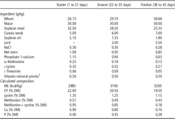
Birds were reared according to the Polish Local Ethical Commission (No 22/2012. 21.06.2012) and in accordance with the animal welfare recommendations of European Union directive 86/609/EEC, in an experimental poultry house Drobex-Agro (Solec Kujawski, Poland) that provided good husbandry conditions (e.g. stocking density, litter, ventilation). Animals were fed ad libitum the standard commercial feed mixtures (Table 1): starter (day 1 to 21), grower (day 22 to 35), finisher (day 35 to 42). Along the rearing period, chickens were weighed and counted within each cage. BW gain (BWG), FI, feed conversion ratio (FCR=FI/BW on pen basis) and mortality were calculated.
Composition and nutrient content of diets

ME=metabolizable energy; DM=dry matter.
1 Provided per kilogram of diets: vitamin A 12 500 IU, vitamin D3 4500 IU, vitamin E 45 mg, vitamin K3 3 mg, vitamin B1 3 mg, vitamin B2 6 mg, vitamin B6 4 mg, pantothenic acid 14 mg, nicotinic acid 50 mg, folic acid 1.75 mg, choline 1.6 g, vitamin B12 0.02 mg, biotin 0.2 mg, Fe 50 mg, Mn 120 mg, Zn 100 mg, Cu 15 mg, J 1.2 mg, Se 0.3 mg, phytase 500 FTU.
Statistical analyses
Growth performance, FI, FCR and mortality data were evaluated by ANOVA (SPSS/PC1Statistics 18.0, SPSS Inc., Chicago, IL, USA, 2010). Differences among groups were determined by contrasts (prebiotic contrast 1: 3×μ C−μ DN−μ BI−μ RFO=0; contrast 2: 2×μ DN−μ BI−μ RFO=0; contrast 3: μ BI−μ RFO=0. Mode of prebiotics’ administration contrast 1: 3×μ C−μ T1−μ T2−μ T3=0; contrast 2: 2×μ T1−μ T2−μ T3=0; contrast 3: μ T2−μ T3=0; µ=overall mean). Hatchability, number of Bifidobacteriaceae and Lactobacillaceae were analyzed by one-way ANOVA using the same statistical package. Differences among the means were determined with Scheffe’s test. The cage was considered the experimental unit.
Results
Trial 1: dose optimization of DiNovo® and Bi2tos for in ovo injection
Hatchability results are presented in Table 2. Hatchability scored for tested doses of 0.18, 0.88 and 3.5 mg of prebiotics/embryo was similar (P>0.05) among control, DN and BI groups. For both prebiotics, the highest tested dose (7.0 mg/embryo) decreased hatchability, resulting in 71.4% hatched chicks for BI (P>0.05) and 56.5% for DN (P<0.05). However, optimal doses of prebiotics were defined as the highest one which did not reduce hatchability (as compared with a control group) and were determined as 0.88 mg/embryo for DN and 3.5 mg/embryo for BI.
Dose effect of prebiotics delivered in ovo on chicks hatchability

a,bMeans within a row with different letters are significantly different (P<0.05).C=Control, in ovo injection of physiological saline; DN=DiNovo®; BI=Bi2tos.
Significance: ns=P>0.05; **P<0.01.
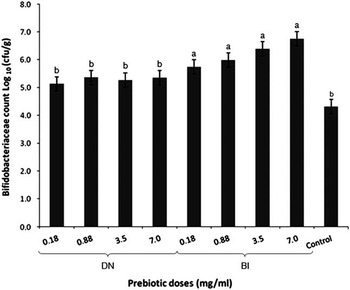
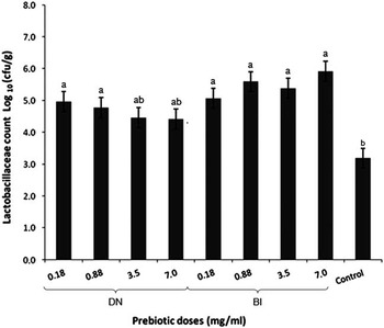
Figures 1 and 2 illustrate impact of prebiotics DN and BI delivered in ovo on the number of Bifidobacterium and Lactobacillus. Effect of DN on bifidobacteria count was not statistically significant compared with the control group (P>0.05). Bifidobacteria count increased in all BI groups (P<0.05) (Figure 1). In case of lactobacilli, differences (P<0.05) were scored between control group and DN for lower doses (0.18 and 0.88 mg/embryo). BI increased (P<0.05) the number of Lactobacillus at all doses compared with the control group (Figure 2).
Impact of prebiotics delivered in ovo on Bifidobacteriaceae count in feces of 1-day-old chicks. DN=DiNovo® L; BI=Bi2tos; Control=in ovo injection of physiological saline. Prebiotic effect: P<0.01. a,bMeans with different letters are significantly different (P<0.05).

Impact of prebiotics delivered in ovo on Lactobacillae count in feces of 1-day-old chicks. DN=DiNovo® L; BI=Bi2tos; Control=in ovo injection of physiological saline. Prebiotic effect: P<0.01. a,bMeans with different letters are significantly different (P<0.05).

Trial 2: comparison between in ovo, in-water and in ovo+in-water routes of prebiotics delivery
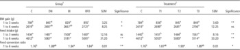
Effects of DN, BI and RFO prebiotics and three different routes of delivery (T1, T2 and T3) on BWG, FI and FCR of broiler chickens are presented in Table 3. Mortality of the chickens during this trial was low (ranged from 2.2% to 5.0%) and not dependent on type of prebiotics or route of delivery.
Effects of prebiotics delivered in ovo, in-water and in ovo combined with in-water on performance traits of broiler chickens

a,bMeans within a row with different letters are significantly different (P<0.05).
1 Group: C=Control, in ovo injection of physiological saline; DN=DiNovo®; BI=Bi2tos; RFO=raffinose family oligosaccharides.
2 Treatment: C=Control, in ovo injection of physiological saline; T1=in ovo; T2=in ovo+in-water; T3=in-water.
Significance: ns=P>0.05; *P<0.05; **P<0.01.
All prebiotics (DN, BI and RFO) significantly improved BWG within the first 3 weeks of life, irrespective of route of delivery (T1, T2 or T3), as compared with the control group (P<0.05). During this period, chickens from RFO group were heavier than those of BI group (P<0.05). Considering route of delivery, BWG was similar among treated groups (P>0.05) but higher compared with control group (P<0.05). During the whole rearing period (1 to 6 weeks), BWG was higher in RFO group in comparison with the control group (P<0.05), whereas DN and BI groups did not differ from control (P>0.05). On the other hand, no effect of administration mode was found for BWG in this period (P>0.05).
Type of prebiotics injected in ovo did not have any significant effect on FI during first 3 weeks of life; whereas, birds of T3 group (in-water) showed higher FI compared with the other experimental groups (P<0.05). Considering the whole rearing period (1 to 6 weeks), prebiotics-treated groups were characterized by higher FI compared with the control group (P<0.05), with the highest values for DN and BI. Similarly, route of prebiotic administration had effect on FI showing higher value for all the prebiotic-treated groups (T1, T2 and T3) compared with control group (P<0.05). However, there were no differences between T1, T2 or T3.
Chickens from the control group showed overall better FCR compared with BI group (1.76 v. 1.94, respectively; P<0.05), with intermediate values for DN and RFO (P>0.05). Moreover, RFO group had lower FCR than BI (P<0.05). The combination of in ovo and in-water administration of prebiotics (T2) resulted in a significantly higher FCR as compared with the chickens of control group (P<0.05); whereas T1 and T3 groups had intermediate FCR values (P>0.05).
Discussion
In this study, we have optimized conditions of prebiotics in ovo delivery and validated this method for broiler chickens production by comparison with in-water supplementation. Prebiotics were sourced from different materials, that is, marine algae (DN), cow milk (BI) or plant seeds (RFO), and differing in biological composition and bioactive properties.
DN is an extract of Laminaria spp., a seaweed containing laminarin. A major active compound of a Laminaria seaweed extract used in this study is a low-molecular-weight polysaccharide containing beta-glucans, effective pro-immunological modulator working through the gut (Vetvicka and Oliveira, Reference Vetvicka and Oliveira2014). Preparations of a soluble polysaccharide laminarin (β-(1,3)-(1,6)-β-glucan), supplemented in-feed, have showed a positive effect on productive traits in monogastrics. In fact, an improvement of the oxidative stability of broiler meat (Ahmed et al., Reference Ahmed, Mun, Islam and Yang2014) and a reduction of the inflammatory factors that improved the morphology of intestine (Heim et al., Reference Heim, Sweeney, O’Shea, Doyle and O’Doherty2015) have been reported. Beta-glucans have been shown to exhibit prebiotic properties by increasing numbers of intestinal Bifidobacterium and Lactobacillus spp. (Jaskari et al., Reference Jaskari, Kontula, Siitonen, Jousimies-Somer, Mattila-Sandholm and Poutanen1998). More recently, Laminaria spp.-derived laminarin has been shown to increase intestinal Lactobacilli numbers in weaned pigs and also reduce coliforms (Murphy et al., Reference Murphy, Dal Bello, O’Doherty, Arendt, Sweeney and Coffey2013). Fucoidan has also been shown to have prebiotic effects in the porcine monogastric model (Lynch et al., Reference Lynch, Sweeney, Callan, O’Sullivan and O’Doherty2010; Sweeney et al., Reference Sweeney, Dillon, Fanning, Egan, O’Shea, Figat, Gutierrez, Mannion, Leonard and O’Doherty2011).
BI belongs to GOS prebiotics, that are produced from lactose by enzymatic digestion with glycoside hydrolases (Torres et al., Reference Torres, Goncalves, Teixeira and Rodrigues2010). GOS used in this study (BI) was synthesized using enzymes from Bifidobacterium bifidum NCIMB 41 171, which is a common intestinal human bacteria. GOS generated with those enzymes have stronger affinity to stimulate growth of bifidobacteria and as such expressed better prebiotic activities based on in vitro studies in pigs and in humans (Tzortzis et al., Reference Tzortzis, Goulas, Gee and Gibson2005; Depeint et al., Reference Depeint, Tzortzis, Vulevic, I’Anson and Gibson2008; Tzortzis, Reference Tzortzis2009). GOS have been known to increase bacterial populations in poultry when supplemented in-feed (Jung et al., Reference Jung, Houde, Baurhoo, Zhao and Lee2008). This prebiotic was also reported to mitigate harmful effects of heat stress in chickens and in mice by stabilizing intestinal integrity in jejunum and alleviating associated inflammatory responses (Akbari et al., Reference Akbari, Braber, Alizadeh, Verheijden, Schoterman, Kraneveld, Garssen and Fink-Gremmels2015; Varasteh et al., Reference Varasteh, Braber, Akbari, Garssen and Fink-Gremmels2015).
RFO are synthesized from sucrose in plants, where they are used for storage and transportation as well as they were reported to play a role in draught tolerance (Sprenger and Keller, Reference Sprenger and Keller2000; Hincha et al., Reference Hincha, Zuther and Heyer2003). As a prebiotic, RFO have been used in chickens in combination with in ovo method of delivery, also in combination with probiotic bacteria. One of the recently studied properties of RFO is protective mechanism against oxidative stress and related liver damage in mice (Zhang et al., Reference Zhang, Zhao, Sun, Lu and Yang2013). Studies in which a model of the chicken embryo is used to compare the biological activity of various prebiotics require determination of the optimal dose for in ovo injection (Schneitz, Reference Schneitz2005). Hereby, we demonstrated two simple screening tests proved efficient to determine doses of prebiotics; that is, hatchability percentage and basic microbiological screening. For in ovo method to be accepted by the industry, hatchability should be as high as possible. In our study, hatchability was not influenced by in ovo treatment until reaching the critical dose, which caused a drop in hatchability, either numerically (3.5 mg of DN and 7.0 mg/embryo BI) or statistically (7.0 mg/embryo of DN; P<0.05). However, many papers report decrease in overall hatchability as a result of in ovo injection of different solutions, for instance glucose (Salmanzadeh, Reference Salmanzadeh2012), royal jelly extracts (Moghaddam et al., Reference Moghaddam, Borji and Komazani2014) or avian influenza vaccines (Cai et al., Reference Cai, Song, Ye, Shao, Padmanabhan, Sutton and Perez2011). The growing embryo is susceptible to homeostatic disturbances and such factors as site of injection, embryo age, solution sterility and immune response to the bioactive factor. Our study shows that the optimal dose of prebiotic solution delivered at day 12 of incubation assures proper growth conditions for the growing embryo.
Second criterion used for prebiotic dose selection in Trial 1 (bacteria count) represents the measure of the overall biological activity. In case of prebiotics, the latter one is defined as the level of intestinal colonization by beneficial bacteria. In Trial 1, both prebiotics injected in ovo (DN and BI) stimulated number of lactobacilli and bifidobacteria in chicken feces. This observation confirmed our earlier findings, that a single in ovo prebiotic (oligosaccharides) injection into 12-day-old chicken embryo leads to an increased number of bifidobacteria at the moment of hatching; it also ensures the long-term maintenance of a high level of intestinal bifidobacteria (Villaluenga et al., Reference Villaluenga, Wardenska, Pilarski, Bednarczyk and Gulewicz2004; Pilarski et al., Reference Pilarski, Bednarczyk, Lisowski, Rutkowski, Bernacki, Wardenska and Gulewicz2005). The mechanism of this process is of considerable interest, particularly in the light of the commonly held opinion that embryonic development of the chicken happens in a sterile environment (Amit-Romach et al., Reference Amit-Romach, Sklan and Uni2004). However, in contrary to the existing paradigm, some authors have indicated that the gastrointestinal tract of the embryo is not sterile. In fact, Deeming (Reference Deeming2005) found that microorganisms might be internalized from yolk at the 18 day of embryonic development. Pedrosa (Reference Pedrosa2009) discovered that the embryo’s intestinal tract is far from being sterile and the pioneer microbial community demonstrates signs of evolution in the last 4 to 5 days before the hatch.
In summary, results of Trial 1 showed that the number of beneficial bacteria increased with the doses of the both (BI and DN) applied prebiotics. As a consequence, and taking into account both criteria (the number of bacteria and the hatching results) we decided to choose the dose of 0.88 mg/embryo for DN and 3.5 mg/embryo for BI for the Trial 2. RFO dose had been optimized earlier, therefore it was not included in this trial.
Results of Trial 2 regarding BWG indicated a significant increase in all prebiotic-treated groups compared with control at week 3. These results provide further support for the hypothesis concerning well-established growth promoting effect of dietary prebiotics, attributed to their ability to strongly bind the pathogenic bacteria and decoy pathogens away from the intestinal lining. Positive effects of the in-feed prebiotic supplementation of broiler chickens on the BW was observed by Dizaji et al. (Reference Dizaji, Hejazi and Zakeri2012). However, statistically significant differences were detected no earlier than 42 day of age (finisher period). We find our results in line with values obtained by Shahir et al. (Reference Shahir, Afsarian, Ghasemi and Tellez2014) for BW and FI that increased significantly through whole rearing period in broilers supplied with oligosaccharides at 0.1%/ton of feed. Jung et al. (Reference Jung, Houde, Baurhoo, Zhao and Lee2008) reported significant promotion of Bifidobacteria in intestine after supplementing GOS at 3/25 kg feed but no change in BW, FI and FC. Similar effect of trans-galacatooligosaccharides on broiler performance is shown by Biggs et al. (Reference Biggs, Parsons and Fahey2007). However, in laying hens, supplementation with RFO in blue lupin seed meal at 20% promoted daily egg productivity comprised by decrease in BW and FI (Zdunczyk et al., Reference Zdunczyk, Jankowski, Rutkowski, Sosnowska, Drazbo, Zdunczyk and Juskiewicz2014). The third tested prebiotic, beta-glucan is well established in aquaculture, and treatment with 1% to 2% w/w feed allowed for significant improvement in FCR and increased BW in marine species (Kuhlwein et al., Reference Kuhlwein, Merrifield, Rawling, Foey and Davies2014). In poultry, addition of 0.025% modified beta-glucan did not improve broiler performance apart from increased BWG in starter period of rearing (Jozefiak et al., Reference Jozefiak, Kaczmarek and Rutkowski2008). Whereas, the inclusion of laminarin and fucoidan beta-glucans as prebiotics improved growth parameters in weaned pigs (O’Doherty et al., Reference O’Doherty, Dillon, Figat, Callan and Sweeney2010). In our study, prebiotic-treated chickens show trend for increased FCR. It has been suggested that the effect of prebiotics on chicken growth performance, could be related to metabolism modification linked to an increase in the digestive enzymes activity (Pruszynska-Oszmalek et al., Reference Pruszynska-Oszmalek, Kolodziejski, Stadnicka, Sassek, Chalupka, Kuston, Nogowski, Mackowiak, Maiorano, Jankowski and Bednarczyk2015), the decrease in bacterial enzymes activity and ammonia production along with the improved FI and digestion (Kabir, Reference Kabir2009). Our results indicate a positive stimulation of the broiler BW expressed as soon as in the starter period (1 to 21 days), which might be explained by early supplementation of chicken embryos with prebiotics using in ovo method.
Tendency of the increased FI and FCR in the prebiotics-treated groups could be due to the stimulation of the intestinal microbiota expansion in the chicken guts by the injection of prebiotics during the in ovo development. The enhanced colonization of the intestines with the microflora, apart from significant effects on the host’s growth performance, gut immunity and development, can increase the energy requirements for the maintenance of the bacterial population inhabiting the chickens’ gastrointestinal tract. It was also demonstrated by Mountzouris et al. (Reference Mountzouris, Tsitrsikos, Palamidi, Arvaniti, Mohnl, Schatzmayr and Fegeros2010) that the inclusion of the growing dosages of probiotics into broilers’ feed caused the proportional increase in the FCR, especially in the grower and finisher growth phases. It is generally accepted that the bacterial species inhabiting the gut compete for the nutrients with the host, which results in an higher energy uptake (Furuse and Yokota, Reference Furuse and Yokota1985). Moreover, intestinal microflora requires increased mucus secretion and epithelial cells turnover in the guts, which is accompanied by an extremely high rate of metabolism and protein synthesis, resulting in 23% to 36% of the whole body energy expenditure (Dibner and Richards, Reference Dibner and Richards2005).
Conclusions
In summary, our study has established an elegant protocol for stimulation of the intestinal microflora populations in broiler chickens. It was achieved using a single in ovo prebiotics delivery during embryonic development. We have demonstrated dose optimization method using hatchability and microbiological screening. Moreover, we have validated this method by comparing it with well-established in-water supplementation with prebiotics. By all means, in ovo prebiotics delivery proved to be no different than in-water supplementation or the two methods combined. At the same time, the amount of the prebiotic used was at least 10 times lower in case on in ovo method (3.5 mg BI/embryo in ovo v. 40 mg BI/chick in-water). As such, in ovo method should be further recommended to the poultry industry. The prebiotic that resulted in the best performance traits improvement was RFO. All prebiotics increased FI and FCR compared with control, which is a typical trade-off of the energy and nutrients use by a better developed intestinal microflora; however, as known, the gut health is also associated with a better immune status of the animals. In this basis, we propose that in ovo route of prebiotic delivery can replace prolonged and costly in-water supplementation of the broiler chickens with those bioactive compounds.
Acknowledgements
The research leading to these results has received funding from the European Union’s Seventh Framework Programme managed by REA Research Executive Agency (http://ec.europa.eu/research/rea) (FP7/2007-2013) under grant agreement number 315198. This research was undertaken as part of a project entitled ‘Thrive-Rite: Natural Compounds to enhance Productivity, Quality and Health in Intensive Farming Systems’. Further details are provided on the consortium’s website (www.thriverite.eu) and the EU Commission’s webpage (http://cordis.europa.eu /project/rcn/104395_en.html). The authors thank Czesław Frischke and Anna Frischke-Szulc (Drobex-Agro Sp. z.o.o.) for enabling them to conduct the experiments in production settings and the authors appreciate help of Dr Krajewski (Vetdiagnostica, Sp. z.o.o.) in bacteriological evaluation.