Ascending aortic pseudoaneurysm is a rare and potentially fatal complication after cardiac and thoracic surgeries. The pathology of pseudoaneurysms is due to the weakness of either layer (intima, media, or adventitia) of the aortic wall resulting in wall damage. Reference Razzouk, Gundry and Wang1,Reference Mulder, Van Bockel, Maas, van den Akker and Hermans2,Reference Malvindi, Van Putte, Heijmen, Schepens and Morshuis3 Additionally, any traumatic or infectious event can be associated with the development of ascending aortic pseudoaneurysms. Reference Quevedo, Trinidad, Castellanos, Atianzar, Anwar and Rafeh4 Spontaneous formation of ascending aortic pseudoaneurysms, where no triggering factors were found, were also reported. The incidence of ascending aortic pseudoaneurysms has been reported to be as low as 0.5%, however, higher incidence rate (up to 13%) were reported in a surveillance imaging series of patients after cardiac or aortic root surgeries. Reference Quevedo, Trinidad, Castellanos, Atianzar, Anwar and Rafeh4 In up to 60% of aortic surgeries, ascending aortic pseudoaneurysms occur at the level of the suture line after surgery. Aortic root procedures, such as Bentall, are the most associated procedures, accounting for 55% of cases. Reference Razzouk, Gundry and Wang1,Reference Malvindi, Van Putte, Heijmen, Schepens and Morshuis3
The complication can develop early, delayed, or even late. The presentation varies from myocardial ischaemia due to coronary compression, dysphagia due to compression of the oesophagus, pulsatile mass over the thorax, ulceration of the skin with bleeding, or infection in pseudoaneurysm. Like all pseudoaneurysms, those of the aorta is prone to rupture, thrombosis, distal embolisation, and fistula formation. Reference Agarwal, Ray and Pallavi5
With the technical advancement in CT scan and MRI, it is now possible to determine the precise location, origin, dimension, and extension of the pseudoaneurysm. Reference Agarwal, Ray and Pallavi5 Although there has been a significant improvement in surgical, anaesthetic, cardiopulmonary bypass technique, and post-operative management, the outcomes of surgical management of pseudoaneurysm remain the same. Reference Mulder, Van Bockel, Maas, van den Akker and Hermans2,Reference Agarwal, Ray and Pallavi5 Pseudoaneurysms larger than 55 mm in diameter need emergent intervention. Reference Quevedo, Trinidad, Castellanos, Atianzar, Anwar and Rafeh4 However, those associated with sepsis carry a poor prognosis. Median sternotomy is particularly hazardous, requiring technical modification to prevent complications associated with exposure of the pseudoaneurysm. Various interventional methods to address this condition has been tried with varied success rates. Reference Agarwal, Ray and Pallavi5,Reference Komanapalli, Burch, Tripathy, Slater and Song6,Reference Lin, Bush, Tong, Chaikof, Martin and Lumsden7
We are presenting a case of giant pseudoaneurysm of ascending aorta, and aortopulmonary fistula 22 years after modified BlalockTaussig shunt managed with the placement of Atrial Septal Occluder device.
Case report
A 38-year-old female patient had undergone right modified BlalockTaussig shunt, as a palliative procedure for congenital cyanotic heart disease in the form of tricuspid atresia, 37 years ago. At the age of 16 years, she had also undergone another shunt in the form of a venous graft between the aorta and the right pulmonary artery.
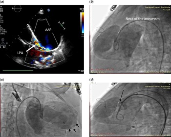
She presented recently with skeletal deformity in the form of kyphoscoliosis, in addition to a left-sided retrosternal chest pain, right upper abdominal pain, and shortness of breath dyspnoea (NYHA functional class III). During the presentation, the patient was a febrile; oxygen saturation on room air was 80%, prominent upper sternal and left thoracic pulsation, enlarged tender liver, and audible continuous murmur. Chest X-ray showed a large mass at the left sternal region with a calcified wall. An echocardiogram revealed a 10 cm echo lucent mass with calcified wall arising from the ascending aorta compressing the main pulmonary artery and left pulmonary branch with a small fistula in between (Fig 1a). The native anatomy was tricuspid atresia, normal relation of the great vessels, single atrium, moderate size muscular VSD, enlargement of left ventricle (internal diameter at end-diastole was 69 mm in diameter) with mild mitral regurgitation. The patient was scheduled for an urgent follow-up CT scan. The scan showed a giant, voluminous bilobed with mural calcification, ascending aortic pseudoaneurysm arising from the anterior aortic aspect with the largest diameter measuring 11 cm. The leaking hole measured 12 mm and the distance from the origin of the neck was 4.5 cm from the right coronary origin. Anterior aspect of the pseudoaneurysm was directly posterior to the sternum and was adherent to the retrosternal region. The pseudoaneurysm had a large cavity extending anteriorly and to the left of the upper ascending aorta. Also adding pressure on both main pulmonary artery and the left pulmonary branch. The giant psudoanurysm was fistulating into the left pulmonary artery and the fistula diameter measured about 4 mm. The patent venous graft was connected between the proximal descending thoracic aorta, directed from left to right then descended vertically to join the right pulmonary branch.
Because of the two previous surgeries, the complex cardiac history, and close the proximity of the pseudoaneurysm to the sternum which carries a high risk of haemorrhage (due to its extension into the soft tissues with the likelihood of impending rupture), surgical treatment was not preferred by the heart team. Therefore, planning an alternative therapeutic approach was considered by performing occlusion of pseudoaneurysm opening with the Atrial Septal Occluder device. The potential of using Septal Occluder device was preferred because of some similarities of anatomical consideration in percutaneous closure of atrial septal defect. That is, somewhat parallel anatomical configuration of a gap in a thin-walled structure with two large chambers on each side.
A written informed consent after explanation of the approach was obtained from the patient.
Procedure description
The procedure was performed under general endotracheal anaesthesia. Right femoral artery access was obtained with 6 Fr introducer sheath and 5000 units of unfractionated heparin were injected intravenously. An ascending aortic angiography was performed with a pigtail catheter 6 Fr and multiple angiograms to define the location of the aneurysm and the angle of take-off of the defect (Fig 1b and c). Left lateral projection was selected because it minimised the overlap of the ascending aorta and the cavity of the aneurysm (Fig 1b). Invasive measurement of pulmonary artery pressure was 40/15 mmHg. A right coronary angiographic catheter (Judkins Right Coronary) was used to engage the orifice of the pseudoaneurysm. The catheter was exchanged with extra stiff guide wire 260 cm (Cook Medical, Bloomington, IN, USA) with 1 cm soft tip which was positioned in the small lobe of the aneurysm. A 20 mm Atrial Septal Defect Occluder device (Lifetech Scientific (Shenzhen) Co., Ltd, China) was selected based on the size of the neck which measured 14 mm, with 10 Fr 80 cm Atrial Septal Defect delivery sheath (Lifetech Scientific (Shenzhen) Co., Ltd, China) and the tip of the delivery system was positioned in the pseudoaneurysm and we try to minimise contact with the wall of the cavity. The insertion dilator was pulled back, the device was inserted, with retraction of the delivery catheter, the left atrial disc was deployed into the cavity with good apposition against the rim of the aortic tissue at the edge of the defect, The delivery catheter was pulled back exposing the centre and right atrial disc The device position was confirmed by fluoroscopy and completion aortogram demonstrated satisfactory positing of the occluder (Fig 1d). The device was released, and the delivery system was removed. The patient tolerated the procedure without immediate complication.
( a ) Transthoracic echocardiogram (parasternal long-axis view) showing a giant ascending aorta pseudoaneurysm fistulating into the left pulmonary artery. ( b ) An ascending aortic angiography in left lateral view has minimised overlap of the ascending aorta and the cavity of the AAP. ( c ) Angiography in RAO projection revealed the large AAP extending to the left of the upper ascending aorta with its cavity edges marked by arrows. ( d ) The bulkiness of the device (arrow) was the result of oversizing needed to stabilise the left atrial disc in AAP cavity.

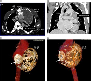
A follow-up echocardiographic examination showed the device in a proper place with closure of the opening between the pseudoaneurysm and the ascending aorta. Three weeks later after the procedure, the patient suffered from dizziness on mild effort and a drop in oxygen saturation to 60% in the supine position. An emergency CT scan revealed that the occluder was well fixed into the neck of the aneurysm with complete exclusion of the thrombosed aneurysm (Fig 2a,c,d), also a closure of the pulmonary fistula with patent pulmonary vasculature without evidence of pulmonary embolism, but still adding pressure on the main pulmonary artery and the left pulmonary branch. The modified BlalockTaussig shunt was still patent (Fig 2b). It seemed that the previous fistula provided some pulmonary blood flow, this explains the drop in oxygen saturation. After discussion with the heart team, where they recommended creation of another shunt to augment pulmonary flow, and excision of the aneurysm. Unfortunately, the patient passed away during the surgery.
Post-operative tomography scan. ( a ) Axial view demonstrates exclusion of the ascending aortic pseudoaneurysm with an Atrial Septal Occluder (ASD occluder). ( b ) Patent venous graft (BT shunt) between the aorta and right pulmonary artery. ( c and d ) A volumetric three-dimensional CT images after device occlusion of pseudoaneurysm. Device was seen in situ (arrow).

Comment
Pseudoaneurysms of the ascending aorta complicate less than 1% of cardiac operation Reference Razzouk, Gundry and Wang1 These lesions arise at the site of aortic wall manipulation during procedures as aortotomy, proximal anastomosis of venous grafts in coronary bypass grafting, needle puncture for measurement or administration of cardioplegic solution, or aortic cannulation to institute cardiopulmonary bypass Reference Quevedo, Trinidad, Castellanos, Atianzar, Anwar and Rafeh4 (as in the case presented). While surgical management is the standard treatment, alternative management should always be considered particularly when the general condition of the patient does not permit surgery. Various case reports have shown promising, immediate, and short-term outcomes. Intervention techniques including coil embolisation and thrombin injection in the sac of pseudoaneurysm have been tried with some complications. Reference Lin, Bush, Tong, Chaikof, Martin and Lumsden7 On the other hand, Septal Occluder device used to occlude the neck of the aneurysm has shown better results. Reference Agarwal, Ray and Pallavi5,Reference Kanani, Neilan, Palacios and Garasic8,Reference Kpodonu, Wheatly, Ramaiah, Rodriguez-Lopez, Strumpf and Diethrich9,Reference Hussain, Strumpf, Wheatley and Diethrich10 No fatal complications have been reported in any case. The potential complication of the procedure could be rupture of pseudoaneurysm during wire placement or delivery of the devices as the wall of the aneurysm is formed by one or two stretched layers of the vessel or haematoma.
The prerequisites of successful device occlusion would be the presence of a relatively narrow communication between the aorta and the pseudoaneurysm, a distance from important vessels (to ensure placement of the device without obstructing the origin of branches) and a spacious sac of the pseudoaneurysm (to allow positioning of the wire and the sheath and to accommodate the disc of the device).
Conclusion
This case highlights the importance, after cardiac surgery routine echocardiographic re-evaluation of ascending aorta integrity; even though the presentation of this sequelae can be very much delayed as in our case. Pseudoaneurysm of the ascending aorta following cardiac surgery can be managed by transcatheter closure using an Atrial Septal Occluder in the presence of narrow neck.
Supplementary material
To view supplementary material for this article, please visit https://doi.org/10.1017/S1047951121003358.
Acknowledgements
The authors are grateful for the support received from the staff at the National Heart Institute.
Financial support
This research received no specific grant from any funding agency, commercial, or not-for-profit sectors.
Conflicts of interest
None.
Ethical standards
The Ethics Commitee of the National Heart Institute approved the study. A Written informed consent was obtained from the patient.