

Introduction
The development of pharmacological treatments that may slow or halt the Alzheimer's disease (AD) process has served as an impetus for the early detection of dementia and the accurate identification of prodromal dementia states, including mild cognitive impairment (MCI). Recent work has focused on the identification of biomarkers for early detection, and structural magnetic resonance imaging (MRI) has proven to be a reliable biomarker for predicting future decline (Jack et al., Reference Jack, Knopman, Jagust, Shaw, Aisen, Weiner and Trojanowski2010). Hippocampal volume and other indices of medial temporal lobe (MTL) atrophy are championed as promising MR biomarkers (Craig-Schapiro, Fagan, & Holtzman, Reference Craig-Schapiro, Fagan and Holtzman2009), but hippocampal atrophy may have multiple etiologies (Dhikav & Anand, Reference Dhikav and Anand2012) and early gray matter (GM) changes can be detected beyond the MTL in prodromal AD and MCI (Dickerson et al., Reference Dickerson, Bakkour, Salat, Feczko, Pacheco, Greve and Buckner2009; Wang et al., Reference Wang, Goldstein, Veledar, Levey, Lah, Meltzer and Mao2009). Dickerson and colleagues (Reference Dickerson, Bakkour, Salat, Feczko, Pacheco, Greve and Buckner2009) identified several cortical regions in which thickness is a potential biomarker for AD, referred to as “AD-signature” cortical thickness. This measure has predicted symptom severity, amyloid binding in asymptomatic older adults, conversion from questionable AD to mild AD (Bakkour, Morris, & Dickerson, Reference Bakkour, Morris and Dickerson2009), and progression to AD 10 years after initially identifying cortical thinning in “AD-signature” areas among cognitively normal individuals (Dickerson et al., Reference Dickerson, Stoub, Shah, Sperling, Killiany, Albert and Detoledo-Morrell2011).
Despite strong neuropathological evidence of white matter (WM) changes in AD (Brun & Englund, Reference Brun and Englund1986), the role of WM in the development of AD and its utility as a structural MR biomarker has been largely overlooked. With the advent of diffusion tensor imaging (DTI), numerous studies have demonstrated changes in regional WM integrity in AD (Salat et al., Reference Salat, Tuch, van der Kouwe, Greve, Pappu, Lee and Rosas2010; Stricker et al., Reference Stricker, Schweinsburg, Delano-Wood, Wierenga, Bangen, Haaland and Bondi2009), and decreases in WM integrity are also reliably found before the diagnosis of clinical dementia (Delano-Wood et al., Reference Delano-Wood, Bondi, Jak, Horne, Schweinsburg, Frank and Salmon2010; Fellgiebel et al., Reference Fellgiebel, Muller, Wille, Dellani, Scheurich, Schmidt and Stoeter2005; Parente et al., Reference Parente, Gasparetto, da Cruz, Domingues, Baptista, Carvalho and Domingues2008; Wang et al., Reference Wang, Goldstein, Veledar, Levey, Lah, Meltzer and Mao2009). WM changes may complement GM changes, explaining unique variance in the disease process. For example, the combination of DTI and cortical thickness measures has higher sensitivity and specificity to differentiate amnestic MCI (aMCI) from healthy control participants than either measure in isolation (Wang et al., Reference Wang, Goldstein, Veledar, Levey, Lah, Meltzer and Mao2009), highlighting the need to examine both imaging modalities in MCI. DTI studies of MCI have shown changes in WM integrity, most reliably within medial temporal and parietal regions (Bosch et al., Reference Bosch, Arenaza-Urquijo, Rami, Sala-Llonch, Junque, Sole-Padulles and Bartres-Faz2012; Huang et al., Reference Huang, Fan, Weiner, Martin-Cook, Xiao, Davis and Diaz-Arrastia2012; Huang & Auchus, Reference Huang and Auchus2007; Jacobs, Van Boxtel, Jolles, Verhey, & Uylings, Reference Jacobs, Van Boxtel, Jolles, Verhey and Uylings2012; Medina et al., Reference Medina, DeToledo-Morrell, Urresta, Gabrieli, Moseley, Fleischman and Stebbins2006; Rose et al., Reference Rose, McMahon, Janke, O'Dowd, de Zubicaray, Strudwick and Chalk2006; Scola et al., Reference Scola, Bozzali, Agosta, Magnani, Franceschi, Sormani and Falini2010; Zhuang et al., Reference Zhuang, Wen, Zhu, Trollor, Kochan, Crawford and Sachdev2010) believed to underlie episodic memory (Buckner, Andrews-Hanna, & Schacter, Reference Buckner, Andrews-Hanna and Schacter2008; Walhovd et al., Reference Walhovd, Fjell, Amlien, Grambaite, Stenset, Bjornerud and Fladby2009).
The relative importance of GM versus WM changes in MCI remains unclear. Although GM changes have traditionally been viewed as most important to the AD pathological process and clinical expression, recent research has challenged this assumption and there is increasing evidence of the potential utility of WM integrity as an early biomarker, with longitudinal evidence that it uniquely predicts cognitive decline relative to cerebrospinal fluid (CSF) biomarkers and GM volume (Scola et al., Reference Scola, Bozzali, Agosta, Magnani, Franceschi, Sormani and Falini2010; Selnes et al., Reference Selnes, Aarsland, Bjornerud, Gjerstad, Wallin, Hessen and Fladby2013; Zhuang et al., Reference Zhuang, Sachdev, Trollor, Kochan, Reppermund, Brodaty and Wen2012). In addition, there is evidence that changes in WM integrity may have unique effects that independently or synergistically contribute to effects of GM atrophy; for example, we have previously shown evidence that WM changes persist when statistically controlling for GM measures in AD (Salat et al., Reference Salat, Tuch, van der Kouwe, Greve, Pappu, Lee and Rosas2010; Stricker et al., Reference Stricker, Schweinsburg, Delano-Wood, Wierenga, Bangen, Haaland and Bondi2009). To our knowledge, only three studies have tested whether WM changes persist when controlling for GM in MCI (Bosch et al., Reference Bosch, Arenaza-Urquijo, Rami, Sala-Llonch, Junque, Sole-Padulles and Bartres-Faz2012; Delano-Wood et al., Reference Delano-Wood, Stricker, Sorg, Nation, Jak, Woods and Bondi2012; Selnes et al., Reference Selnes, Aarsland, Bjornerud, Gjerstad, Wallin, Hessen and Fladby2013), with varying methods and results. Bosch and colleagues controlled for total GM volume, which may be insensitive to early AD-related changes, and argued that a majority of their WM findings were explained by GM atrophy. Delano-Wood and colleagues demonstrated that decreased WM integrity of the posterior cingulum persisted after controlling for hippocampal volume or whole-brain volume. Using methods similar to the present study, Selnes and colleagues demonstrated multiple regions with decreased WM integrity in MCI, the majority of which remained significant after controlling for local cortical thickness. The current study will determine whether WM changes persist after controlling for widespread cortical thinning in regions empirically validated as particularly sensitive to the AD disease process. Demonstrating unique effects of WM integrity in MCI, independent from AD-signature cortical thinning, would add to the growing support for an independent contribution of WM integrity to AD by demonstrating this effect in an at-risk MCI sample.
Similarly, preliminary evidence suggests that the relationship between WM integrity and cognition is independent of GM changes (Bosch et al., Reference Bosch, Arenaza-Urquijo, Rami, Sala-Llonch, Junque, Sole-Padulles and Bartres-Faz2012; Delano-Wood et al., Reference Delano-Wood, Stricker, Sorg, Nation, Jak, Woods and Bondi2012; Grambaite et al., Reference Grambaite, Reinvang, Selnes, Fjell, Walhovd, Stenset and Fladby2011), although most studies investigating the relationship between cognition and WM integrity have not controlled for GM. Studies investigating specific cognitive domains have provided initial evidence that decreased WM integrity correlates with memory (Bosch et al., Reference Bosch, Arenaza-Urquijo, Rami, Sala-Llonch, Junque, Sole-Padulles and Bartres-Faz2012; Goldstein et al., Reference Goldstein, Mao, Wang, Ni, Lah and Levey2009; Walhovd et al., Reference Walhovd, Fjell, Amlien, Grambaite, Stenset, Bjornerud and Fladby2009) and executive functioning in MCI (Chen et al., Reference Chen, Chen, Cheng, Hua, Liu and Chiu2009; Grambaite et al., Reference Grambaite, Reinvang, Selnes, Fjell, Walhovd, Stenset and Fladby2011). WM integrity within posterior brain regions, most notably the posterior cingulate, is also related to memory (Delano-Wood et al., Reference Delano-Wood, Stricker, Sorg, Nation, Jak, Woods and Bondi2012; Fellgiebel et al., Reference Fellgiebel, Muller, Wille, Dellani, Scheurich, Schmidt and Stoeter2005; Rose et al., Reference Rose, McMahon, Janke, O'Dowd, de Zubicaray, Strudwick and Chalk2006; Walhovd et al., Reference Walhovd, Fjell, Amlien, Grambaite, Stenset, Bjornerud and Fladby2009). The importance of parietal WM to executive functioning in MCI has also been suggested (Chen et al., Reference Chen, Chen, Cheng, Hua, Liu and Chiu2009; Jacobs et al., Reference Jacobs, Van Boxtel, Jolles, Verhey and Uylings2012; Kim et al., Reference Kim, Park, Ahn, Seo, Lee, Kim and Na2011). Because multiple studies show strong relationships between cortical thickness and cognition (Chang et al., Reference Chang, Bondi, Fennema-Notestine, McEvoy, Hagler, Jacobson and Dale2010; Fjell et al., Reference Fjell, Walhovd, Amlien, Bjornerud, Reinvang, Gjerstad and Fladby2008; Walhovd et al., Reference Walhovd, Fjell, Amlien, Grambaite, Stenset, Bjornerud and Fladby2009), it is important to determine whether relationships between cognition and WM integrity are independent of cortical thinning.
The current study applied retrospective, neuropsychologically based MCI criteria to a sample of normally aging older adults using the “comprehensive” criteria described by Jak, Bondi, et al. (Reference Jak, Bangen, Wierenga, Delano-Wood, Corey-Bloom and Bondi2009) to examine morphometric group differences in GM and WM integrity. The primary aim of this study was to examine whether decreased WM integrity in MCI would persist when controlling for AD-signature cortical thickness. A secondary aim was to determine whether WM integrity contributes to variance in cognitive performance over and above that explained by AD-signature cortical thickness. We hypothesized that, relative to a normal control (NC) group, participants with neuropsychologically defined MCI would exhibit decreased: (1) hippocampal volume, (2) AD-signature cortical thickness, and (3) WM integrity (i.e., lower fractional anisotropy) in a priori selected regions of interest (ROIs), particularly within medial temporal and parietal regions. Importantly, we predicted that differences in WM integrity across groups would be independent of cortical thinning. We also hypothesized that WM integrity of the medial temporal and parietal regions would be significantly related to poorer memory performance, and that WM integrity in parietal regions would be significantly related to poorer executive functioning, over and above AD-signature cortical thinning.
Method
Participants
One hundred thirteen participants underwent neuropsychological testing and structural MRI. Participants were recruited from two overlapping studies conducted at VA Boston Healthcare System. Twenty-nine participants represent a subset (based on their agreement to undergo structural MRI) of a larger sample recruited from the community through the Harvard Cooperative Program on Aging (HCPA) Claude Pepper Older American Independence Center (OAIC) in response to an advertisement appearing in the HCPA newsletter asking for healthy community-dwelling older African Americans. Eighty-four participants were part of the Understanding Cerebrovascular and Alzheimer's Risk in the Elderly (UCARE) program, recruited through the Boston University Alzheimer's Disease Center (BUADC) based on the criteria of being neurologically healthy and having a first-degree family relative with AD. Participants were excluded for the following: history of head trauma of “mild” severity or greater (Holm, Cassidy, Carroll, & Borg, Reference Holm, Cassidy, Carroll and Borg2005; e.g., within our sample loss of consciousness did not exceed 15 min), more than one head injury, any neurological disorder including dementia (i.e., Parkinson's disease, AD, vascular dementia), severe psychiatric illness, or brain surgery. All participants were literate with at least a ninth-grade education. The VA Boston Healthcare System's institutional review board approved the study according to the Helsinki Declaration, and informed consent was obtained from each participant.
MCI Criteria
We applied the “comprehensive” criteria (Jak, Bondi, et al., Reference Jak, Bondi, Delano-Wood, Wierenga, Corey-Bloom, Salmon and Delis2009) for retrospective diagnosis of MCI based on neuropsychological test scores. This required that at least two performances within a cognitive domain fell one standard deviation (SD) or more below normative expectations for that domain to contribute to MCI classification. A cutoff for impairment of 1 SD below normative data was adopted to strike a balance between reliability and sensitivity to detect mild impairment (Heaton, Miller, Taylor, & Grant, Reference Heaton, Miller, Taylor and Grant2004; Jak, Bondi, et al., Reference Jak, Bangen, Wierenga, Delano-Wood, Corey-Bloom and Bondi2009). Participants were excluded if they had MMSE < 24 (Caucasians) or MMSE < 23 (African Americans; Pedraza et al., Reference Pedraza, Clark, O'Bryant, Smith, Ivnik, Graff-Radford and Lucas2012). Participants were classified as aMCI if memory was impaired (either alone or with additional domains impaired) and as nonamnestic MCI (naMCI) if domain(s) other than memory were impaired. Participants were classified as normal controls (NC) if no cognitive domains were impaired (performance on no more than one measure within a cognitive domain fell more than 1 SD below normative data). Using these criteria, 32 participants met criteria for MCI (14 aMCI, 18 naMCI), and 81 participants were classified as NC.
APOE ε4 Genotyping
Genomic DNA was prepared directly from peripheral blood samples using the Pure Gene (Gentra Systems, Minneapolis, MN) DNA extraction kit, with minor modifications. The DNA was stored and an aliquot removed for APOE genotyping. PCR analysis was carried out essentially as described by Wenham, Price, & Blandell (Reference Wenham, Price and Blandell1991).
Neuropsychological Measures
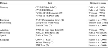
All participants completed a neuropsychological battery assessing cognitive functioning in four domains: memory, attention/processing speed, language, and executive functions. Publically available standard scores were used for group classification (see Table 1). Although the full range of neuropsychological information was necessary for determining MCI diagnosis through use of standard scores, after group classification was completed all analyses used raw scores. Only measures of memory and executive functioning were considered when examining relationships with morphometric measures. Self-report measures included the Geriatric Depression Scale (GDS; Yesavage et al., Reference Yesavage, Brink, Rose, Lum, Huang, Adey and Leirer1982) and the Lawton and Brody instrumental activities of daily living (IADL) questionnaire (Lawton & Brody, Reference Lawton and Brody1969).
Neuropsychological measures and normative sources used for MCI classification

*Participants from the OAIC sample completed Logical Memory but did not complete Visual Reproduction (both were completed in the UCARE sample). Performance on Visual Reproduction has been shown to be more sensitive than Logical Memory for prediction of stable memory deficits in MCI (Teng et al., Reference Teng, Tingus, Lu and Cummings2009) and to predict subsequent AD (Albert et al., Reference Albert, Moss, Tanzi and Jones2001; Griffith et al., Reference Griffith, Netson, Harrell, Zamrini, Brockington and Marson2006; Salmon et al., Reference Salmon, Thomas, Pay, Booth, Hofstetter, Thal and Katzman2002). Therefore we used Visual Reproduction whenever available for group classification, and substituted Logical Memory when it was unavailable (n = 29).
CVLT-II = California Verbal Learning Test—Second Edition; WMS-III = Wechsler Memory Scale—Third Edition; VR = Visual Reproduction; SS = scaled score; WAIS-III = Wechsler Adult Intelligence Scale—Third Edition; Ruff 2&7 = Ruff 2&7 Selective Attention Test; COWAT = Controlled Oral Word Association Test; BNT = Boston Naming Test; WCST = Wisconsin Card Sorting Test.
Mean Arterial Blood Pressure
Blood pressure (BP) was measured in a seated position with the arm at rest at heart level by a laboratory technician using a standard sphygmomanometer after five minutes of rest, and five minutes later. Average systolic and diastolic pressures were computed across the two measurements. Systolic and diastolic blood pressure were considered together to create a mean arterial blood pressure (MABP) using the following formula: MABP: 1/3 (Systolic – diastolic) + diastolic. MABP is a metric commonly used in clinical settings to obtain an accurate metric of overall blood pressure. MABP is believed to indicate perfusion pressure, particularly in body organs, and therefore may be more directly related to brain structure. In addition, prior studies have found significant relationships between MABP and cognition and brain structure in older adults (Brown et al., Reference Brown, McMorris, Longman, Leigh, Hill, Friedenreich and Poulin2008; Guo et al., Reference Guo, Pantoni, Simoni, Bengtsson, Bjorkelund, Lissner and Skoog2009; Leritz, Salat, Milberg, et al., Reference Leritz, Salat, Milberg, Williams, Chapman, Grande and McGlinchey2010). We controlled for this variable in all analyses.
Neuroimaging Protocol
Two participants were scanned using a Siemens 1.5 Tesla Sonata system, with the following parameters: MPRAGE; T1 = 1000 ms, repetition time (TR) = 2.73 s, echo time (TE) = 3.39 ms, flip angle = 7°, slice thickness = 1.33 mm, 128 slices, field of view (FOV) = 256 × 256 mm; DTI: TR = 9000 ms, TE = 68 ms, 60 slices total, acquisition matrix = 128 × 128 (FOV = 256 × 256 mm), slice thickness = 2 mm (for 2 mm3 isotropic voxels) with 0 mm gap, with a b value = 700 s/mm2, 10 T2 and 60 diffusion weighted images, and one image, the T2-weighted “low b” image with a b-value = 0 s/mm2 as an anatomical reference volume. The remaining 111 participants were scanned on the upgraded Siemens 1.5 Avanto System, with slightly different parameters; MPRAGE: T1 = 1000 ms, TR = 2.73 s, TE = 3.31 ms, flip angle = 7°, slice thickness = 1.3 mm, 128 slices, FOV = 256 × 256 mm; DTI: TR = 7200 ms, TE = 77 ms, 60 slices total, acquisition matrix = 128 × 128 (FOV = 256 × 256 mm), slice thickness = 2 mm (for 2 mm3 isotropic voxels) with 0 mm gap, with a b value = 700 s/mm2, 10 T2 and 60 diffusion weighted images, and one image, the T2-weighted “low b” image with a b-value = 0 s/mm2 as an anatomical reference volume.
Image Processing
T1 image processing
Cortical thickness measurements were obtained by first conducting cortical reconstruction using the FreeSurfer image analysis suite, which is freely available for download online (http://surfer.nmr.mgh.harvard.edu/). Methods have been previously described in detail (Dale, Fischl, & Sereno, Reference Dale, Fischl and Sereno1999; Fischl, Sereno, & Dale, Reference Fischl, Sereno and Dale1999); see our previous work for a summary of this process (Leritz, Salat, Williams, et al., Reference Leritz, Salat, Williams, Schnyer, Rudolph, Lipsitz and Milberg2010).
Dickerson et al. (Reference Dickerson, Bakkour, Salat, Feczko, Pacheco, Greve and Buckner2009) used an exploratory map of cortical thinning in mild AD to define a priori ROIs for use in other analyses, termed “AD-signature” cortical thickness. We applied the AD-signature cortical thickness map to our subjects and derived a mean AD-signature cortical thickness value. We used average AD-signature cortical thickness because Bakkour and colleagues (Reference Bakkour, Morris and Dickerson2009) previously demonstrated that this measure best predicted conversion from questionable AD dementia to mild AD dementia relative to hippocampal volume and the MTL thickness ROI.
DTI image processing
Diffusion data were processed using a multistep procedure involving the FreeSurfer image analysis suite and FSL (http://www.fmrib.ox.ac.uk.fsl/) processing streams, specifically Dtifit from FMRIB's Diffusion Toolbox (Behrens et al., Reference Behrens, Woolrich, Jenkinson, Johansen-Berg, Nunes, Clare and Smith2003) and TBSS (Tract-Based Spatial Statistics, Smith et al., Reference Smith, Jenkinson, Johansen-Berg, Rueckert, Nichols, Mackay and Behrens2006), part of FSL (Smith et al., Reference Smith, Jenkinson, Woolrich, Beckmann, Behrens, Johansen-Berg and Matthews2004). See our previous work (Leritz, Salat, Milberg, et al., Reference Leritz, Salat, Milberg, Williams, Chapman, Grande and McGlinchey2010; Salat et al., Reference Salat, Tuch, van der Kouwe, Greve, Pappu, Lee and Rosas2010) for additional details.
ROIs limited to the TBSS skeleton were created using T1-based WM parcellations automatically created during the FreeSurfer processing stream (Salat et al., Reference Salat, Lee, van der Kouwe, Greve, Fischl and Rosas2009). These regional measures were based on gyral folding patterns (Desikan et al., Reference Desikan, Segonne, Fischl, Quinn, Dickerson, Blacker and Killiany2006), which were subsequently diffused from the cortex into the subjacent WM, resulting in a WM parcellation for each gyral label, unique to each individual's anatomy (see Salat et al., Reference Salat, Williams, Leritz, Schnyer, Rudolph, Lipsitz and Milberg2012). Registration of the T1 image to the low b volume was performed using the FreeSurfer bbregister tool (Greve & Fischl, Reference Greve and Fischl2009), a procedure that uses tissue contrast (gray/white matter) as the basis of the registration cost function. Use of the FreeSurfer derived segmentations provided definition of WM regions directly beneath the cortex (Fischl et al., Reference Fischl, Salat, Busa, Albert, Dieterich, Haselgrove and Dale2002; Salat et al., Reference Salat, Lee, van der Kouwe, Greve, Fischl and Rosas2009). ROIs were selected to include those within medial temporal and parietal regions known to be affected early in the AD process, with additional preference given to WM regions underlying those regions that contributed to the AD-signature cortical thickness ROIs (Dickerson et al., Reference Dickerson, Bakkour, Salat, Feczko, Pacheco, Greve and Buckner2009). ROIs included WM underlying the following cortical regions: entorhinal, parahippocampal (combined for some analyses into average medial temporal); posterior cingulate, precuneus, supramarginal, superior parietal (combined for some analyses into average parietal); transverse temporal, superior temporal, middle temporal, inferior temporal, temporal pole, and superior frontal. Fractional anisotropy (FA), a representation of the degree of intravoxel coherence, is a commonly used metric of WM integrity, particularly in studies of MCI and AD. Lower FA indicates lower white matter integrity. Average FA was derived for each WM ROI. An average FA of these ROIs was also created to provide an average measure of FA comparable to the AD-signature cortical thickness summary measure (no empirically derived FA-signature ROI has yet been created). White matter ROI values were extracted from voxels limited to the TBSS skeleton to reduce the influence of partial volume contamination. The ROI-segmented mean skeleton was deprojected from TBSS standard space to each participant's native diffusion volume using the inverse of the participant's transform to standard space to extract native values. Statistics were then extracted from each participant's native diffusion maps, with each segmentation comprised of voxels representing the center of WM tracts within each region. Right and left hemisphere ROIs were combined.
Statistical Analyses
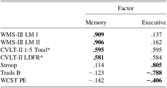
Group comparisons were performed with analysis of covariance (ANCOVA) to test for differences (NC vs. MCI) in morphometric variables, controlling for age, education, and MABP. In addition, similar ANCOVAs were performed for morphometric variables showing significant group differences, also controlling for AD-signature cortical thickness. To examine the relationship between ROIs and cognition, factor scores were created to reduce the number of neuropsychological variables. Raw scores for each variable contributing to the Memory and Executive domains for the MCI classification criteria were submitted into a principal components analysis using varimax rotation with two fixed factors; the minimum eigenvalue for extraction was set at 1. The two factors explained a total of 65% of the variance. The first factor extracted was called “Memory,” and explained 50% of the variance. Because California Verbal Learning Test - Second Edition (CVLT-II) variables showed comparable loadings on both factors, they were assigned to the Memory factor based on our conceptual understanding of these measures (see Table 2). The second factor was called “Executive,” explaining 15% of the variance. The demonstrated factor solution corresponded with the domains used for MCI classification. Factor scores were saved. Hierarchical multiple regressions were completed to determine the relative contribution of WM integrity and AD-signature cortical thickness to the Memory and Executive factors scores. Age, education, and MABP were entered into the first step, AD-signature cortical-thickness was entered into the second step, and WM integrity was entered in the third step. Six hierarchical regressions were run in total, three for each cognitive factor score, with average FA, medial temporal FA, and parietal FA entered in the last step of each model. A significance level of p < .05 was used for all analyses and effect sizes (partial eta squared) are reported for all primary analyses. All analyses were conducted in SPSS (Version 19.0).
Pattern matrix from factor analysis of Memory and Executive Function variables
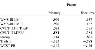
*Factor loadings for both CVLT-II variables were similar for the Memory and Executive Factors. They were considered to be part of the Memory factor because of its conceptual relationship to other variables on this factor.
LM = Logical Memory; LDFR = long delay free recall; PE = perseverative errors; WMS-III = Wechsler Memory Scale—Third Edition; CVLT-II = California Verbal Learning Test—Second Edition; WCST = Wisconsin Card Sorting Test.
Results
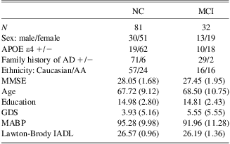
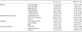
The MCI and NC groups were comparable on age (t = −0.39; p = .70), education (t = −0.29; p = .77), sex (χ2 = 0.13; p = .72), MABP (t = 1.54; p = .13), GDS (t = −1.06; p = .29), Lawton and Brody IADL score (t = 1.68; p = .10), and MMSE (t = 1.61; p = .11). There was a greater proportion of African Americans in the MCI group, although this difference only approached significance (χ2 = 4.16; p = .05). Due to targeted recruitment, a large percentage (88%) had a self-reported positive family history of AD, and this percentage was comparable across MCI and NC groups (χ2 = 0.06; p = .81). Twenty-six percent of the sample was apolipoprotein ε4 positive. There were a higher percentage of APOE ε4 positive participants in the MCI group (31.25%) than in the NC group (23.5%), although this difference was not significant (χ2 = 1.60, p = .21). These values exceed what has been documented in the general population (15%; Strittmatter & Roses, Reference Strittmatter and Roses1995), which may be attributable to the greater proportion of individuals with a family history of AD (see Table 3). See Table 4 for performance on neuropsychological measures across groups.
Sample characteristics by MCI status
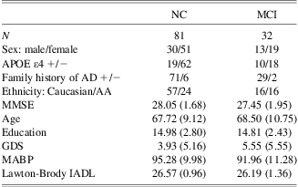
Note. No significant differences between groups for variables represented above.
NC = normal controls; MCI = mild cognitive impairment; APOE = apolipoprotein E; AD = Alzheimer's disease; MMSE = Mini Mental State Examination; GDS = Geriatric Depression Scale; MABP = mean arterial blood pressure; IADL = instrumental activities of daily living.
Mean (SD) for Raw Score Performance on Neuropsychological Measures by MCI Status

Note. Independent sample t-tests showed that all comparisons were significant at p < .05.
a Scores for these measures were not collected from the whole sample, rather they represent performance of 61 NC and 23 MCI participants.
b Sum of Automatic Detection Speed and Controlled Search Speed raw scores.
NC = normal controls; MCI = mild cognitive impairment; CVLT = California Verbal Learning Test—Second Edition; WMS-III = Wechsler Memory Scale—Third Edition; WAIS-III = Wechsler Adult Intelligence Scale—Third Edition; BNT = Boston Naming Test; WCST = Wisconsin Card Sorting Test; DS = digit span; LDFR = long delay free recall; LM = logical memory; PE = perseverative errors.
Group Comparisons
Group comparisons of AD-signature cortical thickness, hippocampal volume and FA, controlling for age, education, and MABP
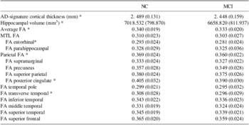
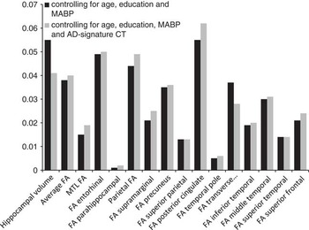
Several measures demonstrated lower values in the MCI group relative to the NC group, including hippocampal volume (F 4,108 = 6.270; p = .014), average FA (F 4,108 = 4.223; p = .042), and average parietal FA (F 4,108 = 5.018; p = .027), and this difference approached significance for AD-signature cortical thickness (F 4,108 = 3.336; p = .071; η 2 = 0.030). Average medial temporal FA was not significantly different across groups (p = .198). Within individual ROIs, lower FA was demonstrated in the MCI group relative to the NC group in WM underlying entorhinal (F 4,108 = 5.572; p = .02), posterior cingulate (F 4,108 = 6.234; p = .014), and transverse temporal (F 4,108 = 4.184; p = .043) cortical regions, and this difference approached significance in WM underlying middle temporal (p = .069) and precuneus (p = .051) cortical regions. No significant differences in FA were found in WM underlying parahippocampal, supramarginal, temporal pole, inferior temporal, superior temporal, superior frontal, or superior parietal cortical regions (all p's > .05) (see Table 5 and Figure 1).
Means (SD) for morphometric MRI data by MCI status

*p < 0.05.
AD = Alzheimer's disease; NC = normal controls; MCI = mild cognitive impairment; AD = Alzheimer's disease; MTL = medial temporal lobe; FA = fractional anisotropy.
Partial eta squared is provided for all group comparison analyses, shown with and without controlling for Alzheimer's disease (AD) -signature cortical thickness.

Group Comparisons Controlling for AD-Signature Cortical Thickness
Group comparisons controlling for age, education, MABP, and AD-signature cortical thickness
The MCI group had lower hippocampal volume (F 5,107 = 4.585; p = .035), parietal FA (F 5,107 = 5.457; p = .021), and average FA (F 5,107 = 4.417; p = .038) than the NC group when controlling for AD-signature cortical thickness. Within individual ROIs, the MCI group had lower FA than the NC group in WM underlying entorhinal (F 5,107 = 5.586; p = .020) and posterior cingulate (F 5,107 = 7.050, p = .009) cortical regions when controlling for AD-signature cortical thickness, and this difference only approached significance in WM underlying transverse temporal (F 5,107 = 3. 072; p = .082) cortical region (see Figure 1).
Association of Morphometric and Neuropsychological Variables
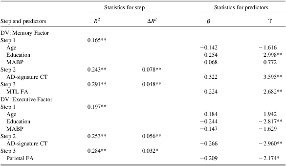
Hierarchical multiple regression analyses demonstrated that AD-signature cortical thickness explained a significant amount of variance (p = .001) in the Memory factor over and above age, education, and MABP. Medial temporal FA explained a significant amount of variance (p = .008) in the Memory factor over and above age, education, MABP, and AD-signature cortical thickness. Average FA and parietal FA did not explain additional unique variance (beyond AD-signature cortical thickness) in the Memory Factor (p > .05). AD-signature cortical thickness explained a significant amount of variance (p = .005) in the Executive factor over and above age, education, and MABP. Parietal FA explained a significant amount of variance (p = .032) in the Executive factor, over and above age, education, MABP, and AD-signature cortical thickness. Average FA and parietal FA did not explain additional unique variance (beyond AD-signature cortical thickness) in the Memory Factor (p > .05) (see Table 6).
Hierarchical regression models
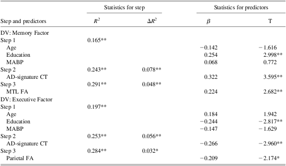
Note. Statistics for predictors represent values for full model.
*p < .05.
**p < .01.
DV = dependent variable; MABP = mean arterial blood pressure; AD = Alzheimer's disease; CT = cortical thickness; MTL FA = average medial temporal lobe fractional anisotropy.
Discussion
In this sample of healthy older adults with a high rate of family history of Alzheimer's disease, an MCI group characterized using comprehensive neuropsychological criteria (Jak, Bondi, et al., Reference Jak, Bondi, Delano-Wood, Wierenga, Corey-Bloom, Salmon and Delis2009) exhibited decreased hippocampal volume and WM integrity relative to normal controls. Nearly all differences in WM integrity were statistically independent of AD-signature cortical thinning. Furthermore, WM integrity was related to cognition, independent of cortical thinning.
Consistent with our hypotheses, the current results demonstrated decreased hippocampal volume in the MCI group. Despite the well-known finding of hippocampal volume loss in MCI (Shi, Liu, Zhou, Yu, & Jiang, Reference Shi, Liu, Zhou, Yu and Jiang2009), this is only the second study to demonstrate this when defining MCI through a neuropsychological approach (Jak, Bangen, et al., Reference Jak, Bangen, Wierenga, Delano-Wood, Corey-Bloom and Bondi2009). We also demonstrated a trend toward widespread cortical thinning (i.e., decreased AD-signature cortical thickness) in MCI.
As hypothesized, the present study demonstrated decreased WM microstructural integrity as manifested in lower average FA (of our selected ROIs) and lower parietal FA among the MCI group. Parietal WM degeneration has been previously documented in MCI (Bosch et al., Reference Bosch, Arenaza-Urquijo, Rami, Sala-Llonch, Junque, Sole-Padulles and Bartres-Faz2012; Chua, Wen, Slavin, & Sachdev, Reference Chua, Wen, Slavin and Sachdev2008; Delano-Wood et al., Reference Delano-Wood, Stricker, Sorg, Nation, Jak, Woods and Bondi2012; Medina et al., Reference Medina, DeToledo-Morrell, Urresta, Gabrieli, Moseley, Fleischman and Stebbins2006; Rose et al., Reference Rose, McMahon, Janke, O'Dowd, de Zubicaray, Strudwick and Chalk2006; Scola et al., Reference Scola, Bozzali, Agosta, Magnani, Franceschi, Sormani and Falini2010; Shim, Yoon, Shon, Ahn, & Yang, Reference Shim, Yoon, Shon, Ahn and Yang2008; Shu, Wang, Qi, Li, & He, Reference Shu, Wang, Qi, Li and He2011; Zhuang et al., Reference Zhuang, Wen, Zhu, Trollor, Kochan, Crawford and Sachdev2010). Within ROIs comprising our measure of posterior WM, FA in the posterior cingulate was significantly decreased. Decreased WM integrity in the posterior cingulate has emerged as a highly replicable finding across DTI studies of MCI and AD (Catheline et al., Reference Catheline, Periot, Amirault, Braun, Dartigues, Auriacombe and Allard2010; Chua et al., Reference Chua, Wen, Chen, Kochan, Slavin, Trollor and Sachdev2009, Reference Chua, Wen, Slavin and Sachdev2008; Delano-Wood et al., Reference Delano-Wood, Stricker, Sorg, Nation, Jak, Woods and Bondi2012). These results suggest that alterations within posterior WM regions, particularly the posterior cingulate, may serve as a potential imaging biomarker of early AD-related brain changes. The results of the present study extend the existing literature by demonstrating that parietal WM changes remained significant even when controlling for AD-signature cortical thickness, suggesting that posterior WM changes are independent of grey matter atrophy within this susceptible population.
The lack of decreased WM integrity in our average medial temporal WM ROI is unexpected and inconsistent with our hypotheses. Within individual ROIs, the MCI group showed decreased FA in WM underlying the entorhinal, but not parahippocampal, cortex. Other studies have identified decreased WM integrity in parahippocampal WM in MCI (Liu et al., Reference Liu, Spulber, Lehtimaki, Kononen, Hallikainen, Grohn and Soininen2011; O'Dwyer et al., Reference O'Dwyer, Lamberton, Bokde, Ewers, Faluyi, Tanner and Hampel2011; Rose et al., Reference Rose, McMahon, Janke, O'Dowd, de Zubicaray, Strudwick and Chalk2006; Zhang et al., Reference Zhang, Schuff, Jahng, Bayne, Mori, Schad and Weiner2007). One possible explanation for this discrepancy is that our sample may represent early MCI, as these are individuals who were neurologically normal per history, identified only by neuropsychological status. Another possible explanation for the lack of parahippocampal WM differences in our study may be due to collapsing across aMCI and naMCI groups. Zhuang and colleagues (Reference Zhuang, Wen, Zhu, Trollor, Kochan, Crawford and Sachdev2010) demonstrated a differing pattern of results in a large sample of aMCI, naMCI, and healthy control subjects; although widespread differences relative to control subjects were found in both MCI groups, significant differences in temporal WM were only found in the aMCI group. Of note, however, is that they found no significant differences when directly comparing aMCI and naMCI groups. Our results are consistent with Grambaite and colleagues (Reference Grambaite, Reinvang, Selnes, Fjell, Walhovd, Stenset and Fladby2011); their investigation also demonstrated decreased integrity in the WM underlying the entorhinal, but not parahippocampal, cortex in aMCI relative to a “less-advanced” MCI sample. AD neuropathology is present in the entorhinal cortex in the earliest stages of the disease process, even before the hippocampus and parahippocampal gyrus (Braak & Braak, Reference Braak and Braak1996; Gomez-Isla et al., Reference Gomez-Isla, Price, McKeel, Morris, Growdon and Hyman1996), and a similar pattern of findings has been documented in morphometry studies focused on GM alterations (Desikan et al., Reference Desikan, Cabral, Hess, Dillon, Glastonbury, Weiner and Fischl2009; Devanand et al., Reference Devanand, Bansal, Liu, Hao, Pradhaban and Peterson2012; Fennema-Notestine et al., Reference Fennema-Notestine, Hagler, McEvoy, Fleisher, Wu, Karow and Dale2009). Similarly, the more anterior (entorhinal) findings would be expected given that in AD the degenerative changes in the parahippocampal WM seem to follow an anterior to posterior gradient (Salat et al., Reference Salat, Tuch, van der Kouwe, Greve, Pappu, Lee and Rosas2010). WM underlying the entorhinal cortex has not been frequently included as an ROI in past DTI studies. Our methodological approach that applies T1-based cortical parcellations to the underlying WM facilitated inclusion of this ROI. Potential advantages of this method include correspondence between WM and cortical thickness ROIs and a clearly defined method by which to replicate analyses. Given our finding of decreased WM integrity in the WM underlying the entorhinal cortex in this mixed aMCI and naMCI sample, which persisted even when controlling for AD-signature cortical thickness, this region deserves further investigation in future MCI studies.
The current results provide support for the relative independence of decreased WM integrity in MCI from GM atrophy as measured by widespread cortical thinning in regions known to be sensitive to AD. These results are consistent with those of Selnes et al. (Reference Selnes, Fjell, Gjerstad, Bjornerud, Wallin, Due-Tonnessen and Fladby2012) who also demonstrated that most of the MCI group differences in WM integrity persisted despite controlling for localized cortical thinning. Collectively, our findings also suggest that changes in WM integrity explain unique variance and may precede, or have additive or synergistic effects on, GM atrophy in individuals at risk for AD. This interpretation is consistent with Brun and Englund's (Reference Brun and Englund1986) neuropathological findings that indicate WM changes in AD are not likely to be solely due to Wallerian degeneration. Further support for the early importance of white matter changes was recently demonstrated by Selnes and colleagues (Reference Selnes, Aarsland, Bjornerud, Gjerstad, Wallin, Hessen and Fladby2013) in a longitudinal study. They showed that measures of WM integrity better predicted decline in cognition and MTL atrophy over 2–3 years than did CSF biomarkers in a sample of patients with subjective cognitive complaints and MCI and proposed that changes in WM may precede GM atrophy in the Alzheimer's pathological cascade model (Jack et al., Reference Jack, Knopman, Jagust, Shaw, Aisen, Weiner and Trojanowski2010). The current study adds to a growing body of literature that suggests that WM integrity may serve as an independent MR biomarker in studies examining individuals at risk for AD. Furthermore, including measures of WM integrity in future biomarker studies may have significant practical importance. For example, some investigators have proposed that vascular processes may precede or accelerate AD neuropathology (de la Torre, Reference de la Torre2002; Leszek, Sochocka, & Gasiorowski, Reference Leszek, Sochocka and Gasiorowski2012). Vascular risk factors are easily modifiable, thus, could possibly prevent development of dementia later in life through early preventative efforts or could potentially slow cognitive decline (Richard & Pasquier, Reference Richard and Pasquier2012). Further determining white matter's contribution to early detection by regularly including DTI in conversion studies and in clinical trials may significantly impact future treatment and prevention efforts.
Analyses investigating associations with cognition highlighted that the clinical significance of WM changes may also be independent from gray matter changes. Overall, these results show that across a sample of healthy older adults and individuals with MCI, WM integrity explains variance in cognition over and above cortical thickness in regions shown to be sensitive to early AD-related changes. Analyses within specific cognitive domains partially supported our hypotheses. As predicted, medial temporal FA was significantly related to memory performance. Furthermore, consistent with results of Grambaite and colleagues (Reference Grambaite, Reinvang, Selnes, Fjell, Walhovd, Stenset and Fladby2011), we demonstrated that this relationship was independent of cortical thinning. Unlike their study wherein they locally controlled for cortical thinning, we demonstrate that this relationship persists when accounting for widespread cortical thickness in a pattern known to be sensitive to early AD neuropathological changes (Dickerson et al., Reference Dickerson, Stoub, Shah, Sperling, Killiany, Albert and Detoledo-Morrell2011). Also consistent with Grambaite and colleagues, we did not find a relationship between parietal WM integrity and memory performance. Other studies have demonstrated a relationship between parietal WM regions such as the posterior cingulate (Fellgiebel et al., Reference Fellgiebel, Muller, Wille, Dellani, Scheurich, Schmidt and Stoeter2005; Rose et al., Reference Rose, McMahon, Janke, O'Dowd, de Zubicaray, Strudwick and Chalk2006), but these studies did not control for gray matter changes. Results also revealed a relationship between parietal WM integrity and executive functioning, consistent with findings from other investigators (Chen et al., Reference Chen, Chen, Cheng, Hua, Liu and Chiu2009; Jacobs et al., Reference Jacobs, Van Boxtel, Jolles, Verhey and Uylings2012; Kim et al., Reference Kim, Park, Ahn, Seo, Lee, Kim and Na2011). The current results extend prior findings by demonstrating that this relationship is independent of cortical thinning. Additional work applying a voxelwise approach is needed to more extensively examine which WM regions correlate with memory and executive functioning.
This study has several limitations. Investigation of the relationship between WM and cognition collapsed across groups to avoid arbitrarily restricting range and to maximize power. Furthermore, because the same measures used for the Memory and Executive domains in MCI diagnosis were used for the Memory and Executive Factors for these analyses, inclusion of group status in the regression model or limiting analyses to subgroups may be viewed as partially dependent. Therefore, results from our regression analyses must be interpreted with caution, as results suggest that there is a relationship between these cognitive domains and FA, supporting potential clinical implications of variation in white matter integrity over and above cortical thinning, but do not suggest specificity of these relationships to MCI. Because a subjective or objective change in cognition over time was not required for MCI diagnosis in the current study, and follow-up data is not available, it remains possible that the current results could reflect normal variability in morphology that is also related to cognitive strengths and weaknesses, rather than an early Alzheimer's process. The elevated prevalence of family history and APOE ε4 rates offer some argument against this, but future work is needed with prospectively diagnosed MCI to selectively include MCI presumed to be due to AD (Albert et al., Reference Albert, DeKosky, Dickson, Dubois, Feldman, Fox and Phelps2011). Longitudinal studies are needed to examine the relative ability of WM integrity versus measures of GM to predict conversion from MCI to Alzheimer's dementia and to replicate the current results in a sample destined to develop AD. The current sample, while at risk for AD, may reflect a variety of etiologies for MCI. While the elevated rate of family history of AD and the APOE ε4 allele relative to the general population increases the likelihood of preclinical AD within this sample, it may limit the generalizability of our results. In addition, the difference in rate of MCI in African American and Caucasian participants approached significance. Although we used normative data with corrections for ethnicity whenever possible (Heaton et al., Reference Heaton, Miller, Taylor and Grant2004), not all measures included in this study had this option. Our group is currently working on a separate manuscript to examine potential differences across our African American and Caucasian participants. Due to small sample sizes and an effort to limit the number of analyses, we chose not to analyze aMCI and naMCI subgroups separately given evidence that naMCI frequently represents early AD (Fischer et al., Reference Fischer, Jungwirth, Zehetmayer, Weissgram, Hoenigschnabl, Gelpi and Tragl2007; Rountree et al., Reference Rountree, Waring, Chan, Lupo, Darby and Doody2007; Schneider, Arvanitakis, Leurgans, & Bennett, Reference Schneider, Arvanitakis, Leurgans and Bennett2009). However, this may have affected our ability to detect medial temporal lobe effects (Zhuang et al., Reference Zhuang, Wen, Zhu, Trollor, Kochan, Crawford and Sachdev2010). Ongoing studies will expand our MCI sample, allowing for us to more specifically investigate morphometric differences in amnestic and non-amnestic MCI.
In summary, we found evidence that neuropsychologically defined MCI is associated with reduced gray and white matter integrity, and group differences in WM integrity persisted even when controlling for cortical thickness in regions sensitive to the AD process. This is the first study to demonstrate that decreased integrity in entorhinal and parietal WM in MCI may be independent of widespread gray matter changes. Moreover, results suggest that changes in WM integrity are clinically significant and cannot be fully attributed to gray matter atrophy. Future work will explore whether similar results can be demonstrated in prospectively diagnosed MCI and further examination of potential differences by MCI subtype is needed. Improved understanding of the pattern of WM changes that develop in early AD and prodromal AD states such as MCI has important implications for early detection and monitoring treatment effects in clinical trials, and the current study suggests a prominent and preferential role of parietal and temporal lobe WM integrity and relationships to cognition during this critical stage.
Acknowledgments
This work was supported by the National Institute of Neurologic Disorders and Stroke (grant numbers K23NS062148); the National Institute of Nursing Research (grant number R01NR010827), the National Institute on Aging (grant numbers P60AG08812, P01AG004390); and by Medical Research Service VA Merit Review Awards to William Milberg and Regina McGlinchey. This research was conducted while Nikki Stricker was a Gilbert Foundation/AFAR Research Grant recipient. The authors thank Marge Ahlquist for her assistance with BP collection and phlebotomy on all participants. The authors report no conflicts of interest.