Introduction
Adenoid cystic carcinoma is an uncommon tumour that accounts for less than 1 per cent of all head and neck malignancies, and 10 per cent of all salivary gland neoplasms.Reference Witt1, Reference Kim, Sung, Chung, Rhee, Park and Kim2 It occurs more often in minor salivary glands than major salivary glands.Reference Wiseman, Popat and Nestor3 Adenoid cystic carcinoma is a slowly progressive, relentless tumour that has a tendency to recur locally and metastasise distantly.Reference Haddad, Enepekides, Manolidis and Black4, Reference Spiro, Huvos and Strong5 One study found perineural spread in approximately 60 per cent of cases, with a high incidence of local recurrence (approximately 63 per cent).Reference Takagi, Fukuda, Furuta, Yagi, Hamma and Nagahashi6 Recurrence and metastasis can occur even decades after treatment of the primary tumour.Reference Yousem, Gad and Tufano7 The sinonasal tract is the second most common site for adenoid cystic carcinoma (26.8 per cent), after the oral cavity (70.1 per cent).Reference Spiro8
The diagnosis of sinonasal tract malignancy is often delayed due to the similarity of symptoms to those of inflammatory disease. Cranial nerve paralysis, facial pain, trismus, headache and eye symptoms may indicate advanced disease, and occur once tumour growth has extended beyond the paranasal sinuses or the nasal cavity.
Clinical reports of sinonasal adenoid cystic carcinoma are still rare. Therefore, the present study was conducted to evaluate the pathophysiology, clinical characteristics and treatment outcome of this tumour.
Materials and methods
We performed a retrospective analysis of four consecutive patients with adenoid cystic carcinoma of the sinonasal tract treated at a tertiary, teaching hospital.
We reviewed patients' complete medical records, including out-patient clinic records, operative notes, and reports for imaging and histopathology, in order to evaluate their clinical features, tumour behaviour and treatment outcome. Patients' clinical and staging parameters are summarised in Table I, including age at diagnosis, sex, primary site, tumour-node-metastasis (TNM) staging, histopathological findings, treatment modalities, duration of follow up, distant metastases and treatment outcome. The minimum follow up was five years. The follow-up period was defined as the time from the first therapeutic intervention to the most recent clinic review or death.
Patients' clinical, pathological and treatment data

*At diagnosis. Pt no = patient number; y = years; TNM = tumour-node-metastasis classification; Post-op RT = post-operative radiotherapy; FU = follow up; mth = months; Dist mets = distant metastasis; Rec = recurrence; F = female; R = right; ant = anterior; −ve = negative; EER = endoscopic endonasal resection; post = posterior; M = male; +ve = positive; CT = chemotherapy; L = left; EACFR = endoscopic assisted craniofacial resection
Descriptive statistical analysis was performed due to the small study population.
Results
The mean age at presentation was 55.75 years (age range, 46–62 years), with an equal male:female ratio. The most common primary site was the ethmoid sinus (three patients; cases one, two and four), followed by the sphenoid sinus (one patient, case three).
All patients presented at an advanced stage: two had TNM staging of T3 N0 M0 (cases one and two) and two were staged as T4 N0 M0 (cases three and four). All tumours were of the cribriform type.
All patients underwent planned surgical resection of the affected region via endoscopic endonasal resection or endoscopic assisted craniofacial resection, followed by adjuvant radiotherapy. Two patients (cases three and four) had aggressive local infiltration resulting in perineural spread, limited surgical resection and positive surgical margins.
Patient follow up ranged from 60 to 94 months, with a mean duration of 77 months. One patient (case three) developed an enlarged neck node a year after radiotherapy, and at 44 months follow up developed extensive locoregional metastases, which were treated with chemotherapy to reduce the tumour stage. This patient also developed distant metastases. At the end of the study period, three patients were still alive without recurrence, and were receiving ongoing follow up (cases one, two and four). Case three died at 60 months post-surgery due to severe pneumonia.
Discussion
Minor salivary gland tumours of the sinonasal tract are uncommon. More than 80 per cent of minor salivary gland tumours are malignant.Reference Bradley9 Of all malignant paranasal sinus tumours, 5 to 15 per cent are adenoid cystic carcinomas.Reference Kraus, Roberts, Medendorp, Levine, Wood and Tucker10, Reference Miller and Calcaterra11 Of all patients with adenoid cystic carcinoma of the head and neck, those whose disease originates in the paranasal sinuses or nasal cavity have the best prognosis.Reference Wiseman, Popat and Nestor3 (These findings were described more than 40 years ago by Leafstedt et al. in a review from the Roswell Park Cancer Institute.)Reference Wiseman, Popat and Nestor3, Reference Leafstedt, Gacta and Sako12
In the present series, patients presented between the fourth and sixth decades of life, similarly to other studies.Reference Miller and Calcaterra11, Reference Rhee, Won, Lee, Min, Sung and Kim13, Reference Sung, Kim, Kim, Min, Seong and Roh14 Earlier series reported that adenoid cystic carcinoma was more common in females, but a recent literature review identified a slight male preponderance.Reference Witt1, Reference Kim, Sung, Chung, Rhee, Park and Kim2, Reference Rhee, Won, Lee, Min, Sung and Kim13, Reference Sung, Kim, Kim, Min, Seong and Roh14 The current series had an equal male:female ratio, perhaps due to the small sample size.
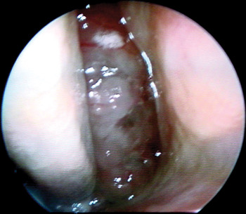
Sinonasal adenoid cystic carcinoma mostly arises from the maxillary sinus (>60 per cent), followed by the nasal cavity (20–30 per cent), nasopharynx (8–10 per cent) and ethmoidal sinus (8–10 per cent).Reference Kraus, Roberts, Medendorp, Levine, Wood and Tucker10, Reference Rhee, Won, Lee, Min, Sung and Kim13, Reference Sung, Kim, Kim, Min, Seong and Roh14 In our series, the most common site was the ethmoid sinus, found in three patients (Figure 1); the fourth patient had a sphenoid sinus tumour. No patient had a maxillary sinus tumour. Unsurprisingly, these frequencies of primary site involvement vary from those reported in the literature, due to the latter's larger sample population.
Endoscopic view of right nasal cavity of case one, showing a friable intranasal mass arising from the ethmoid sinus.

Adenoid cystic carcinoma can also arise from the nasopharyngeal area. In Malaysia, the most common nasopharyngeal malignancy is nasopharyngeal carcinoma (NPC).Reference Prasad and Rampal15 It is important to distinguish this tumour from adenoid cystic carcinoma as its treatment modalities and prognosis differ.
In the present series, intermittent epistaxis was the main presenting symptom in three of the cases. The other patient presented with unilateral tinnitus without nasal symptoms. Both epistaxis and tinnitus are also common symptoms of NPC.
Since early manifestations of adenoid cystic carcinoma can mimic inflammatory diseases such as chronic rhinitis and rhinosinusitis, and since the paranasal sinuses are air-filled spaces, diagnosis is often delayed, with patients often presenting with advanced stage tumour. In our series, two patients presented with a T3 tumour and two with a T4 tumour.
The reported incidence of lymph node metastases in adenoid cystic carcinoma is low: 3–5.5 per cent.Reference Miller and Calcaterra11, Reference Rhee, Won, Lee, Min, Sung and Kim13–Reference Prasad and Rampal15 The incidence of nodal metastasis in sinonasal adenoid cystic carcinoma cases is lower still, compared with NPC, which might be due to the sparse lymphatic distribution in the sinonasal tract and surrounding structures.Reference Rhee, Won, Lee, Min, Sung and Kim13 No elective neck dissection was performed in any of the patients in the present series, as none had cervical metastases at the time of diagnosis. One of our patients developed ipsilateral neck node swelling a year after radiotherapy. Such neck node swelling is associated with a poor prognosis. The same patient also developed co-morbidities due to extensive locoregional infiltration.
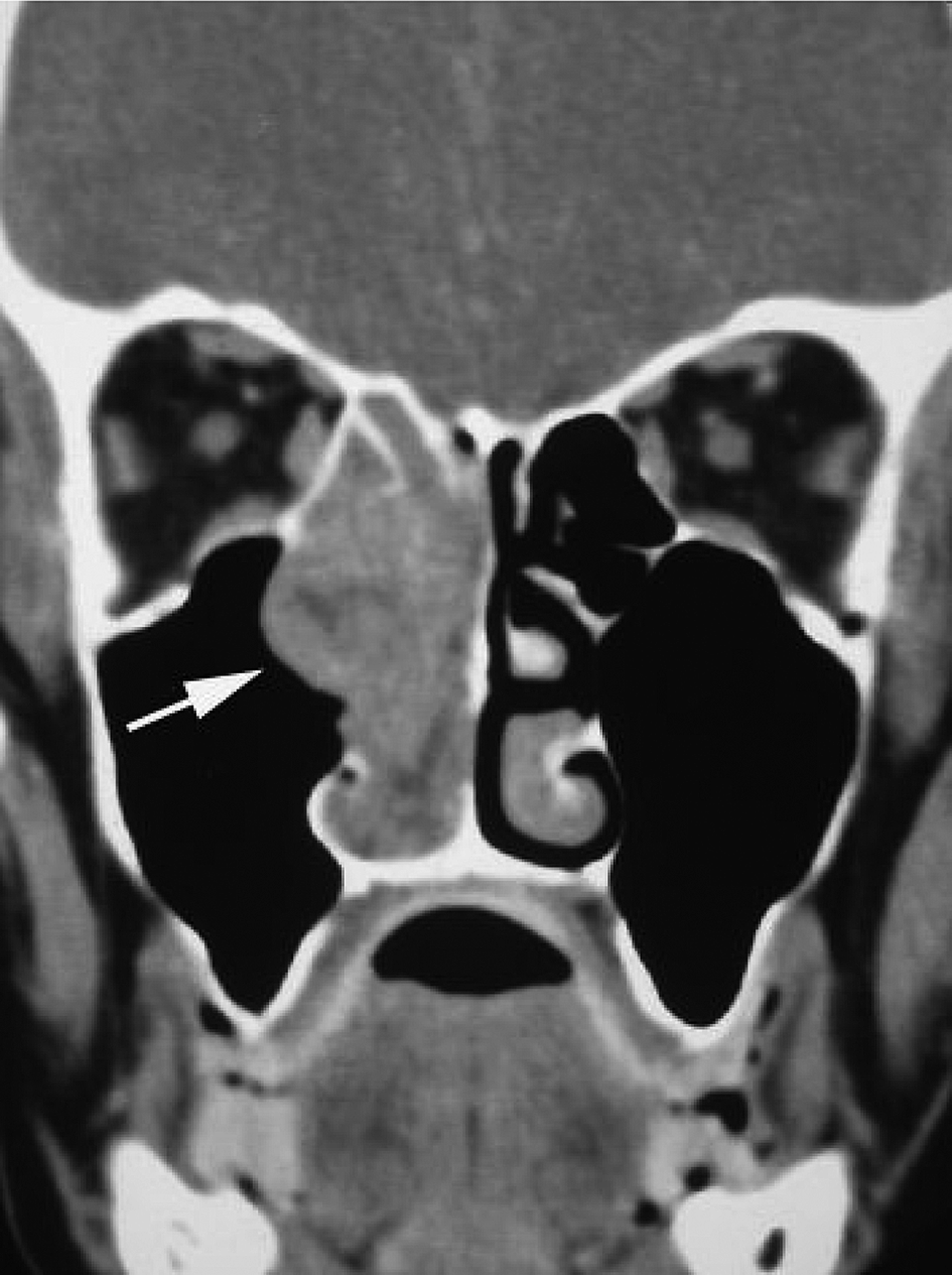
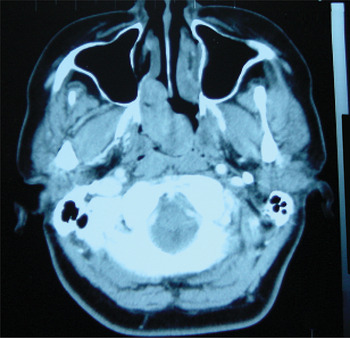
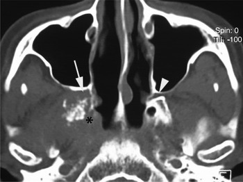
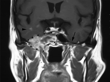
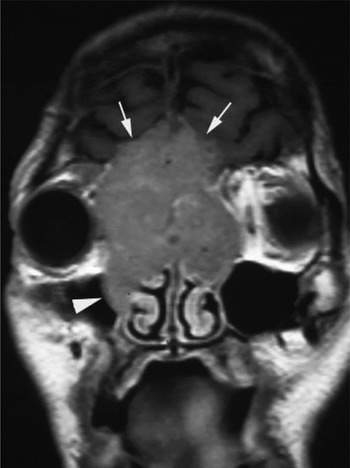
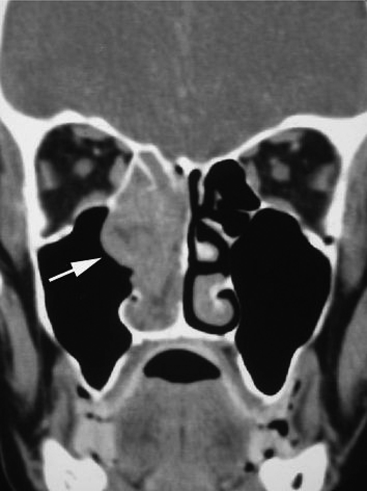
Pre-operative imaging is essential to delineate the extent of such tumours, due to the anatomical complexity of the sinonasal tract. Both computed tomography (CT) and magnetic resonance imaging are helpful, and complement each other in assessing anterior and posterior skull base involvement. Computed tomography is the best imaging modality for defining effects on the surrounding bone, as shown in Figures 2, 3 and 4. Magnetic resonance imaging is the best imaging modality for evaluating skull base invasion, perineural spread, degree of orbital involvement and intracranial extension, as shown in Figures 5 and 6. Adenoid cystic carcinoma is a notoriously neurotropic neoplasm, with frequently perineural spread (Figure 5).Reference Yousem, Gad and Tufano7 Perineural invasion by adenoid cystic carcinoma is considered one of the determining factors facilitating locoregional recurrence and distant metastasis.Reference Rhee, Won, Lee, Min, Sung and Kim13, Reference Sung, Kim, Kim, Min, Seong and Roh14, Reference Garden, Weber and Morrison16 On CT, perineural spread is demonstrated by enlargement of the neural foramina and/or by soft tissue infiltration of the fat adjacent to or within the neural foramina.Reference Yousem, Gad and Tufano17 In our two patients with perineural spread, one had extensive locoregional metastases.
Coronal, contrast-enhanced computed tomography scan of patient one, showing a right anterior ethmoidal mass extending inferiorly into the nasal cavity. Lateral invasion past the medial right maxillary wall into the maxillary antrum (arrow) is seen. The intracranial cavity and skull base are intact. No breach of the nasal septum or hard palate is seen.
Axial, contrast-enhanced computed tomography scan of patient two, showing a right posterior ethmoidal mass extending to the right posterior nasal space, crossing the midline and involving the left posterior nasal space. Anteriorly, the mass extends into the right nasal cavity.

Axial computed tomography scan of patient three showing widening of the right pterygopalatine fossa (arrow), providing further evidence of perineural spread involving the pterygopalatine ganglion. The arrowhead indicates the normal contralateral pterygopalatine fossa. Destruction of the right pterygoid plate and widening of the right vidian canal (asterisk) indicates perineural spread along the vidian nerve.

Coronal, T1-weighted, gadolinium-enhanced magnetic resonance imaging scan of patient three, showing an extensive right sphenoid sinus mass (indicated by black dots) infiltrating laterally into the masticator space, pterygopalatine fossa and middle cranial fossa. Invasion of the right cavernous sinus is evident. Metastatic parenchymal nodules are seen within the right temporal lobe (arrowhead). Perineural spread along the mandibular branch of the right trigeminal nerve is inferred from widening of the right foramen ovale. Note the normal left foramen ovale (arrow). The ipsilateral Meckel's cave is intact.

Coronal, T1-weighted, gadolinium-enhanced magnetic resonance imaging scan of patient four, showing an avidly enhancing mass involving both anterior ethmoid sinuses, extending superiorly into the cribriform plates and invading the parenchyma of both frontal lobes (arrows). Lateral encroachment into both orbital cavities and the right maxillary ostium (arrowhead) is noted. Involvement of both middle turbinates is seen (part of the normal right middle turbinate can be appreciated).

There are three conventional histological types of adenoid cystic carcinoma, namely tubular, cribriform and solid.Reference Seifert and Sobin18–Reference Perzin, Gullane and Clairmont20 In 1990, Batsakis et al. proposed the concept of three grades of adenoid cystic carcinoma, grade I being predominantly tubular, grade II being cribriform and grade III being solid.Reference Batsakis, Luna and El-Naggar21 All patients in the present series had a cribriform growth pattern, which carries a better prognosis than solid types.Reference Szanto, Luna, Tortoledo and White19, Reference Perzin, Gullane and Clairmont20
Adenoid cystic carcinoma with high grade transformation is a further histological type, which does not fit into the conventional histological typing system. A recent literature review reported that 57.1 per cent of adenoid cystic carcinoma cases with high grade transformation showed a marked propensity for lymph node metastasis.Reference Raja, Jennifer, Zubair, Virginia and Barnes22 This finding should prompt further discussion on the role of elective neck dissection in patients with adenoid cystic carcinoma of the sinonasal tract.
In a recent study, high local recurrence rates were seen for advanced stage (i.e. T3 and T4) tumours despite post-operative radiation therapy; therefore, complete resection of the primary tumour, with an adequate tumour-free margin, is recommended.Reference Rhee, Won, Lee, Min, Sung and Kim13 In the present series, our management included radical surgery followed by adjuvant radiotherapy. Three patients underwent endoscopic endonasal resection, and one received endoscopic assisted craniofacial resection due to dural involvement.
A complete ethmoidectomy was performed in two patients undergoing endoscopic endonasal resection (cases one and two) and also in the one patient receiving endoscopic assisted craniofacial resection (case four). In the first two cases, procedures involving the ethmoid sinus comprised complete removal of the superior turbinate, complete resection of the middle turbinate, wide middle meatus antrostomy (anteriorly up to the naso-lacrimal duct region and posteriorly up to the sphenopalatine artery region), and removal of the inferior turbinate with preservation of one-third of its anterior portion. In case four (receiving endoscopic assisted craniofacial resection), procedures involving the paranasal sinuses comprised ethmoidectomy, sphenoidectomy, wide middle meatus antrostomy and frontal sinusotomy. Endoscopic assisted craniofacial resection is routinely performed in our centre by the neuro-ENT team for paranasal sinus tumours involving the dura.Reference Gendeh, Salina, Selladurai and Jegan23
Our third case underwent right sphenoidectomy and posterior ethmoidectomy with middle meatus antrostomy, with partial clearance of tumour primarily from the sphenoid sinus with extension into the cavernous sinus.
Following surgery, no patient had any evidence of disease at the primary site except for patient three, in whom clearance had been only partial due to involvement of the cavernous sinus. Proximity of the sinonasal tract to vital structures limits wide local resection. Adenoid cystic carcinomas are more difficult to clear surgically compared with other cancers of the head and neck, and this often results in positive surgical margins.Reference Garden, Weber and Morrison16 In the present series, two patients had positive margins, one of whom developed locoregional infiltration and nodal metastasis. Haddad et al. have suggested that positive margins may be a reflection of disease which is more aggressive, invasive and resistant, rather than a reflection of inadequate resection.Reference Haddad, Enepekides, Manolidis and Black4
• Four cases of advanced adenoid cystic carcinoma of the sinonasal tract, undergoing endoscopic nasal surgery, are presented
• Three cases originated from the ethmoid sinus
• Surgery involved endonasal endoscopic resection and endoscopic assisted craniofacial resection
• This treatment, plus post-operative radiotherapy, generally produced good outcomes
• Cervical node metastasis is a poor prognostic factor associated with locoregional recurrence
Surgical resection with adjuvant radiotherapy is the mainstay of treatment for patients with adenoid cystic carcinoma of the paranasal sinuses or nasal cavity.Reference Wiseman, Popat and Nestor3 Adjuvant post-operative radiotherapy is given in cases of incomplete surgical resection, positive surgical margins (upon histological analysis) and advanced TNM staging (i.e. T3 and above). Adenoid cystic carcinoma is considered to be a radiosensitive but not radiocurable tumour.Reference Leafstedt, Gacta and Sako12 According to Spiro et al., post-operative radiotherapy probably delays rather than prevents local recurrence.Reference Spiro, Huvos and Strong24 In agreement with previous studies, our findings suggest that a combination of adequate endonasal endoscopic clearance and adjuvant radiotherapy is effective in achieving local control.Reference Kim, Sung, Chung, Rhee, Park and Kim2, Reference Haddad, Enepekides, Manolidis and Black4, Reference Rhee, Won, Lee, Min, Sung and Kim13, Reference Sung, Kim, Kim, Min, Seong and Roh14 In the present study, the five-year survival rate was 100 per cent, compared with 50 per cent reported in other studies.Reference Rhee, Won, Lee, Min, Sung and Kim13 Our better results could be explained by the fact that most of our patients underwent complete resection with adjuvant radiotherapy, except for one case with cavernous sinus involvement and distant metastasis.