Introduction
The “vascular depression” hypothesis proposes that cardiovascular disease is an important etiological factor in depression, particularly late-life depression (Alexopoulos et al., Reference Alexopoulos, Meyers, Young, Campbell, Silbersweig and Charlson1997; Sheline et al., Reference Sheline2010). This is supported by clinical observations which confirm that stroke and cardiovascular disease increase the likelihood of depression (Thomas et al., Reference Thomas, Kalaria and O'Brien2004), and that specific cardiovascular risk factors may predispose to depression. Neuroimaging findings have been important in the development of the vascular depression theory. Magnetic resonance imaging (MRI) has shown that white-matter hyperintensities are more common in patients with late-life depression compared to controls (Herrmann et al., Reference Herrmann, Le Masurier and Ebmeier2008) and a vascular basis for white-matter hyperintensities has been suggested (Thomas et al., Reference Thomas2002; Reference Thomas, Perry, Kalaria, Oakley, McMeekin and O'Brien2003; Chen et al., Reference Chen, Chen, Kuo, Chiang, Ko and Lin2006; Smith et al., Reference Smith2009). White-matter hyperintensities are particularly prevalent within frontal regions (Sheline et al., 2008; Dalby et al., Reference Dalby2010).
Further investigation of white-matter pathology may yield important information on the effects of raised cardiovascular risk on brain structure. Changes in fiber tracts on a macro- or microscopic scale have been associated with vascular disease and can have major implications for brain function (Roman, Reference Roman1996). In this paper we describe a diffusion tensor imaging (DTI) protocol, which has been able to detect subtle changes in white matter (Sexton et al., Reference Sexton, Mackay and Ebmeier2009). This method measures the direction of water diffusion within brain tissue. Restricted diffusion is expected within white-matter tracts (anisotropic diffusion), where water molecules are constrained by myelin sheaths and the parallel organization of fibers. Measuring the direction of diffusion allows investigation of orientation, as well as integrity of white matter. The sensitivity of this method is important, given that any changes to structural white-matter connectivity are likely to be subtle, given the absence of major cardiovascular disease in our sample.
There are emerging studies which consider the effect of specific cardiovascular risk factors on brain structure. Hypertension appears to confer a vulnerability to depression which may be mediated through micro-vascular brain injury (Hajjar et al., Reference Hajjar2011) and reduced white-matter integrity in frontal-striatal regions (Hoptman et al., Reference Hoptman2009). The Framingham Stroke Risk Profile (FSRP) has been associated with the volume of white-matter hyperintensities in those with depression (Smith et al., Reference Smith2010) and those without (Jeerakathil et al., Reference Jeerakathil2004), although previous research has not focused on associations between FSRP and changes in white-matter connectivity. The FSRP is widely used to predict individual risk of stroke (D'Agostino et al., Reference D'Agostino, Wolf, Belanger and Kannel1994; Reference D'Agostino2008; Wilson et al., Reference Wilson, D'Agostino, Levy, Belanger, Silbershatz and Kannel1998) and uses data that can be readily acquired in clinical settings. Given the strength of evidence linking vascular disease and depression (Alexopoulos et al., Reference Alexopoulos, Meyers, Young, Campbell, Silbersweig and Charlson1997), it is plausible that the FSRP will also predict structural brain changes in those with depression.
This paper describes an exploratory case-control study which considers the correlation between elevated cardiovascular risk as identified by the FSRP and white-matter connectivity measured using DTI. Reductions in fractional anisotropy (FA) in late-life depression are well documented (Sexton et al., Reference Sexton, Mackay and Ebmeier2009). If vascular pathology contributes to reductions in white-matter integrity, in accordance with the vascular hypothesis of depression, we would expect a correlation between FSRP and regions where FA is reduced in depression. In the same regions in controls, where white-matter integrity is relatively preserved, vascular risk is not expected to be significantly correlated with FA.
Clinically, “vascular depression” is particularly relevant to those with late-life depression, an area of increasing importance given that the proportion of the population aged over 65 is rising significantly (Office for National Statistics, 2008; Christensen et al., Reference Christensen, Doblhammer, Rau and Vaupel2009). With a greater prevalence of people with chronic cardiovascular disease (Crimmins, Reference Crimmins2004), better understanding of the effects of such disease on brain structure and mental health will be vital in preventing and treating depression.
Hypothesis
Depressed participants with high FSRP will show reduced white-matter integrity, whereas controls matched for vascular risk will show minimal white-matter changes. Specifically, depressed participants will show reduced white-matter integrity with higher FSRP; controls matched for mean vascular risk and overall variance will show minimal covariance with white-matter changes.
Methods
Sample
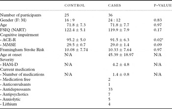
The sample comprised 36 participants with major depression (mean age 71.83 ± 7.71 years) recruited through primary and secondary care services in Oxfordshire and Buckinghamshire in the UK, and 25 controls (mean age 71.76 ± 7.30 years) recruited from the community. The mean 10-year Framingham Stroke Risk score was 10.33 ± 7.64 in cases and 10.08 ± 7.74 in controls; other baseline characteristics are shown in Table 1.
Baseline characteristics of the sample

Values presented are mean ± standard deviation.
*statistical significance, p < 0.05.
FSIQ (NART) = Full-Scale Intelligence Quotient (National Adult Reading Test); ACE-R = Addenbrooke's Cognitive Examination Revised; MMSE = Mini-Mental State Examination; HAM-D = Hamilton Depression Rating Scale.
Inclusion and exclusion criteria
For inclusion in this study all participants were required to be more than 60 years old, with no potentially confounding comorbid medical, psychiatric, or neurological conditions. Those recruited to the depressed group met the criteria for DSM-IV major depression (American Psychiatric Association, 1994), ascertained through clinical interview with a psychiatrist, but were not necessarily currently depressed. Controls were required to have no history of memory impairment or psychiatric illness, determined by administering the Structured Clinical Interview for DSM-IV Axis 1 Disorders Non-Patient Edition (First et al., Reference First, Spitzer, Gibbon and Williams2007). Participants with implanted metallic devices were excluded, as required by standard MRI safety protocols.
Clinical interview
All participants underwent clinical interview and examination. The clinical assessment was based on a semi-structured proforma designed for this study. Participants with depression were interviewed to determine age at onset of depression, defined as the age at which an individual experienced their first episode of major depression. Personal testimony was corroborated with information from hospital notes, where available. The severity of depressive symptoms experienced was further classified according to the 17-item Hamilton Depression Rating Scale (HAM-D; Hamilton, Reference Hamilton1967) and the 15-item Geriatric Depression Scale (GDS; Yesavage et al., Reference Yesavage1982). Medication status was ascertained through personal testimony and hospital notes, classified under the following headings: antidepressant, antipsychotic, anticonvulsant, lithium salts and anxiolytics.
The FSRP was calculated for all participants and was based upon the following predictors: age, systolic blood pressure, diabetes mellitus, cigarette smoking, prior cardiovascular disease, atrial fibrillation, left ventricular hypertrophy, and use of hypertensive medication (D'Agostino et al., Reference D'Agostino, Wolf, Belanger and Kannel1994). These were obtained from personal testimony, together with hospital notes. Clinical examination with a standard sphygmomanometer was used to assess blood pressure.
Cognitive impairment was assessed in all participants using standardized neuropsychological measures including the Addenbrooke's Cognitive Examination Revised (ACE-R; Mioshi et al., Reference Mioshi, Dawson, Mitchell, Arnold and Hodges2006), which includes the Mini-Mental State Examination (MMSE; Folstein et al., Reference Folstein, Folstein and McHugh1975). The Full-Scale Intelligence Quotient (FSIQ) was estimated from the number of errors on the National Adult Reading Test (McGurn et al., Reference McGurn2004), and the number of years of full-time education was determined from personal testimony.
MRI acquisition
All participants underwent a 60-direction DTI on a 3.0 Tesla Trio Siemens scanner, performed at the University of Oxford Centre for Clinical Magnetic Resonance Imaging (OCMR), with a 12-channel head-coil. Whole-brain DTI was acquired using an echoplanar imaging sequence (TR = 7900/7800 ms, TE = 98 / 82 ms, field of view = 240 mm, voxel size = 2.5 mm isotropic, b value = 1000, number of directions = 60, number of acquisitions = 2). T2-weighted images were also acquired to characterize WMH (TR = 6000 ms, TE = 91 ms, field of view = 220 mm, voxel size = 0.7 × 0.7 × 4.0 mm).
Ethical approval
This study received a favorable ethical opinion from the Local Research Ethics Committee. All participants gave written, informed consent prior to participation in this study.
Image analysis
Image analysis was performed using FSL tools (www.fmrib.ox.ac.uk/fsl) (Smith et al., Reference Smith2004) to assess the correlation between fractional anisotropy (FA) and FSRP. First, we performed voxelwise statistical analysis of the FA data using Tract Based Spatial Statistics (TBSS) (Smith et al., Reference Smith2006) across the whole TBSS skeleton. DTI data were checked manually to remove artifacts before eddy current correction was run to correct for eddy current distortions and head motion. FA images were created by fitting a tensor model to the raw diffusion data using FMRIB's diffusion toolbox, and then brain-extracted using Brain Extraction Tool (BET) (Smith, Reference Smith2002). These data were aligned to common space using FMRIB's non-linear image registration tool (FNIRT) (Andersson et al., Reference Andersson, Jenkinson and Smith2007a; Reference Andersson, Jenkinson and Smith2007b). The mean FA image was created and thinned to create a mean FA skeleton representing the centers of all tracts common to the group. Each participant's aligned FA data were then projected onto this skeleton before applying voxelwise cross-subject statistics.
Statistics were performed using “randomize” – a permutation-based inference tool for nonparametric statistical thresholding, with age included as a confound regressor (Nichols and Holmes, Reference Nichols and Holmes2002). The significance threshold for correlations was set at p < 0.05, using threshold-free cluster-enhancement (Smith and Nichols, Reference Smith and Nichols2009). Group differences in FA were investigated across the whole skeleton; tracts of interest were based on the ICBM-DTI-81 white-matter labels atlas and the JHU white-matter tractography atlas within the FSL atlas tool.
Second, we performed partial correlation analysis between FSRP and mean values of DTI, on a tract-by-tract basis, including only the voxels that were significantly different between groups. Partial correlation analysis used age and gender as covariates, and investigated significant differences between groups in each tract of interest in (i) depressed participants, and (ii) control participants. Group differences between depressed and control groups have been reported elsewhere (Sexton et al., in press).
Results
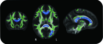
Using TBSS, participants in the depressed group showed significant correlations between FSRP and FA (Figure 1) across widespread regions of the brain, indicating significant impairment in white-matter integrity, in line with the original hypothesis. For controls there was no correlation between FSRP and FA.
Correlations between Framingham Stroke Risk Profile and fractional anisotropy (FA), in depressed group. Green: mean FA skeleton; blue: reduction in FA (p < 0.05)

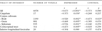
Using partial correlation analysis, participants in the depressed group showed significant correlations between FSRP and FA (r = -0.673, p < 0.001), whereas in controls, there was no significant relationship (r = −0.373, p = 0.08) (Table 2). This was highly significant (p < 0.01) within the body of corpus callosum (r = −0.520, p = 0.002), genu of corpus callosum (r = −0.468, p = 0.005), splenium of corpus callosum (r = −0.536, p = 0.001), cortico-spinal tract (r = −0.473, p = 0.005), and also significant (p < 0.05) within the cingulum (r = −0.373, p = 0.03). In the control group, there was one significant correlation (p < 0.05) within the body of corpus callosum (r = −0.473, p = 0.023).
Correlations between Framingham stroke risk profile and fractional anisotropy, for voxels significantly different between groups

*p < 0.05, **p < 0.01
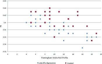
Figure 2 illustrates the mean FA in voxels significantly correlated with FSRP across the depressed group. This shows that whilst the regression slope was similar for both groups, controls showed greater variance in FA values.
Correlations between Framingham stroke risk profile (FSRP) and fractional anisotropy (FA).
LLD = ; CON =

Discussion
The FSRP was associated with reduced white-matter integrity in participants with depression. Our results suggest that white-matter tracts, including the corpus callosum, cortico-spinal tract and cingulum, warrant further investigation in order to investigate the mechanisms underlying vascular depression. These white-matter tracts with significant correlations were examined in more detail to compare FSRP and FA between depressed and control groups. Greater variability in FA values was evident in controls (Figure 2), a particularly interesting finding given that both groups were matched for vascular risk, as measured by FSRP. This leads to the hypothesis that the greater white-matter integrity in controls may provide resilience against the effects of vascular risk, and the subsequent development of depression. Though there was one area of regional significance in controls (body of corpus callosum), this was less significant than in the depressed group.
High vascular burden of disease is more likely with advancing age and thus “vascular depression” is especially relevant to those with late-onset depression. Vascular disease may precipitate both relapse in those who have had depression in the past, and first-onset in later life. The knowledge that vascular illness and elevated risk factors might have a specific effect on brain structure is important in understanding depression, and raises further questions about the links between depression and cognitive impairment. Both conditions not only share common risk factors, but also structural changes such as hippocampal atrophy (McKinnon et al., Reference McKinnon, Yucel, Nazarov and MacQueen2009) and reduced brain volume (Dotson et al., Reference Dotson, Davatzikos, Kraut and Resnick2009). Also, neuropsychological deficits in executive function and processing speed are well documented in depression (Herrmann et al., Reference Herrmann, Goodwin and Ebmeier2007). Impaired white-matter integrity within pre-frontal regions would be a plausible explanation linking structural and functional changes in both conditions (Buckner, Reference Buckner2004; Park and Reuter-Lorenz, Reference Park and Reuter-Lorenz2009; Shimony et al., Reference Shimony2009).
A key strength of this study was the MRI acquisition process which entailed two repeats of 60-direction DTI. This exceeds the suggested minimum of 30 directions estimated as needed for robust estimation of FA (Jones, Reference Jones2004) and is higher than many published studies (Sexton et al., Reference Sexton, Mackay and Ebmeier2009). There are, however, limitations to this study, particularly in the recruitment of the sample which was not systematically ascertained. Furthermore, most of the depressed participants had early-onset disease (n = 23), before the age of 60, rather than late-onset disease (n = 13). Although age at onset was not the focus of this study, the literature suggests that the impact of vascular risk on mood is greater for those developing depression over the age of 60, and it will be of interest to explore the effect of age at onset in more detail within a larger sample.
Participants were not excluded on the basis of medication. Whilst this means that our results are more representative of those who are routinely seen in specialist mental healthcare settings, it is also possible that medication may lead to structural brain changes that could influence our results. Given the age of our sample, where many have comorbid health problems, and the nature of the sample (recruitment of those with major depressive disorder), excluding people on the basis of medication would have not only made recruitment much more difficult, but would also have introduced new bias to the study.
Framingham scores and spread were similar in both groups, yet there was no correlation with FA in the control group. There are several possible explanations for this, relating to both cases and controls. It may be that those people becoming “cases” have pre-existing impairments in white-matter integrity which provide a biological vulnerability to depression, not uniquely related to vascular disease. Another explanation is that depression itself might induce changes within white-matter structure. Whilst this cross-sectional study does not provide information about the causality of vascular brain changes, this could be addressed through future longitudinal studies. On the other hand, controls may have specific protective factors which offer them resilience against white-matter change and developing depression. These are most likely to be biological; for example, functional differences with increased recruitment of other parts of the brain, particularly in frontal regions (Buckner, Reference Buckner2004; Park and Reuter-Lorenz, Reference Park and Reuter-Lorenz2009). Medication may also be a protective factor (greater use of statins could be one possibility). Finally, it may mean that the level of cardiovascular risk as measured by FSRP does not correlate accurately with structural brain changes, and that alternative measures of vascular risk need to be investigated. Although this study was not designed to address these questions, they provide interesting scope for future research.
In summary, this exploratory study combines the FSRP, a well-validated, multi-factorial, cardiovascular risk algorithm, with white-matter changes quantified using DTI. Our results suggest that even in the absence of major vascular disease, elevated FSRPs in those with depression are associated with significant changes in white-matter connectivity, to a degree not observed within the control group. This is apparent across widespread regions of the brain, most significantly within the corpus callosum and cortico-spinal tract. Greater understanding of the mechanisms by which vascular risk may contribute to pathophysiology in depression is important in order to prevent and treat this common illness. These results support the vascular depression hypothesis, and should lead to further research within larger, systematically (and preferably prospectively) acquired cohorts.
Conflict of interest
None.
Description of authors' roles
CLA undertook clinical interviews and analyzed the data. CES coordinated the study, undertook recruitment and data collection, and analyzed the data. UGK and LMM undertook recruitment and data collection. CEM was involved in design, supervised the project and advised on analysis. KPE designed the study, undertook clinical interviews and supervised data collection and analysis. CLA wrote the first draft of the paper and CES, UGK, LMM, MK, ASM, CEM and KPE reviewed and rewrote it, and approved the final version.
Acknowledgments
The authors thank all those who have helped with this study, particularly the clinical staff who referred patients; Dr Kevin Bradley, Consultant Radiologist, who reviewed brain scans; and, most importantly, all the participants and their relatives. Funding was provided by the Gordon Edward Small's Charitable Trust (Scottish Charity Register: SC008962). CLA had support from Oxford University Clinical Academic Graduate School (OUCAGS@medsci.ox.ac.uk).