Introduction
Cognitive alterations are common in Parkinson's disease (PD; Litvan et al., Reference Litvan, Aarsland, Adler, Goldman, Kulisevsky, Mollenhauer and Weintraub2011; Troster, Reference Troster2011). Even in the earliest stage, approximately 24 to 36% of PD patients show cognitive impairments (Foltynie, Brayne, Robbins, & Barker, Reference Foltynie, Brayne, Robbins and Barker2004; Muslimovic, Post, Speelman, & Schmand, Reference Muslimovic, Post, Speelman and Schmand2005), while 3 to 8 years after onset, this proportion is 50% to 80% (Aarsland, Bronnick, Larsen, Tysnes, & Alves, Reference Aarsland, Bronnick, Larsen, Tysnes and Alves2009; Muslimovic, Post, Speelman, De Haan, & Schmand, Reference Muslimovic, Post, Speelman, De Haan and Schmand2009; Williams-Gray, Foltynie, Brayne, Robbins, & Barker, Reference Williams-Gray, Foltynie, Brayne, Robbins and Barker2007). For an overview of longitudinal studies of cognitive changes in PD, see (Muslimovic, Schmand, Speelman, & de Haan, Reference Muslimovic, Schmand, Speelman and de Haan2007; Williams-Gray et al., Reference Williams-Gray, Foltynie, Brayne, Robbins and Barker2007).
Cognitive changes affect multiple domains (Dubois & Pillon, Reference Dubois and Pillon1997; Taylor & Saint-Cyr, Reference Taylor and Saint-Cyr1995), but executive dysfunction appears to be most profoundly impaired (Zgaljardic, Borod, Foldi, & Mattis, Reference Zgaljardic, Borod, Foldi and Mattis2003). It is assumed to result from the primary pathology in PD, that is, dopamine depletion, which disrupts fronto-striatal functioning (Alexander, DeLong, & Strick, Reference Alexander, DeLong and Strick1986; Owen, Reference Owen2004). Executive deficits in PD typically resemble symptoms seen in patients with frontal lobe damage, that is, problems with working memory (Lewis, Slabosz, Robbins, Barker, & Owen, Reference Lewis, Slabosz, Robbins, Barker and Owen2005), deficits in planning, initiating, and monitoring (Morris et al., Reference Morris, Downes, Sahakian, Evenden, Heald and Robbins1988), impaired inhibition (Gauggel, Rieger, & Feghoff, Reference Gauggel, Rieger and Feghoff2004), and difficulties with set shifting (Cools, Barker, Sahakian, & Robbins, Reference Cools, Barker, Sahakian and Robbins2001b; Monchi et al., Reference Monchi, Petrides, Doyon, Postuma, Worsley and Dagher2004). Patients are impaired on various executive functioning tasks, such as phonemic and semantic fluency, Wisconsin Card Sorting Test, Stroop interference test, and Trail Making Test part B (for an overview, see Kudlicka, Clare, & Hindle, Reference Kudlicka, Clare and Hindle2011).
Despite a plethora of studies on cognitive impairments in PD, it is still unclear to what extent cognitive functions are affected. While most studies reported impaired executive performance (Zgaljardic et al., Reference Zgaljardic, Borod, Foldi and Mattis2003), some did not find executive deficits (e.g., Fournet, Moreaud, Roulin, Naegele, & Pellat, Reference Fournet, Moreaud, Roulin, Naegele and Pellat1996). Part of this discrepancy might be explained by dopaminergic medication, which improves some aspects of executive functioning, such as set shifting, but impairs others, such as reversal learning (Cools, Barker, Sahakian, & Robbins, Reference Cools, Barker, Sahakian and Robbins2001a). Second, although there is ample cross-sectional evidence for executive impairment in PD, and deterioration of executive functions over time has been shown beyond doubt (Kudlicka et al., Reference Kudlicka, Clare and Hindle2011), executive decline appears to be less prominent than memory decline (Muslimovic et al., Reference Muslimovic, Schmand, Speelman and de Haan2007).
Memory impairments are frequently reported in non-demented PD patients (Stebbins, Gabrieli, Masciari, Monti, & Goetz, Reference Stebbins, Gabrieli, Masciari, Monti and Goetz1999). Because this often concerns difficulties with the retrieval of information, memory deficits are seen as secondary to the executive impairments (Dubois & Pillon, Reference Dubois and Pillon1997; Dujardin, Defebvre, Grunberg, Becquet, & Destee, Reference Dujardin, Defebvre, Grunberg, Becquet and Destee2001). However, subtle deficits in recognition memory also exist in PD (Whittington, Podd, & Kan, Reference Whittington, Podd and Kan2000), which cannot be fully explained by executive dysfunction (Bronnick, Alves, Aarsland, Tysnes, & Larsen, Reference Bronnick, Alves, Aarsland, Tysnes and Larsen2011). Thus, memory impairments, in particular impaired delayed recall and recognition, might be more closely related to hippocampal atrophy (Beyer et al., Reference Beyer, Bronnick, Hwang, Bergsland, Tysnes, Larsen and Apostolova2013). PD patients show impairments in other domains as well, such as visuospatial functions (Kerai, Bracewell, Hindle, & Leek, Reference Kerai, Bracewell, Hindle and Leek2012) and psychomotor speed (Muslimovic et al., Reference Muslimovic, Post, Speelman, De Haan and Schmand2009).
Notwithstanding the growing number of studies on cognition in PD, the course of decline in newly diagnosed patients still remains unclear. First, most studies were cross-sectional, conducted in convenience samples of prevalent cases. Second, the relatively scarce longitudinal studies have been hindered by methodological issues, such as the lack of control groups or the use of limited neuropsychological test batteries (Muslimovic et al., Reference Muslimovic, Schmand, Speelman and de Haan2007). Few studies have followed a cohort of newly diagnosed patients over a substantially long period of time (Aarsland, Andersen, Larsen, Lolk, & Kragh-Sorensen, Reference Aarsland, Andersen, Larsen, Lolk and Kragh-Sorensen2003; Williams-Gray et al., Reference Williams-Gray, Evans, Goris, Foltynie, Ban, Robbins and Barker2009). Another problem, which makes it difficult to describe the course of PD, is the heterogeneous nature of the disease (Taylor & Saint-Cyr, Reference Taylor and Saint-Cyr1995). The characteristic pathology is dopamine depletion in the midbrain, but other pathological factors may appear, such as the accumulation of Lewy bodies in the cortex (Braak et al., Reference Braak, Del Tredici, Rub, de Vos, Jansen Steur and Braak2003), and changes in other neurotransmitter systems, such as the cholinergic system (Jellinger, Reference Jellinger2001, Reference Jellinger2012).
To gain better insight into the cognitive changes of PD, we started a study that focused on the course of cognitive decline in a large group of newly diagnosed patients and a control group (Muslimovic et al., Reference Muslimovic, Post, Speelman, De Haan and Schmand2005, Reference Muslimovic, Post, Speelman and Schmand2009). Both groups were presented with a range of neuropsychological tests at baseline and 3 years later. At baseline, 24% of the patients showed cognitive impairments, most prominently in the executive and memory domains. After 3 years, the percentage with impairments had doubled. The deficits were most prominent in attention and psychomotor speed.
The present study further explores cognitive decline with a 5-year follow-up of this sample. Our aims were to (1) determine the pattern and severity of cognitive impairments, (2) describe the frequency of cognitive decline at an individual level, and (3) find prognostic markers for cognitive decline in the individual patient. We also explored the effects of selective attrition.
Methods
Subjects
The current study is part of the larger CARPA project (Comorbidity and Aging in Rehabilitation Patients with sequelae of poliomyelitis, osteoarthritis, and PD: the influence on Activities). Patients were recruited from neurology outpatient clinics. Between 2002 and 2005, 145 newly diagnosed PD patients were referred for inclusion in the study. The inclusion criterion was a diagnosis of idiopathic PD, established at baseline by movement disorder specialists (J.D. or B.P.) according to standard criteria (Gelb, Oliver, & Gilman, Reference Gelb, Oliver and Gilman1999), and checked twice during the study (by J.D. or B.P.). Patients were excluded if they were older than 85 years, had a score <24 on the Mini-Mental State Examination (MMSE; Folstein, Folstein, & McHugh, Reference Folstein, Folstein and McHugh1975), insufficient knowledge of the Dutch language, or suffered from a somatic illness with a life-expectancy of less than a year. None of the patients had a history of stroke, suffered from psychiatric illness, or any other neurological comorbidity.
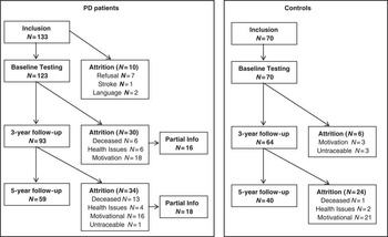
Eight patients were excluded because their referral diagnosis was incorrect, that is, did not satisfy the standard PD criteria. Two patients did not speak Dutch, one patient appeared to have been diagnosed with PD 2 years earlier, and one patient withdrew consent. Thus, 133 patients were included in the CARPA project. The majority of patients were born in Europe, that is, the Netherlands (N = 123), Germany (N = 1), Austria (N = 1), and Portugal (N = 1). Three patients were born in Indonesia, two patients were born in Suriname, one patient was born in Aruba, and one patient was originally from Turkey. Throughout the study, the diagnosis changed in four patients (Lewy body dementia, multiple system atrophy, dystonic tremor, progressive supranuclear palsy). At baseline, 123 patients underwent neuropsychological assessment. In the present 5-year follow-up, 59 (48%) patients participated. See Figure 1 for the reasons of loss-to-follow-up.
Overview of patients (left) and controls (right) who participated in the present study and those who were lost to neuropsychological follow-up.

Healthy controls were spouses, relatives, or friends of the patients. Controls were excluded if they had impairments in hearing or visual acuity, a history of major psychiatric disorder, head injury with loss of consciousness in excess of 1 hr, cerebrovascular or other central nervous system illness, drug or alcohol abuse, or if they were taking psychoactive medication. At baseline, 70 healthy control subjects participated in the neuropsychological study; 5 years later, 40 (57%) controls agreed to participate (Figure 1).
The institutional review boards of the participating hospitals approved the study, and written informed consent was obtained from all participants.
All patients used dopaminergic medication. At 5-year follow-up, 13 patients (22%) were on levodopa or dopamine agonist alone, while the remaining 46 patients (78%) were on combination therapies. We converted all medication to a levodopa equivalent dose (LED; Esselink et al., 2004). Six patients (10%) used benzodiazepines, two (4%) antidepressants, and one (2%) antipsychotics (quetiapine).
Procedures
Approximately 1 month before neuropsychological testing, a trained nurse practitioner collected detailed information about the disease course, symptoms, medical history, medication use, and response to pharmacotherapy using a semi-structured interview. The nurse also administered the MMSE and scales related to disease severity and functional status (described below).
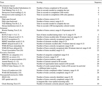
A clinical neuropsychologist (D.M. or M.B.) administered cognitive tests, either in the hospital or at the participant's home. Patients were tested in their optimal motor state, and we tried to keep distractions to a minimum. Tests were presented in a fixed order (see Appendix A) with short breaks every half hour. The duration of the testing session lasted between 2 and 3 hr. This was not sufficient for some patients to complete all tests; in such cases, the last three or four tests were omitted.
Instruments
We evaluated six cognitive domains: psychomotor speed, attention, language, memory, executive functioning, and visuospatial abilities (see Appendix).
Stage of disease was rated with the Hoehn & Yahr scale (Hoehn & Yahr, Reference Hoehn and Yahr1967). The motor section of the Unified Parkinson's Disease Rating Scale (UPDRS; Fahn & Elton, Reference Fahn and Elton1987) was used to assess severity of motor dysfunction. Functional status was determined with the Schwab and England scale (SE; Schwab & England, Reference Schwab and England1969), Barthel's Activities of Daily Living Index (BADL; Collin, Wade, Davies, & Horne, Reference Collin, Wade, Davies and Horne1988), and the Functional Independence Measure (FIM; van der Putten, Hobart, Freeman, & Thompson, Reference van der Putten, Hobart, Freeman and Thompson1999). Higher scores indicate a more advanced stage of PD (Hoehn & Yahr scale), more severe extrapyramidal symptoms (UPDRS), and better functional status (SE, BADL, and FIM). Mood and anxiety were measured with the Hospital Anxiety and Depression Scale (HADS; Zigmond & Snaith, Reference Zigmond and Snaith1983), and the Montgomery-Åsberg Depression Rating Scale (MADRS; Montgomery & Asberg, Reference Montgomery and Asberg1979). Higher scores indicate possible presence of mood or anxiety disorders.
Data Imputation
In the PD group, 8.9% neuropsychological data points were missing, and 2.8% in the control group. For the PD group, most missing data were from Wechsler Memory Scale-III (WMS-III) facial recognition (38%), Tower of London (33%), Wechsler Adult Intelligence Scale (WAIS) Similarities (45%), and Groningen Intelligence Test (GIT) spatial task (37%). These were tests administered at the end of the session. Without these tests, 1.3% data points were missing. However, due to the importance of these tests, we chose to include them in the analyses. Additional analyses (not described here) showed that patients who performed all tests were younger, had a lower age of disease onset, and less axial impairments than patients who did not complete the full neuropsychological examination. No other differences in disease characteristics were observed between the two groups.
For tests that a subject attempted but was unable to perform, we replaced missing scores with the lowest score obtained within that group. For tests that could not be completed due to fatigue, lack of motivation or time constraints, missing values were replaced with the groups’ mean score. We also explored imputation by estimates of missing scores with regression analyses. This method gave comparable results (data not shown; available on request).
Statistical Analysis
Continuous variables were visually inspected and statistically tested by Kolmogorov–Smirnov test for normality of the distribution. Demographic and clinical variables were analyzed with t tests, χ 2-tests, or Mann-Whitney tests, where appropriate. All p-values <.05 were considered statistically significant unless stated otherwise. Because of the explorative nature of our analyses, we did not correct for multiple comparisons (other than the correction inherent to multivariate analyses; see below) (Rothman, Reference Rothman1990).
Pattern and Severity of Cognitive Impairments
First, to examine group differences at baseline, multivariate analyses of covariance (MANCOVAs) were performed for each cognitive domain with baseline neuropsychological test scores as independent variables, group (patients vs. controls) as factor, and age, sex, and education as covariates. In case of significant main effects in a cognitive domain, subsequent analyses of covariance (ANCOVAs) were performed for each test separately.
Next, longitudinal data were analyzed using Linear Mixed Models (LMMs) with maximum likelihood estimation to examine cognitive decline. In these models, we accounted for the correlations between repeated measurements. For each neuropsychological test measure, we performed a LMM with age, sex, and education as covariates, group (patient vs. controls) and time as factors, and raw test scores at the three time points as dependent variables. First, we tested whether group differences existed, that is, main effect of group. Thereafter, we added time × group to the model to examine interaction effects between time and group.
We aimed to study cognitive functions as purely as possible by controlling for motor slowness in analyses of tests with speeded motor components. Therefore, we calculated a composite measure for motor processing speed by averaging standard scores on the Trail Making Test part A and Stroop CWT word reading. These are the two subtests with the strongest motor components, while they do not heavily tax cognitive functions. Instead, they depend on highly automatic processes that require few cognitive resources and are quite robust against brain diseases.
Frequency of Individual Cognitive Decline
Patients exhibiting cognitive decline were identified by multivariate normative comparison (MNC; Huizenga, Smeding, Grasman, & Schmand, Reference Huizenga, Smeding, Grasman and Schmand2007). This method is more sensitive than the conventional clinical approach, which uses normative scores in a univariate way. In neuropsychological evaluations a patient usually completes several tests, the results of which are then compared to normative samples. However, these conventional univariate comparisons have a considerable type I error risk, and are insensitive to patterns in deviation from the norm. The MNC method, based on Hotelling's T2 statistic, compares the pattern of change over the follow-up period of each individual patient to the pattern of change in the control group. This method has an increased power compared to Bonferroni corrected univariate comparisons in groups of this size and with at least moderate correlations between tests. For this MNC method, we used raw change scores for each neuropsychological test measure, that is, 5-year follow-up score – baseline score.
Prognostic Factors of Decline
To identify prognostic factors of decline, we compared demographic characteristics, baseline clinical variables, and baseline neuropsychological test scores of the patients with cognitive decline as identified by MNC to those of patients without decline. Separate scores were created from the UPDRS total motor score to assess levodopa responsive motor symptoms (i.e., tremor and bradykinesia; Levy A score) and symptoms that are unresponsive or partially responsive to levodopa (i.e., postural reflexes and speech; Levy B score) (Levy et al., Reference Levy, Tang, Cote, Louis, Alfaro, Mejia and Marder2000).
Next, we entered all variables associated with cognitive decline at p ≤ .15 in a hierarchical logistic regression analysis (forward stepwise). For ease of interpretation, and because not all variables were normally distributed, we dichotomized continuous variables. Demographic and clinical baseline variables were entered in the first block. In a second block, we added the baseline neuropsychological test scores to the model. Goodness of model fit was assessed with Hosmer and Lemeshow test. Effect sizes were expressed as odds ratios (OR) with 95% confidence interval.
PDD
Patients were diagnosed with PD dementia (PDD) according to MDS criteria (Dubois et al., Reference Dubois, Burn, Goetz, Aarsland, Brown, Broe and Emre2007; Emre et al., Reference Emre, Aarsland, Brown, Burn, Duyckaerts, Mizuno and Dubois2007). We diagnosed a patient with PDD if the PD diagnosis was made before the onset of dementia, the patient had a MMSE score <26, cognitive deficits that were severe enough to interfere with daily living, and showed impairments on at least two of the following test items: MMSE serial sevens, MMSE pentagon copying, MMSE 3-word recall, and the clock drawing test. Cognitive impairment severe enough to interfere with daily life was defined as a score ≤5 on at least one of the FIM items 14 to 18. These items examine to what extent cognitive dysfunction affects functional independence in everyday life; they cover language comprehension and expression, social interaction, problem solving, and memory.
Loss to Follow-up
Two sets of secondary analyses were conducted to examine attrition in detail. First, we compared baseline clinical characteristics and test performance of patients who were lost to follow-up with those of patients who continued to participate. Next, we compared partial information on neurological and cognitive status at 5-year follow-up from patients lost to neuropsychological follow-up with data from the patients who were still fully participating.
Results
Demographic and Clinical Variables
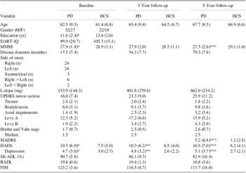
Patients had 1 year less education, higher HADS and MADRS scores, and lower MMSE scores than controls (Table 1). The follow-up interval was slightly longer for controls (66 months) than for patients (62 months). At 5-year follow-up, 22 patients suffered from on–off fluctuations (median duration in off: less than 25% of the day).
Demographic and clinical characteristics of PD patients (N = 59) and healthy controls (N = 40). Mean (SD)

*<.05 compared to controls at baseline.
**<.05 compared to controls at 3-year follow-up.
***<.05 compared to controls at 5-year follow-up.
PD = patients with Parkinson's disease; HCS = healthy control subjects; DART-IQ = Dutch Adult Reading Test; MMSE = Mini-Mental State Examination; MADRS = Montgomery and Asberg Depression Rating Scale; HADS = Hospital Anxiety and Depression Rating Scale; UPDRS = Unified Parkinson Disease Rating Scale; Levy A = Motor impairments that are responsive to levodopa; Levy B = Motor symptoms that are partially responsive or unresponsive to levodopa; SE-ADL = Schwab and England Activities of Daily Living; BADL = Barthel Activities of Daily Living Index; FIM = Functional Independence Measure.
Pattern and Severity of Cognitive Impairments
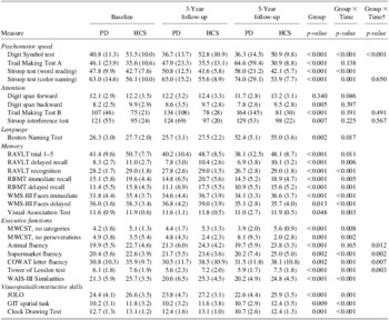
At baseline, MANCOVAs showed significant differences in performance for each cognitive domain, except visuospatial skills, between patients and controls, that is, psychomotor speed (p < .001); attention (p = .001); language (p = .043); memory (p < .001); executive function (p = .001); visuospatial skills (p = .090) (Table 2). Note that the language domain consisted of one test and therefore we only performed an ANCOVA. Additional ANCOVAs showed that the groups significantly differed from each other on all tests, except for Digit Span forward, Visual Association Test, Modified Wisconsin Card Sorting Test (MWCST) perseverations, Controlled Oral Word Association Test (COWAT) letter fluency, GIT matrices, and Clock Drawing Test. The vast majority of controls obtained normal scores, that is, between −2 and +2 SD from the age, sex, and education corrected mean. Seven controls scored below −2 SD on one or two tests.
Neuropsychological test findings (raw scores) of 59 PD patients and 40 healthy controls (HCS) at baseline, 3- and 5-year follow-up

Note. Values given as mean (SD).
†Linear Mixed Model with UPDRS item 31 (bodybradykinesia) as covariate, in addition to age, sex, and years of education. PD = Parkinson's disease; HCS = Healthy control subjects; RAVLT = Rey Auditory Verbal Learning Test; RBMT = Rivermead Behavioral Memory Test; WMS = Wechsler Memory Scale; MWCST = Modified Wisconsin Card Sorting Test; COWAT = Controlled Oral Word Association Test; WAIS = Wechsler Adult Intelligence Scale; GIT = Groningen Intelligence Test; JOLO = Judgment of Line Orientation.
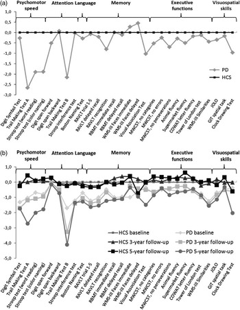
LMMs showed that patients obtained lower scores than controls on all tests, except for Digit Span forward (Table 2). Significant interaction effects of group and time were seen on all tests, except for tests of attention, and Trail Making Test A and animal fluency. On most tests, patients obtained lower scores over time while scores in the control group were relatively stable, showing that cognition in PD declines faster than in normal healthy aging. When we added the motor composite score as an extra covariate, only Stroop CWT color naming lost its statistical significance, whereas the interaction effect for animal fluency became significant. The profile of test scores is visualized in Figure 2a.
a: Standardized neuropsychological test scores for PD patients and healthy controls at baseline, 3-year follow-up, and 5-year follow-up. b: Standardized change scores between baseline and 5-year neuropsychological follow-up for PD patients and healthy controls. Standardized test scores in Figure 2a are Z-scores obtained by dividing the difference between the subject's raw test score and the mean baseline score of the control group by the standard deviation of scores in the control group, i.e. (raw test score minus mean score of control group) / SD of scores of the control group. Standardized change scores in Figure 2b are Z-scores obtained by dividing the difference between the subject's change score and the mean change score of the control group by the standard deviation of change scores in the control group, i.e. (change score minus mean change score of control group) / SD of change scores of the control group. Negative Z-scores indicate decreased performance compared to baseline performance of controls (Figure 2a) or faster decline than the control group (Figure 2b). PD = Parkinson's disease; HCS = Healthy control subjects; RAVLT = Rey Auditory Verbal Learning Test; RBMT = Rivermead Behavioral Memory Test; WMS = Wechsler Memory Scale; MWCST = Modified Wisconsin Card Sorting Test; COWAT = Controlled Oral Word Association Test; WAIS = Wechsler Adult Intelligence Scale; GIT = Groningen Intelligence Test; JOLO = Judgment of Line Orientation.

Frequency of Cognitive Decline
Next, we examined decline at the individual level with MNC using raw change scores. By this statistical criterion, 31 patients [53% of 59; 95% confidence interval (CI) = 39–66] showed significantly more decline than controls (at p < .05, one-sided). Removing tests that were influenced by motor processing speed did not alter this result. The pattern of change is depicted in Figure 2b.
Prognostic Factors of Cognitive Decline
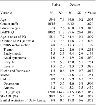
Patients who showed cognitive decline (n = 31) were compared to patients showing no cognitive decline (n = 28), with respect to demographic and clinical characteristics at baseline (Table 3). Significant differences (with p ≤ 0.15) were found on age, years of education, age at onset of PD, UPDRS motor score, bradykinesia, axial symptoms, Levy B, Hoehn & Yahr scale, and LED.
Demographic and clinical variables at baseline for patients who were cognitively stable (N = 28) and those who showed cognitive decline after 5 years (N = 31)

DART-IQ = Dutch Adult Reading Test; UPDRS = Unified Parkinson's Disease Rating Scale; Levy A = Motor impairments that are responsive to levodopa; Levy B = Motor symptoms that are partially responsive or unresponsive to levodopa; MMSE = Mini-Mental State Examination; HADS = Hospital Anxiety and Depression Scale; LED = Levodopa equivalent dose; SE-ADL = Schwab and England Activities of Daily Living.
Patients with cognitive decline had lower test scores than cognitively stable patients on Digit Symbol test (p = .01), Stroop CWT color naming (p = .09) and interference test (p = .03), Trail Making Test part B (p = .03), Auditory Verbal Learning Test immediate (p = .04) and delayed recall (p = .06), Rivermead Behavioural Memory Test stories immediate (p < .01) and delayed recall (p < .01), Visual Association Test (p = .02), MWCST categories (p = .03), COWAT letter fluency (p = .03), and Similarities (p = .09). Age and age at onset of PD were highly correlated. Therefore, only age at onset was used as a predictor variable.
Logistic regression analyses showed that the model significantly improved at each block (block 1: p < .03, block 2: p = .01). In the first block, age of onset (OR = 4.71; 95% CI = 1.49–14.94; p = .01) and Hoehn & Yahr score (OR =3.37; 95% CI = 1.06–10.70; p = .04) were significant predictors of cognitive decline. In the second block where the psychological test variables were entered, Hoehn & Yahr score lost its significance. Age of onset (OR = 4.34; 95% CI = 1.22–16.11; p = .024) and Rivermead Behavioral Memory Test immediate recall (OR = 7.78; 95% CI = 2.10 – 28.86; p = .002) contributed significantly to the prediction of decline. The Hosmer and Lemeshow test showed the model fitted the data (χ 2 = 1.605; p = .90).
PDD Diagnoses
Of the 59 patients who completed all three assessments, 2 patients had PDD at the 3-year follow-up, and another 3 patients at the 5-year follow-up. Thus, after 5 years, 5 of 59 (8.5%) had PDD. Of the 133 patients who were included in the CARPA study, 20 patients (15.0%) had developed PDD.
Loss to Follow-up
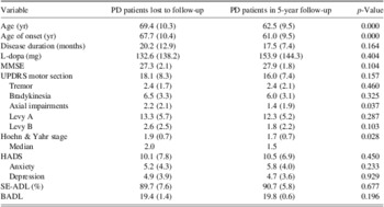
Due to the relatively large number of patients lost to follow-up, the true percentage of patients showing cognitive decline is larger than 53%. We compared patients lost to neuropsychological follow-up (n = 64) with patients who fully participated in the current follow-up (n = 59). Patients who were lost to follow-up were older, had higher age at disease onset, more axial impairments, and scored higher on the Hoehn & Yahr scale (Table 4). Thus, patients who were lost to follow-up had greater disease severity at baseline. Of the 64 patients who were lost to neuropsychological follow-up, 14 patients developed PDD.
Demographic and clinical baseline data of PD patients who were lost to neuropsychological follow-up (N = 64) and patients who fully participated in the 5-year follow-up (N = 59)

Note. Values given as mean (SD).
MMSE = Mini Mental State Examination; HADS = Hospital Anxiety and Depression Rating Scale; UPDRS = Unified Parkinson Disease Rating Scale; Levy A = Motor impairments that are responsive to levodopa; Levy B = Motor symptoms that are partially responsive or unresponsive to levodopa HADS = Hospital Anxiety and Depression Rating Scale; SE-ADL = Schwab and England Activities of Daily Living; BADL = Barthel Activities of Daily Living Index.
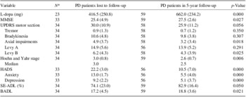
Since this confirmed that attrition led to an underestimation of cognitive decline, we conducted a second set of analyses. We compared the clinical data we were able to obtain from 34 patients, who were lost to the present neuropsychological follow-up but had neurological and functional assessments, with data obtained from patients who continued to fully participate (n = 59; Table 5). Patients who were lost had lower MMSE scores, higher Hoehn & Yahr scores, showed more axial symptoms and more motor symptoms unresponsive or partially responsive to levodopa, had more symptoms of anxiety and depression, and scored lower on the SE-ADL and BADL scales. Again, these results indicate greater disability in patients who were lost to follow-up.
Clinical data at 5-year follow-up of PD patients who were lost to neuropsychological follow-up and patients who fully participated in the 5-year follow-up

Note. Values given as mean (SD).
*Varies due to missing values.
MMSE = Mini-Mental State Examination; HADS = Hospital Anxiety and Depression Rating Scale; UPDRS = Unified Parkinson Disease Rating Scale; Levy A =Motor impairments that are responsive to levodopa; Levy B = Motor symptoms that are partially responsive or unresponsive to levodopa; HADS = Hospital Anxiety and Depression Rating Scale; SE-ADL = Schwab and England Activities of Daily Living; BADL = Barthel Activities of Daily Living Index.
Due to loss to follow-up, reliable data on cognitive decline 5 years after diagnosis are lacking in 50 patients (64 patients lost to follow-up minus 14 patients with PDD). If we assume—as a conservative estimate—the same percentage of decline in this subgroup as in those who continued to participate (i.e., 53%), then approximately 27 patients in this group should have declined. The total number of patients, who show cognitive decline, including PDD, should then be estimated at 73 of 123, or 59%.
Discussion
Our aim was to examine the pattern and severity of cognitive impairments, the frequency of individual cognitive decline, and prognostic markers of cognitive decline in a hospital-based cohort of newly diagnosed PD patients. Overall, patients performed worse than controls on most neuropsychological tests. Impairments were already observed at baseline in most cognitive domains, and patients declined faster than controls in all domains except attention (Table 2). Fifty-three percent of the patients who continued to participate showed more cognitive decline than the 5th percentile of control subjects over the course of 5 years, including five patients (8.5%) with PDD. In total, 15% of the original patient sample had become demented. Attrition led to selection bias; therefore, the actual percentage of patients with cognitive decline is probably even greater. We estimate it at 59%, including PDD. Higher age at onset and weak memory performance at baseline predicted cognitive decline.
These results, that is, impaired performance in patients on the majority of neuropsychological tests, are in line with our previous analyses and with other studies (Muslimovic et al., Reference Muslimovic, Post, Speelman, De Haan and Schmand2005, Reference Muslimovic, Post, Speelman and Schmand2009; Williams-Gray et al., Reference Williams-Gray, Foltynie, Brayne, Robbins and Barker2007). PD patients not only obtain lower baseline scores on neuropsychological testing, but also their cognitive performance declines faster over time than in normal healthy aging. Some neuropsychological tests have strong motor components such as Trail Making Test part B and verbal fluency tests. Correcting for motor processing speed, however, did not greatly alter the results. Thus, after 5 years, patients showed cognitive impairments irrespective of motor processing speed.
The pattern of cognitive decline (Figure 2) is largely similar to our previous 3-year follow-up report with substantial decline in the psychomotor domain. The decline in attention and executive functioning is more restricted to certain tests, that is, Digit Span, and the Modified Wisconsin Card Sorting Test (categories and perseverations). The large score drop on the Trail Making Test part B must be due to motor slowing, since the group by time interaction effect was not significant after correcting for motor speed (Table 2). Decline over the course of 5 years was slightly smaller, but evident in the visuospatial domain. However, this was largely restricted to clock drawing. In the memory domain we observed moderate decline. Figure 2 even shows a slight increase in performance for WMS-III Faces. However, this is due to decreased performance in the control group, while performance in patients was relatively stable.
The decline in memory and visuospatial skills, albeit moderate, could be related to the neuropathological staging of PD. In clinically manifest PD, the essential neuropathology involves the nigrostriatal tracts and the mesocortical pathways (Jellinger, Reference Jellinger2001). Disruption in both pathways has been related to executive dysfunctioning. In later stages, pathology is more widespread, also affecting posterior cortical areas, which correlates with the risk of developing dementia (Braak, Rub, Jansen Steur, Del Tredici, & de Vos, Reference Braak, Rub, Jansen Steur, Del Tredici and de Vos2005). Memory decline and visuospatial impairment may be mediated by these posterior disease processes (Lee et al., Reference Lee, Park, Song, Sohn, Lee and Lee2010; Williams-Gray et al., Reference Williams-Gray, Foltynie, Brayne, Robbins and Barker2007). An alternative explanation is that decline in memory and visuospatial skills is due to executive components in visuospatial and memory tests (Beyer et al., Reference Beyer, Bronnick, Hwang, Bergsland, Tysnes, Larsen and Apostolova2013; Dubois & Pillon, Reference Dubois and Pillon1997; Emre, Reference Emre2007). Neuropsychological tests never exclusively tax a single cognitive domain. The Trail Making Test part B and Stroop interference test, for instance, are frequently seen as measures of executive functioning, but also as tests of attention, while both require a speeded psychomotor response. Thus, the division of cognition into domains, and the allocation of tests to a particular domain, is artificial to a certain extent.
Our final aim was to identify predictors of cognitive decline. Previous studies reported various predictors, such as age, male sex, hallucinations, axial impairment, impaired semantic fluency or pentagon copying, and executive dysfunction (Aarsland et al., Reference Aarsland, Andersen, Larsen, Lolk and Kragh-Sorensen2003; Hobson & Meara, Reference Hobson and Meara2004; Levy et al., Reference Levy, Jacobs, Tang, Cote, Louis, Alfaro and Marder2002; Muslimovic et al., Reference Muslimovic, Post, Speelman, De Haan and Schmand2009; Uc et al., Reference Uc, McDermott, Marder, Anderson, Litvan and Como2009; Williams-Gray et al., Reference Williams-Gray, Evans, Goris, Foltynie, Ban, Robbins and Barker2009). In our study, only age at onset and weak memory performance at baseline were independent prognostic factors of cognitive decline. Age at onset of PD has previously been reported as a predictor of cognitive decline (Muslimovic et al., Reference Muslimovic, Post, Speelman, De Haan and Schmand2009). Memory impairment as a predictor for cognitive decline, however, is a less common finding. In 2005, Hobson and Meara reported that more profound memory impairment at baseline increases the risk of dementia (Hobson & Meara, Reference Hobson and Meara2004). Due to the high age (i.e., mean age of 78) in their cohort, they ascribed this to the development of Alzheimer's disease. However, patients in our cohort were relatively young. Furthermore, Alzheimer's disease pathology in PD is usually rather mild. It has, therefore, been suggested that cognitive decline is more likely attributable to cumulative effects of PD pathology (Braak et al., Reference Braak, Rub, Jansen Steur, Del Tredici and de Vos2005).
The fact that memory impairments at baseline also predict further cognitive decline, and the actual decline in memory that we found, may thus reflect the involvement of other brain areas besides the nigrostriatal system and mesocortical pathways. This notion is in line with another finding in our study, that is, the finding that the primary determinants of disability in PD are motor symptoms not responsive to levodopa and cognitive dysfunction at the time of diagnosis (Velseboer et al., 2013). Although memory impairment in PD most often concerns the retrieval of information, in the current study we also saw decline on a recognition test and visual association test. These tests reflect more posterior, medial temporal lobe dysfunction. This supports the recently postulated dual syndrome hypothesis of PD: (1) a syndrome characterized by frontal-striatal dysfunction and (2) a syndrome with distinctive early visuospatial deficits that are indicative of the involvement of temporal and other posterior cortical areas (Kehagia, Barker, & Robbins, Reference Kehagia, Barker and Robbins2012). Patients with more profound executive impairment at baseline, suggestive of fronto-striatal dysfunction, generally progress slower than those with more profound memory and visuospatial impairments.
Despite the fact that a memory test predicted cognitive decline, we see a moderate decline over time in the memory domain itself. This could be due to the psychometric properties of the neuropsychological tests: it is possible that the reliability of neuropsychological tests is not uniform across the score distribution. Scores in the midst of the distribution, thus in the normal scoring ranges, might be measured more reliably and sensitively than scores in the lower ranges of the distribution. Therefore, decline in the range that patients already score lower than healthy controls, may be more difficult to measure. A second possible explanation for the discrepancy between memory impairment as a prognostic factor and the modest decline in memory functioning, relates to the unpredictable pattern of disease progression. The progression of cognitive decline might be unequally phased. For example, early decline in a cognitive domain may be followed by a relatively stable period without decline. This could be related to the characteristics of disease progression itself, but also by other factors, such as dopaminergic medication. An increase in medication may elevate dopamine levels in the midbrain which could positively affect cognition. These explanations are admittedly speculative.
One of the main problems in follow-up studies is attrition, because this may lead to selection bias. Selection bias is often left implicit, so that the true extent of cognitive decline remains unclear. Fortunately, we had some information about cognitive status and physical functioning of patients who refused to participate in the neuropsychological follow-up. These data corroborated that patients who were lost to follow-up were older, had an older age of onset, and had more severe motor symptoms and more cognitive deficits than the patients who were still participating. Thus, the true extent of cognitive decline in PD is greater than what is shown by our neuropsychological test results. This is also supported by the percentage of patients with PDD after 5 years, which was 8.5% in the neuropsychological part of the study, but twice as high in the total sample.
This study has several strengths. First, at baseline we studied a relatively large sample of newly diagnosed PD patients. This allowed us to monitor disease progression and cognitive decline in detail from the moment of diagnosis. Second, we used a sensitive statistical method (MNC) to detect cognitive change at the individual level. Third, by extending the follow-up duration to 5 years, the power of our analyses was improved. A fourth strength is the large battery of neuropsychological tests. Fifth, the diagnosis of PD was validated twice, at baseline and at 5-year follow-up.
There are also caveats. First, our patient sample was recruited in hospitals. One may argue, therefore, that its representativeness for the PD population at large is not clear. However, the clinical characteristics of our sample are not very different from those in population-based studies (Aarsland et al., Reference Aarsland, Andersen, Larsen, Lolk and Kragh-Sorensen2003; Elgh et al., Reference Elgh, Domellof, Linder, Edstrom, Stenlund and Forsgren2009; Foltynie et al., Reference Foltynie, Brayne, Robbins and Barker2004). Moreover, in the Netherlands virtually all PD patients are sooner or later referred to a neurologist. Second, our study has a high attrition rate. This is not just an issue in the current study, but selection bias should be taken into account in all studies that investigate (cognitive) changes in PD. If patients who are lost to follow-up (or who refuse to participate in cross-sectional studies) are more severely ill, then it is likely that the true extent of cognitive decline is greater than what has been shown until now in PD research. The high attrition rate was partly due to the length of the neuropsychological evaluation, which some patients found too burdensome. Third, another drawback of using a large neuropsychological test battery was that not all patients could complete all tests. We imputed missing values so we could analyze these tests and would not lose power in our analyses, but this might affect the results. Therefore, we repeated the analyses without tests that had a large number of missing values (data not shown), but this did not change our conclusions.
In conclusion, 5 years after the diagnosis of PD, approximately two-thirds of the patients show cognitive decline. The greatest decline was observed in the areas of psychomotor speed and memory. Patients with a higher age at onset and memory impairment in an early disease stage are most at risk for further cognitive decline.
Acknowledgments
The authors thank the reviewers for their valuable comments which helped us to improve this manuscript. We thank the nurse practitioners for their help in data collection. Also, we thank the Medical Center Alkmaar, Meander Medical Center Amersfoort, Slotervaart Hospital Amsterdam, St. Lucas-Andreas Hospital Amsterdam, OLVG Amsterdam, and Academic Medical Center Amsterdam for their help with patient inclusion and for facilitating the neurological and neuropsychological examinations. And most of all, we thank the patients and healthy controls for their cooperation. This work was supported by the Prinses Beatrix fonds [grant PGO01-0138 to BS] and the Michael J. Fox Foundation (grant to BS). It is part of the CARPA project, sponsored by ZonMW. Rob M.A. de Bie received an unrestricted fellowship grant from Medtronic (Minneapolis, MN). All other authors report no conflicts of interest.
Overview of the neuropsychological tests with the scoring ranges and sequence of administration