Structural cardiovascular malformations occur in up to two-fifths of patients with Turner’s syndrome.Reference Gotzsche, Krag-Olsen, Nielsen, Sørensen and Kristensen1–Reference Mazzanti, Prandstraller and Fattori5 According to recent studies, coarctation of the aorta and aortic valves with 2 leaflets are most common, accounting for one-tenth and one-sixth respectively of the cardiovascular malformations to be found in such patients.Reference Gotzsche, Krag-Olsen, Nielsen, Sørensen and Kristensen1–Reference Volkl, Degenhardt, Koch, Simm, Dörr and Singer4 Dilation of the ascending aorta is also relatively common, particularly when associated with coarctation, a bifoliate aortic valve, or systemic arterial hypertension.Reference Mazzanti and Cacciari2, Reference Mazzanti, Prandstraller and Fattori5–Reference Dawson-Falk, Wright, Bakker, Pitlick, Wilson and Rosenfeld8 The prevalence of dilation of the aortic root in patients without the associated predisposing cardiac lesions, however, is currently unknown. In this study, therefore, we sought to assess, first, the prevalence of aortic dilation in a large group of patients with Turner’s syndrome but lacking the associated predisposing cardiac lesions, and second, to evaluate the presence of morphometric remodelling of the aortic root, assessing also its potential relation to the underlying genotype.
Materials and methods
Population of patients
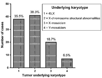
Between March, 2000, and June, 2007, we evaluated echocardiographically at our Institution 152 patients, aged from 0.1 to 35.7 years, with genetically proven Turner’s syndrome. Of these patients, we subsequently excluded 45 (29.6%) because of associated lesions that predisposed them to aortic dilation, specifically an aortic valve with 2 leaflets, a dysfunctional aortic valve, aortic coarctation or arterial hypertension, along with those having other associated congenital cardiac malformations. We carried out magnetic resonance imaging in those patients in whom it proved impossible echocardiographically to clarify the anatomy of the aortic valve or the aortic isthmus. Thus, we were left with 107 patients, with an average age of 19.6+/−8.4 years, with no cardiac lesions predisposing to aortic dilation, nor any other form of congenital cardiac disease. In all these patients, a complete echocardiographic scan was available for review, and all had undergone genetic assessment as previously reported.Reference Mazzanti and Cacciari2 We classified the identified karyotypes on a scale from 1 to 4 as shown in Figure 1. We had performed transthoracic echocardiography on 71 healthy volunteer females during the same period, and these subjects were used as controls. All patients or legal guardians gave written informed consent for participation in the study. The protocol we used conforms to the ethical guidelines of the 1975 Declaration of Helsinki, as reflected by prior approval from our human research committee. We measured the height and weight of all subjects at the time of the study, and used this data to calculate body surface area and body mass index. The least median-of-squares method was used to calculate the scores for standard deviation of the body mass index.Reference Cacciari, Milani and Balsamo9 National growth charts were used as our reference.Reference Cacciari, Milani and Balsamo9 All patients had undergone ambulatory monitoring of blood pressure before being included in the study. Medical records were reviewed for history of replacement of oestrogens, administration of growth hormone, and evidence of arterial hypertension.
Distribution of the underlying genotype in our patients with Turner’s syndrome. We graded the different genotypes on a scale from 1 to 4 as shown in the figure.

Echocardiography
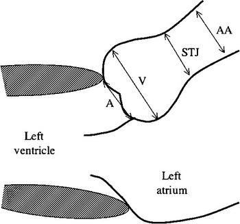
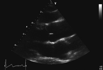
All patients and control subjects underwent transthoracic echocardiography using a Sonos 4500 or 5500 echocardiographic machine manufactured by Philips Medical Systems, from Endover, Maryland, interfaced with a multi-frequency transducer. The anatomy of the aortic valve was assessed using the parasternal short-axis view, while the aortic root was additionally imaged from the parasternal long-axis acoustic window. We made measurements of the diameter of the aortic root at 4 levels (Fig. 2), specifically the level of the basal hinges of the aortic leaflets, the widest level across the sinuses of Valsalva, the sinutubular junction, and the proximal part of the ascending aorta.Reference Roman, Devereux, Kramer-Fox and O’Loughlin10 Measurements were performed perpendicular to the long axis of the aorta using the leading edge technique.Reference Roman, Devereux, Kramer-Fox and O’Loughlin10, Reference Lang, Bierig and Devereux11 We made 3 measurements at each of these levels, using the average in the subsequent analysis. We then indexed the values obtained in each subject for body surface area. For each patient, we also indexed the diameters, in millimetres of the basal diameter of the root, the sinutubular junction, and the ascending aorta against the diameter at the level of the sinuses of Valsalva. We used the guidelines provided by the American Society of Echocardiography to identify and quantify aortic regurgitation and stenosis.Reference Zoghbi, Enriquez-Sarano and Foster12 All measurements were made by the same observer.
The sites of measurement of the diameters of the aortic root were made at the basal attachments of the leaflets of the aortic valve (A), the widest dimensions of the sinuses of Valsalva (V), the sinutubular junction (STJ), and the ascending aorta (AA). Measurements were made at end-diastole from the parasternal long axis acoustic window.

Statistical analysis
Normal distribution of data was tested using the Kolmogorov-Smirnov test prior to any further analysis. Data is then expressed as mean plus or minus the standard deviation. The various diameters of the root in the healthy subjects were plotted against body surface area, permitting the calculation of 99% prediction bands for normality using linear regression. The diameters measured at the corresponding sites in the patients were then plotted on the same graphs to identify those with aortic diameters outside the prediction bands. For each aortic segment, we compared both the diameters and the ratios of the aortic segments in the patients and their control using the two-tailed unpaired t-test. In the controls and the patients, we also sought any associations between age, height, body surface area, and respectively the diameters of the ascending aorta and sinutubular junction using Pearson correlation analysis. The association between the underlying karyotype and the diameters of the ascending aorta and sinutubular junction indexed to body surface area was assessed by Spearmen rank correlation analysis. We used multiple linear regression to study the association these diameters and age, body surface area, body mass index, and karyotype. A value of p of less than 0.05 was considered significant. In 15 patients, the echocardiographic measurements of each aortic segment were repeated by a second investigator, who was unaware of previous results. For each variable, the standard deviation of the difference between the measurements was divided by the mean and expressed as a percentage to provide the coefficient of variability. All calculations were performed using the Graphpad Prism 4.0 software package as manufactured by Graphpad Corporation, from California, United States of America.
Results
At the time of the study, 76 patients (71%) were receiving hormonal treatment with a combination of oestrogens and progesterone, while 92 patients (86%) were receiving, or had received, growth hormone. We found no statistically significant differences in age, weight, and body surface area between the patients and their controls. The control subjects, however, were taller, at 1.53+/−0.30 versus 1.42+/−0.17 metres (p = 0.012), had a lower body mass index at 19.6+/−2.3 versus 22.6+/−4.5 (p < 0.0001), and a lower body mass index at 0.15+/−0.30 versus 1.48+/−3.0 (p = 0.0024) than the patients with Turner’s syndrome.
The coefficients of variability for inter-observer reproducibility of the measurements were 5.8% for the diameter at the level of the hinges of the aortic valvar leaflets, 6.5% for the widest diameter of the sinuses of Valsalva 6.5%, 6.1% for the diameter of the sinutubular .junction 6.1%, and 4.9% for the diameter of the ascending aorta No patient had aortic valvar regurgitation or stenosis.
Prevalence and patterns of aortic dilation
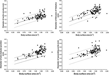
A comparison of echocardiographic data showed that the patients had larger diameters of the aorta at the level of the sinuses of Valsalva, the sinutubular junction, and the ascending aorta than did the control subjects (Table 1). Abnormal dilation of the aorta was frequently seen in the patients (Fig. 3) at the level of the sinuses of Valsalva, being found in 12 patients (11.2%, 95% confidence intervals from 6.4 to 18.7%), and the ascending aorta, found in 9 patients (8.4%, 95% confidence intervals from 4.3 to 15.4%), but especially at the level of sinutubular junction, found in 29 patients (27.0%, 95% confidence intervals from 19.5 to 36.2%).
Comparison of the diameters of the aortic root at the level of the basal attachment of the valvar leaflets, the widest dimension of the sinuses of Valsalva, the sinutubular junction, and the ascending aorta as measured by transthoracic echocardiography in patients with Turner’s syndrome and our control subjects.

Distribution of the aortic diameters observed in our patients with Turner’s syndrome according to their body surface area. The values obtained in the control group, not displayed in the graph, were used to generate 99% prediction bands using linear regression. Aortic diameters lying outside the prediction band were considered abnormal.

Morphometric assessment of the aortic root
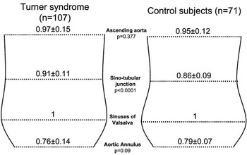
The patients were associated with an abnormal architecture of the aortic root (Fig. 4), the most prominent feature being a relative dilation of the sinutubular junction compared to control subjects (p < 0.0001), with no significant increase in the ratio of the widest diameter of the sinuses of Valsalva relative to the ascending aorta (p = 0.377), leading to loss of the typical shape of the aortic root (Fig. 5).
Comparison of the ratios of the aortic diameters measured in our patients with Turner’s syndrome and in our control subjects.

The typical change in the shape of the aortic root as observed in a patient with Turner’s syndrome, with an increase in the ratio of the diameter at the sinutubular junction relative to the sinuses of Valsalva.

Impact of clinical and demographic characteristics on aortic diameters
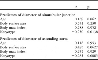
The patients with the underlying karyotype of 45X had larger diameters at the levels of the sinutubular junction and ascending aorta when compared to those with different karyotypes (Table 2), despite having similar body surface areas (p = 0.623). We show correlations between the demographic and clinical characteristics of our cohort and the diameters of the ascending and sinutubular junction in Table 3.
Comparison of the diameters of the aortic root at the level of the basal attachment of the valvar leaflets, the widest dimension of the sinuses of Valsalva, the sinutubular junction, and the ascending aorta as measured by transthoracic echocardiography in those patients with Turner’s syndrome with and without a 45X karyotype.

Results of correlation analysis between aortic diameters and demographic and clinical characteristics of the cohorts studied.

The age when studied, and body surface areas, correlated well with the diameters of the ascending aorta and sinutubular junction in both the patients and their healthy controls. Aortic diameters correlated well with height in the control subjects, but not in the patients. We observed an inverse correlation between the underlying karyotype and the indexed diameters of both the ascending aorta and sinutubular junction, with patients with a 45X karyotype also having the largest aortic diameters. No association was observed between other indexed aortic diameters at any level and the karyotype. At multiple linear regression (Table 4), the underlying karyotype proved the strongest predictor of the diameters at the sinutubular junction and ascending aorta.
Predictors of the diameters of the sinutubular junction and ascending aorta in 107 patients with Turner’s syndrome as assessed using multiple linear regression analysis.

Discussion
Our study shows that, in patients with Turner’s syndrome, the aortic root can be abnormally dilated even in the absence of predisposing associated cardiac lesions, such as aortic valvar dysfunction, an aortic valve with 2 leaflets, aortic coarctation, or arterial hypertension. The prevalence of a dilated ascending aorta in such patients without cardiovascular lesions predisposing them to aortic dilation was 8.4%. The ascending aorta has usually been regarded as the site of expression of the disease, and previous reports have focussed mainly on this area.Reference Ostberg, Brookes, McCarthy, Halcox and Conway6, Reference Van den Berg, Bannink and Wielopolski13 Even though a recent report showed increased diameters at the sinutubular junction when compared to healthy controls,Reference Lanzarini, Larizza, Prete, Calcaterra and Klersy14 as far as we are aware, ours is the first study to show that dilation of the sinutubular junction might be even more common than dilation of the ascending aorta. We have also shown that patients with Turner’s syndrome undergo remodelling of the aortic root, primarily characterized by a dilation of the sinutubular junction that leads to the loss of the normal shape. The sinutubular junction is a critical area in the root, having important structural and mechanical functions. It is known that this junction plays a major role in the ergonomics of the aorta, and its integrity is essential to prevent aortic valvar dysfunction and incompetence.Reference Padial, Oliver, Sagie, Weyman, King and Levine15 After the arterial switch operation for transposition, for example, dilation of the sinutubular junction related to the surgical procedure is associated with the occurrence of aortic regurgitation.Reference Formigari, Toscano and Giardini16 Whether dilation of the sinutubular junction may also predispose patients with Turner’s syndrome to the development of aortic regurgitation later in life is currently unknown. The exclusion of patients with aortic regurgitation from the present study, and the absence of longitudinal follow-up data, prevented us from addressing this question.
The reason for dilation of the sinutubular junction and ascending aorta in patients with Turner’s syndrome in the absence of aortic valvar disease, aortic coarctation, or hypertension is unknown. Patients with bifoliate aortic valve may present progressive dilation of the root that is not completely related to the associated valvar pathology,Reference Nistri, Sorbo, Marin, Palisi, Scognamiglio and Thiene17–Reference Keane, Wiegers, Plappert, Pochettino, Bavaria and Sutton19 with the dilation progressing in some cases even after effective valvar replacement.Reference Yasuda, Nakatani and Stugaard20 Evidence exists that, in those with bifoliate valves, the substrate for progression of aortic dilation is the presence of intrinsic mural abnormalities, such as disruption of the fibrous matrix and loss of smooth muscle cells.Reference Fedak, Verma, David, Leask, Weisel and Butany21, Reference Fedak, David, Borger, Verma, Butany and Weisel22 These lesions are similar in fibrillin-1-deficient patients with Marfan’s syndrome.Reference Bunton, Biery, Myers, Gayraud, Ramirez and Dietz23–Reference Niwa, Perloff and Bhuta25 Identical abnormalities of the aortic wall have been found in patients with Turner’s syndrome.Reference Lin, Lippe and Rosenfeld7, Reference Bordeleau, Cwinn, Turek, Barron-Klauninger and Victor26 The aortic dilation and the morphologic alteration of the aortic root that we have observed in our patients, therefore, may be the result of an intrinsic abnormality in the aortic wall. Patients with Turner’s syndrome are known to be at risk for aortic dissection, even at a young age.Reference Lin, Lippe and Rosenfeld7 Recent reports showed that such a risk of is not limited to those with associated risk factors, such as aortic valvar disease, aortic coarctation, or hypertension.Reference Lin, Lippe and Rosenfeld7, Reference Lin, Lippe and Geffner27 Indeed, up to one-fifth of the patients who developed aortic dissection have been reported to have no macroscopic risk factor except aortic dilation. The mechanism responsible for aortic dilation and dissection in this subpopulation, however, is currently unknown. It is possible that a morphologic remodelling of the aortic root, related to an abnormal aortic wall, might trigger aortic dissection even in the absence of associated risk factors. Even though the aortas of some of our patients were dilated when compared to those of healthy subjects, the overall aortic diameters we observed are below the threshold for surgical intervention because of the risk of aortic dissection. This explains the fact that no patient developed aortic dissection, or required aortic surgery, during the period of follow-up.
The possibility of an association between genotype and phenotype has already been already reported in the setting of Turner’s syndrome with regard to congenital cardiac defects. Indeed, previous studies have shown a higher prevalence of such defects in subjects with the 45X karyotype.Reference Gotzsche, Krag-Olsen, Nielsen, Sørensen and Kristensen1, Reference Ostberg, Brookes, McCarthy, Halcox and Conway6 The possibility that such correlations also exist at the level of the aortic root, however, has not been elucidated fully. An attempt to establish an association between karyotype and aortic phenotype has been made previously.Reference Matura, Ho, Rosing and Bondy28 The group making this observation showed that the presence of an aortic phenotype characterized by aortic dilation was associated with changes to the short arm of chromosome X. Our findings suggest further associations between the type of underlying genetic abnormality and the aortic phenotype, with our patients with the 45X karyotype having the largest aortic diameters. This association was confirmed by direct comparison of aortic diameters in patients with and without the 45X karyotype, and by correlation analysis between aortic diameters and karyotype. Our multiple regression analysis also showed that, in our patients, the karyotype was the main determinant of the diameters of the sinutubular junction and ascending aorta. This association was independent of age and associated conditions. The information is clinically relevant, since it suggests that patients with the 45X karyotype might be more prone to aortic dilation than those with different karyotypes. Further longitudinal study is necessary to confirm that the patients with the 45X karyotype are not at higher risk for aortic complications.
Short stature, combined with relative excessive weight, is a typical feature of patients having Turner’s syndrome. This explains our finding that body surface area was similar in our patients with Turner’s syndrome and their controls despite the noted differences in height. In normal subjects, body surface area is a more important determinant of the size of aortic diameters than either height or weight alone.Reference Sluysmans and Colan29 In keeping with this finding, we noted a close association between aortic dimensions and body surface area for both our patients with Turner’s syndrome and our control subjects. As expected, we also observed a close association between height and aortic diameters in the healthy controls, supporting the concept that shorter healthy individuals should have smaller aortas. We could not find, however, any association between height and aortic diameters in the patients with Turner’s syndrome. This data underscores the importance of aortic dilation in the setting of Turner’s syndrome. Indeed, as has recently been suggested,Reference Matura, Ho, Rosing and Bondy28 the aortas of patients with Turner’s syndrome should be considered even more dilated when compared to those of healthy and taller volunteers of equivalent body surface area when taking account of the shorter stature of those having Turner’s syndrome.
Limitations
A previous study has shown the existence of a linear relation between the diameter of the ascending aorta and age, both in patients with Turner’s syndrome patients and normal subjects.Reference Ostberg, Brookes, McCarthy, Halcox and Conway6 This correlation was less robust in our patients with Turner’s syndrome (r = 0.276). Patients with associated cardiac lesions, for which the damage of the aorta is time-dependent, were assessed in that study,Reference Ostberg, Brookes, McCarthy, Halcox and Conway6 however, and this may well explain the discrepancy between the findings. Treatment with growth hormone treatment is known to have a beneficial influence on the properties of the aortic wall in patients with Turner’s syndrome, with aortic distensibility shown to be lower, and aortic dilation more prominent, in those patients receiving the lowest dose of growth hormone.Reference Van den Berg, Bannink and Wielopolski13 A large proportion of our patients was receiving, or had received, growth hormone treatment at the recommended dosage at the time of our study. Our results, therefore, cannot be ascribed to lack of appropriate treatment with growth hormone.
The genetic heterogeneity underscoring Turner’s syndrome is large, and is likely reflected in the phenotype. For example the percentage of mosaicism can vary greatly and this might influence the clinical expression of the syndrome, including aortic diameters. Even if the grouping of genotypes, and use of a scoring classification, is necessary for statistical purposes, it might be seen as a limitation to our study. It is certainly the case that the classification we adopted, although based on current genetic knowledge of the syndrome, is arbitrary. Simplification of the genetic background of Turner’s syndrome, nonetheless, might explain the level of association we observed between aortic diameters and genotype.
Given the relatively young age of our population, and our exclusion of patients with cardiovascular lesions predisposing to aortic dilation, the absolute aortic diameters we observed are below the threshold for surgical intervention. Our population, nonetheless, provided a valid model with which to explore the effect of the isolated genetic anomaly on the size and shape of the aortic root. The data shown in Figure 3 suggests that the majority of patients with dilation of either the sinutubular junction or the ascending aorta have a body surface area greater than 1.1 m2. We think this is related to the age distribution of the population studied, with the majority of patients also having a body surface area greater than 1.1 m2, so that the absolute number of patients with dilated aortas is larger in this group.
In conclusion, we have shown that dilation of the sinutubular junction is frequent in patients with Turner’s syndrome, being more common than dilation of the ascending aorta. Turner’s syndrome, therefore, is associated with remodelling of the aortic root characterized by prominent dilation of the sinutubular junction. There also seems to be a relation between the observed aortic dilation and the underlying genotype.