INTRODUCTION
Strongyloides ratti has been investigated not only as a model parasite for human strongyloidiasis, but also for its complex life cycle of evolutionary interest (Gemmill et al. Reference Gemmill, Viney and Read1997; Viney et al. Reference Viney, Steer and Wilkes2006; Viney and Lok, Reference Viney and Lok2007). Infective larvae (L3i), which are females genetically (Harvey and Viney, Reference Harvey and Viney2001), penetrate the skin of a host rat, develop to female adults in the upper small intestine, and lay eggs parthenogenetically. Each egg (or hatched 1st-stage larva) excreted in faeces eventually develops into either an infective larva, a free-living female or a free-living male. Although the majority of eggs in the early stage of infection develop into L3i, the direction of development can be influenced by various factors such as ambient temperature (Harvey et al. Reference Harvey, Gemmill, Read and Viney2000; Minato et al. Reference Minato, Kimura, Shintoku and Uga2008), faecal fatty acid concentration (Minematsu et al. Reference Minematsu, Mimori, Tanaka and Tada1989) and the immunological status of hosts (Gemmill et al. Reference Gemmill, Viney and Read1997; Harvey et al. Reference Harvey, Gemmill, Read and Viney2000) etc. For example, proportionally more free-living adults than L3i develop at 25°C compared with 15°C. Free-living females, after mating, produce fertilized eggs and in this case all eggs will become L3i (Harvey and Viney, Reference Harvey and Viney2001).
It was once believed that S. ratti adults parasitized the small intestine only and were expelled completely by host immunity within 3–4 weeks after primary infection (Mimori et al. Reference Mimori, Nawa, Korenaga and Tada1982; Korenaga et al. Reference Korenaga, Nawa, Mimori and Tada1983). The expulsion was reported to be mediated by mucosal mast cells (Onah and Nawa, Reference Onah and Nawa2000). Contrary to the belief, we reported, using a Wistar rat-S. ratti (Nagoya strain) model, that a portion of adults withstood expulsion, gradually shifting the site of infection towards the lower part of the small intestine, and eventually established in the large intestine (caecum and colon) on and after day 19 post-infection (p.i.), where they continued parasitizing for up to approximately 100 days. Eggs laid by large intestinal adults developed to free-living males and females with a sex ratio of approximately 1. In total, adults in the large intestine produced more offspring than those in the small intestine through sexual reproduction and long-lasting parasitism, despite the fact that the former adults were far less numerous than the latter. These observations will support an assumption that the parasitism in the large intestine is not an accidental phenomenon but a survival strategy of the parasite (Kimura et al. Reference Kimura, Shintoku, Kadosaka, Fujiwara, Kondo and Itoh1999). We later confirmed that 32·4% of wild Rattus norvegicus captured in Nagoya city harboured S. ratti adults in the large intestine (Shintoku et al. Reference Shintoku, Kimura, Kadosaka, Hasegawa, Kondo, Itoh and Islam2005). If large intestinal parasitism is an evolutionary characteristic of S. ratti, there must be delicate host-parasite interactions to generate and sustain such a phenomenon. We hypothesized that S. ratti in the small intestine under increasing host immune attacks would acquire adaptability to survive in the large intestine, and therefore we conducted transplantation experiments of adults recovered from the small intestine at different days p.i. into the colon of naive and infection-primed Wistar rats.
Meanwhile, our preliminary study revealed different immunoblot patterns of superoxide dismutase (SOD) between adults in the small and large intestines, suggesting involvement of free radicals in the establishment of large intestinal parasitism. Since butylated hydroxyanisole (BHA), a widely used anti-oxidant for food preservation, was reported to inhibit rejection of Nippostrongylus brasiliensis from rat intestine (Smith and Bryant, Reference Smith and Bryant1989b), the effect of BHA on large intestinal parasitism of S. ratti was also studied.
MATERIALS AND METHODS
Parasites and animals
The S. ratti strain used in the experiments was originally established in 1985 by the Department of Medical Zoology, Nagoya University with the species obtained from wild Rattus captured in Nagoya city, Japan. The parasite was donated to our laboratory in 1990 and has been maintained since. Six-week-old male Wistar rats purchased from Japan SLC (Shizuoka, Japan) were used throughout the present study. All experiments were carried out in Aichi Medical University, Japan, with the approval of the University Animal Care and Use Committee.
Transplantation of adult worms
S. ratti adults used for transplantation were prepared as follows. For experiments with naive rats, 32 rats were infected subcutaneously with 8000–10 000 L3i, and sacrificed with an overdose of ether on day 7 p.i. (12 rats) and day 19 p.i. (20 rats) to obtain Day-7 and Day-19 adults from the small intestine, respectively. For experiments with infection-primed rats, 46 rats were infected similarly and autopsied at days 7 (20 rats) and 19 (26 rats) p.i. Adult worms were collected using a Baermann apparatus by incubation for 2 h at 37°C, and washing 5 times in sterilized saline.
In the transplantation experiments with naive rats, each of 12 rats was inoculated, via laparotomy, with 2000 Day-7 adults into the upper colon, 5–10 mm from the caecum, using a 1 ml syringe fitted with an 18 G needle. Additionally, 12 rats were similarly inoculated with 2000 Day-19 adults. After inoculation, each syringe was washed and checked for remaining worms to estimate the number of transplanted adults. In the transplantation experiments with infection-primed rats, 46 rats were first infected subcutaneously with 3000 L3i and, at day 19 p.i. mebendazole at 75 mg/kg/day was given orally through a gastric tube for 3 days to eliminate existing worms (Grove, Reference Grove1982). Then, 4 days after treatment, each of 20 rats received 2000 Day-7 adults and the other 26 rats received 2000 Day-19 adults/rat as in the experiment with naive rats. At the time of transplantation, the rats were 10 weeks old. A preliminary experiment with mebendazole at 50–100 mg/kg/day for 3 days confirmed that the treatment could clear intestinal adults completely.
Faecal egg excretion by transplanted adults
In 4 experiments with naive and infection-primed rats, and with 2 groups of transplants (Day-7 and Day-19 adults), a 24-h stool sample was collected individually on days 4, 7, 14, 21, 28, 35 and 42 post-transplantation (p.t.) and weighed. The egg count/10 mg of faeces was determined by examining 100 μl of sample from the solution containing 2 g of faeces thoroughly dissolved in 20 ml of 10% formalin. The number of eggs, which actually included hatched 1st-stage larvae, was then expressed as eggs per day (EPD). For this study, the same 5 naive rats/transplant group and 7 immune rats/transplant group kept until day 42 p.t. were used.
Recovery of transplanted adults, measurement of body length and counting the number of intrauterine eggs
For adult worm recovery at different days after transplantation with Day-7 or Day-19 adults, the naive rats were sacrificed at day 1 p.t. (4 rats/group) and day 42 p.t. (8 rats/group) with an overdose of ether. The infection-primed rats were sacrificed similarly on days 1, 7, 21 and 42 p.i. (5–7 rats/group at each time). The intestine was removed, divided into 4 parts (upper and lower small intestines; caecum and colon including rectum), and opened sagittally to wash intestinal contents with mildly running water. Then worms were recovered using a Baermann apparatus for 4 h at 37°C. In the experiments with infection-primed rats, adults recovered from the large intestine were fixed with 70% hot alcohol and the body length was measured with 100 random worms (20/rat) using a computerized video-micrometer (VM-30, Olympus, Tokyo). If the number recovered was less than 100, all worms were checked. Using the same worms, the number of intrauterine eggs was counted as a measure of fecundity.
BHA experiment
Antioxidant BHA (Wako Pure Chemical Industries, Japan) was mixed with powdered rat food at concentrations of 0 (control), 1, 5 and 10 g/kg of food weight. For each BHA dose, 21 rats were allocated. They were given the experimental food ad libitum starting from 7 days prior to subcutaneous infection with 3000 L3i. Faecal egg counts expressed as EPD (1st-stage larvae inclusive) were determined once a week starting from day 7 p.i. with the same 5 rats in each BHA dose until day 42 p.i. Rats were autopsied at days 7, 14, 28 (5 rats/dose) and 42 (6 rats/dose) p.i. The intestine was divided into the 4 parts, and adult worms were recovered with a Baermann apparatus (37°C, 4 h). Using the same rats for the EPD study, the body weight was checked at days 0, 6, 13, 20, 27, 34 and 41 p.i., and the total amount of faeces excreted/day was also checked at days 7, 14, 21, 28, 35 and 42 p.i.
Statistical analyses
Various statistical tests were employed to analyse the data obtained in the present study. They were Fisher's exact test, t-test, two-way repeated measure ANOVA, two-way ANOVA, and Tukey-Kramer test for multiple comparisons. The specific test is indicated in the text whenever needed.
RESULTS
Change of parasitic sites in the intestine after transplantation
Day-7 and Day-19 adults were transplanted into the colon of naive and immune rats. Whether the worms change their ‘gut position’, especially to the small intestine, after transplantation will be important information for analysing the findings. Therefore, the data on the position were summarized first as follows. Of 70 recipient rats, 7 (10·0%) were found to have worms in the lower small intestine. The rate was not different between naive (8·3%, 2/24) and immune rats (10·9%, 5/46) (Fisher's exact test, P=0·55), or between Day-7 (5·7%, 2/35) and Day-19 adults (14·3%, 5/35) (P=0·21). At days 1, 7, 21 and 42 p.t., the rates were 25% (5/20), 20% (2/10), 0% (0/10) and 0% (0/30), respectively. A statistical difference was found only between days 1 and 42 p.t. (Fisher's exact test, P=0·007). Four rats had only 1 worm each, and the other 3 rats had 9, 17 and 28 worms. These numbers account for a small fraction of the total recovery in each rat: less than 1% in 5 rats, and 1·9% and 3·4% in the other 2.
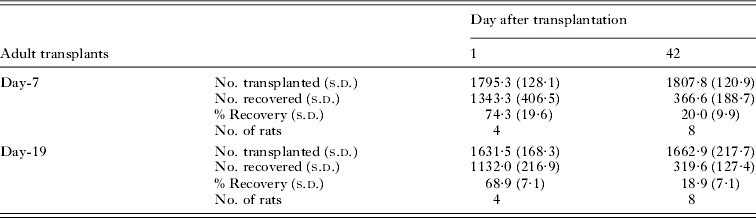
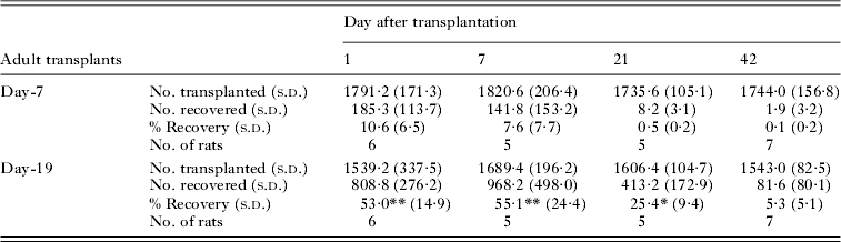
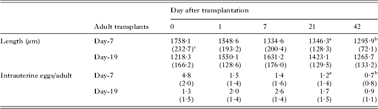
Based on these results, the significance of adults that moved into the small intestine was judged to be negligible. Thus, in the transplantation experiments (Tables 1–3), only adults recovered from the large intestine were considered.
Recovery rate (%) of adults at days 1 and 42 after transplantation of Day-7 or Day-19 adults into the colon of naive rats

Recovery rate (%) of adults at different days after transplantation of Day-7 or Day-19 adults into the colon of immune rats

* P<0.05;
** P<0.01 by Tukey-Kramer test compared with Day-7 adults.
Changes in the mean body length and the number of intrauterine eggs after transplantation of Day-7 or Day-19 adults into the colon of immune rats

c s.d. given in parentheses.
Transplantation into the colon of naive rats
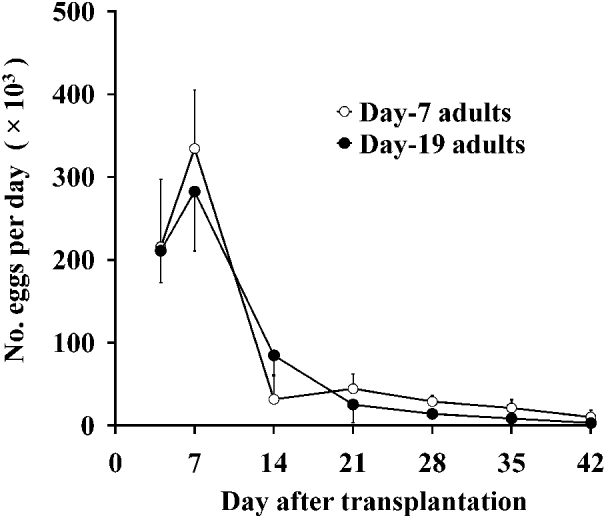
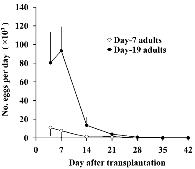
With a group of 5 rats having Day-7 adults, and another group of 5 rats having Day-19 adults, the number of eggs in faeces (EPD) was counted at days 4, 7, 14, 21, 28, 35 and 42 p.t. (Fig. 1). No difference was found in the egg excretion pattern between the groups (two-way repeated measure ANOVA, P=0·31). At days 1 and 42 p.t., animals were autopsied and worms were recovered. The numbers of adults transplanted and recovered, and the recovery rates (%) are shown in Table 1. There was no difference in the average rate of recovery between the Day-7 and Day-19 groups at day 1 p.t. (74·3% vs 68·9%), or at day 42 p.t. (20·0% vs 18·9%) (two-way ANOVA, P=0·58).
The number of Strongyloides ratti eggs (eggs/day) excreted in faeces of rats after transplantation of 2000 Day-7 or Day-19 adults/rat into the colon of naive rats. The transplants were obtained from the small intestine at day 7 (Day-7 adults) or 19 (Day-19 adults) after infection with 3rd-stage infective larvae. Five rats were used in each group. Vertical bar=1 s.d.

Transplantation into the colon of immune rats
Basically the same transplantation experiments as above but with infection-primed rats were carried out. The change in egg output is shown in Fig. 2. The 2 groups with 7 rats each showed a clear difference (two-way repeated measure ANOVA, P<0·001) with significant interaction of days p.t. and transplant groups (P<0·001). The Tukey-Kramer test for multiple comparisons revealed that the Day-19 group excreted significantly more eggs at days 4 and 7 p.t. (P<0·01 for both days). The adults were recovered at days 1, 7, 21, and 42 p.t. with 5–7 animals/group each time (Table 2). The difference in recovery was clear between the groups (two-way ANOVA, P<0·001), with interaction of days p.t. and transplant groups (P<0·001). The Day-19 group resulted in significantly higher recovery rates at days 1, 7 and 21 p.t. (Tukey-Kramer test, P<0·01 for days 1 and 7 p.t., P<0·05 for day 21 p.t.).
The number of Strongyloides ratti eggs excreted in faeces of rats after transplantation of 2000 Day-7 or Day-19 adults/rat into the colon of immunized rats. The transplants were obtained as in Fig. 1. Seven rats were used in each group. Vertical bar=1 s.d.

The body length and the number of eggs in the uterus of recovered adults are shown in Table 3. Before transplantation (day 0), the mean body length of Day-19 adults was 31% shorter than that of Day-7 adults (t-test, P<0·001). With Day-7 adults, the body length reduced significantly from 1758·1 μm before transplantation to 1548·6 μm at day 1 p.t. (one-way ANOVA, P<0·001; Tukey-Kramer test, P<0·01), and further to 1334·6 μm at day 7 p.t. (P<0·01). There was no difference among days 7, 21 and 42 p.t. Contrary to this, the length of Day-19 adults increased significantly from 1218·3 μm before transplantation to 1550·1 μm at 1 day p.t. (one-way ANOVA, P<0·001; Tukey-Kramer test, P<0·01), and further to 1631·2 μm at day 7 p.t. (P<0·01). Thereafter, the length decreased gradually, returning to the day 0 level at day 42 p.t.
The mean number of intrauterine eggs in Day-19 adults was only 27% of that in Day-7 adults before transplantation (t-test, P<0·001) (Table 3). With Day-7 adults, the number reduced quickly from 4·8 at day 0 to 1·5 at day 1 p.t. (one-way ANOVA, P<0·001; Tukey-Kramer test, P<0·01). Between days 1 and 42 p.t., the number of eggs ranged from 0·7 to 1·5, and no difference was found among them. With Day-19 adults, however, the number increased significantly from 1·3 at day 0 to 2·0 at day 1 p.t. (one-way ANOVA, P<0·001; Tukey-Kramer test, P<0·01), which increased further to 2·6 at day 7 p.t. (P<0·05). The number reduced to the day 0 level on and after day 21 p.t.
Egg excretion, adult recovery and gut position in BHA-treated rats
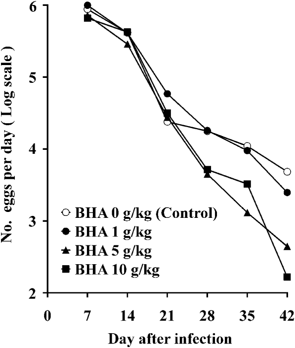
In all BHA dosage groups, EPDs reached approximately 1 million at day 7 p.i., then decreased sharply to the level of<5000 by day 42 p.i. When EPDs were expressed as log (EPD +1), a difference in the pattern of egg reduction became apparent between the groups: in the control group (BHA 0 g/kg), the rate of reduction slowed down after day 21 p.i., producing a ‘quasi-plateau’ of egg excretion, while in the 5 and 10 g/kg dosage groups, egg output continued falling (Fig. 3). When the 4 treatment groups were compared with log-transformed data, between-group difference was significant (two-way repeated measure ANOVA, P=0·001), with significant interaction of days p.i. and BHA doses (P<0·001). Tukey-Kramer test revealed that at day 35 p.i., the egg count in the 5 g/kg group was significantly lower than in the 0, 1 and 10 g/kg groups (P<0·01 in all comparisons), and at day 42 p.i., the egg count in the 10 g/kg group was lower than in the 0 and 1 g/kg groups, and the count in the 5 g/kg group was lower than in the 0 g/kg group (P<0·01 in all comparisons).
The number of eggs excreted in faeces of rats, which were infected subcutaneously with 3000 Strongyloides ratti infective larvae, and treated with different doses of butylated hydroxyanisole (BHA) mixed with powdered rat food. Five rats were used for each dose.

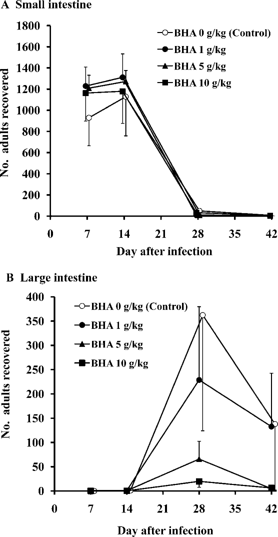
The numbers of adults recovered from the small and large intestines at different days after infection are shown in Fig. 4. In the small intestine (Fig. 4A), there was no difference among dosage groups (two-way ANOVA, P=0·20). A clear difference was noticed in the large intestine, where adult recovery reduced dose dependently (Fig. 4B): two-way ANOVA resulted in a between-group difference (P<0·001) with significant interaction between day p.i. and BHA dose (P<0·001). The Tukey-Kramer test revealed a difference at day 28 p.i. between the control group and the BHA 5 or 10 g/kg group (P<0·01), and between the 1 g/kg and the 10 g/kg groups (P<0·05).
Recovery of Strongyloides ratti adults from the small (A) and large (B) intestines of rats treated with different doses of BHA. Five rats/dose were autopsied at days 7, 14 and 28 after infection, and 6 rats/dose at day 42. Vertical bar=1 s.d.

The body weight of BHA-treated rats increased steadily in all 4 groups and there was no statistical difference among the groups (two-way repeated measure ANOVA, P=0·74). The amount of daily faeces increased gradually reaching a plateau at day 28 p.i., and there was again no difference among the groups (two-way repeated measure ANOVA, P=0·41).
DISCUSSION
Transplantation experiment with naive rats
The transplantation of Day-7 and Day-19 adults into the colon of naive rats resulted in a similar egg excretion pattern despite considerable differences in their parasitic background: the former had not been exposed to strong host immune responses like worm expulsion, whereas the latter had survived it but with profound damage caused by host immunity as observed by electron microscopy (Moqbel and McLaren, Reference Moqbel and McLaren1980). Additionally, compared with Day-7 adults, Day-19 adults were evidently stunted and their fecundity, or intrauterine egg count, was much lower at the time of transplantation. The comparable egg production by these different transplants indicates that the large intestine per se is a permissive site for parasitism. The egg output reached the maximum at day 7 p.t. and then decreased sharply in the following 7 days, which is almost like a worm expulsion pattern from the small intestine observed in a primary infection. Whether mast cells are playing a significant role in the expulsion is not known. In an ordinary subcutaneous infection of L3i with naive rats, mast cell proliferation was observed in the colon with a peak at day 32 p.i., together with a peak of adult worms in the colon (Shintoku, unpublished observation). However, we must keep it in mind that the present experimental procedure of transplanting as many as 2000 adults is unnatural.
The autopsy study revealed that there was no difference between the Day-7 and Day-19 groups in the adult recovery rates at days 1 and 42 p.t., in accordance with the similarity in the egg excretion pattern, and also confirming that damaged, stunted and less fecund Day-19 adults could recover and survive as well as Day-7 adults. The reversibility of damaged S. ratti adults, in terms of body length and fecundity, was reported when they were transplanted into the small intestine of naive rats (Moqbel et al. Reference Moqbel, McLaren and Wakelin1980). In the present study, spillover of worms into the small intestine and their physical recovery in that site can be excluded: at day 1 p.t., with 4 rats in each group, only 1 of 5373 Day-7 adults (0·02%) and 28 of 4528 Day-19 adults (0·62%) were recovered from the lower small intestine.
Transplantation experiment with infection-primed rats
Very different egg excretion patterns were obtained in the transplantation experiment with immune rats: Day-19 adults produced a high peak at day 7 p.t., whereas no peak was observable with Day-7 adults, suggesting that they were expelled quickly. This is confirmed by autopsy results, in which the recovery in the Day-19 group showed 5–7 times higher rates than in the Day-7 group at days 1 and 7 p.t. and >50 times at days 21 and 42 p.t. It is clear that Day-19 adults had adapted to parasitizing the large intestine.
This adaptability was further confirmed by the significant increases in body length and the number of intrauterine eggs in Day-19 adults during the first 7 days after transplantation, whereas Day-7 adults reduced both measurements quickly. Previous studies showed that host immune responses reduced the body length and fecundity of adults in the small intestine (Wilkes et al. Reference Wilkes, Thompson, Gardner, Paterson and Viney2004; Viney et al. Reference Viney, Steer and Wilkes2006; Bleay et al. Reference Bleay, Wilkes, Paterson and Viney2007). Our results will be the first confirmation that these two parameters increased in immune rats. It seems that between days 7 and 19 p.i. in the primary infection, small intestinal adults undergo significant biological and physiological changes, enabling them to continue parasitism in the large intestine of highly immune rats. It is unlikely that there is a subgroup having innate ability to parasitize the large intestine: a trial to select ‘colon-preferring’ adults was not successful after more than 30 generations (Kimura, unpublished data). An attempt to select S. ratti that were resistant to the host immune response was also reported to be unsuccessful (Wilkes and Viney, Reference Wilkes and Viney2004). Other than S. ratti, the reinfection of N. brasiliensis to immune rats produced adapted worms that were resistant to expulsion, and much less affected by passive immunity (Ogilvie, Reference Ogilvie1972). In the case of Trichinella spiralis, 2 populations that prefer different gut positions were successfully selected (Sukhdeo and Bansemir, Reference Sukhdeo and Bansemir1996).
Few studies have focused on the biological/physiological changes induced in parasites after they had been exposed to strong host immunity. The electron microscopical study by Moqbel and McLaren (Reference Moqbel and McLaren1980), with S. ratti adults obtained from the small intestine of rats at 10–25 days p.i., or 7 days after challenge infection, described the formation of a plug around the mouth which may block food intake, the empty gut lumen and loss of glycogen storage granules suggestive of starvation, and the consumption of most of the cellular contents of gut cells possibly through the action of lysosomes. It seems that extensive cellular, tissue and organ remodelling occur as a prerequisite for large intestinal parasitism. It is speculated that autophagy could be involved in the process.
At the same time, a quick morphological adjustment might be required to the new environment of the large intestine, where the mucosa consists of only crypts and is probably less ‘bushy’ than the small intestine for parasites. Thus, at day 1 p.t., the body length of Day-7 adults decreased by 209 μm (12%), and that of Day-19 adults increased by 332 μm (21%), resulting in almost the same length of 1550 μm in both groups. Whether this coincidence could be related to such adjustment or not is interesting and, in this respect, the quick increase/decrease in the number of intrauterine eggs at day 1 p.t. may have some relevance.
BHA study
BHA has various antioxidant mechanisms as a lipid peroxidation inhibitor, reducing agent, metal chelator and scavenger of hydrogen peroxide, superoxide and free radicals (Gülçin et al. Reference Gülçin, Alici and Cesur2005). With N. brasiliensis-infected rats, free radicals produced by host peritoneal leucocytes were shown to damage the parasites directly and cause worm expulsion, and the BHA treatment could counter the radicals and result in longer survival of the parasites (Smith and Bryant, Reference Smith and Bryant1989a,b). Also, reactive oxygen species were reported to directly activate mast cells, and release a variety of mediators in ischemia/reperfusion injury of mesenteric venules (Swindle and Metcalfe, Reference Swindle and Metcalfe2007), implying that BHA might interfere with mast cell functions and thus inhibit S. ratti expulsion. The present study yielded unexpected results: (i) the BHA treatment at 5 and 10 g/kg doses erased the quasi-plateau of the egg excretion curve recognized after day 21 p.i. in the control, and (ii) the treatment inhibited the establishment of large intestinal parasitism in a dose-dependent manner, and almost completely at 10 g/kg dose. The quasi-plateau is the product of adults in the large intestine (Kimura et al. Reference Kimura, Shintoku, Kadosaka, Fujiwara, Kondo and Itoh1999), and result (i) indicates the absence of adults in the large intestine after day 21 p.i. It is possible that free radicals produced by host rats were involved in the process of worm adaptation to the large intestine of immune rats. It has been reported that free radicals are not simply damaging but will work as a signal to induce production of a variety of antioxidant proteins and detoxifying enzymes that will protect cells (Kaspar et al. Reference Kaspar, Niture and Jaiswal2009). Judging from the fact that the establishment of small intestinal parasitism and following worm expulsion were not affected by the treatment, free radicals might be activated to a sufficient level sometime after worm expulsion.
The elimination of large intestinal parasitism may be explained by direct BHA toxicity to adults in the small intestine. However, at least up to day 14 p.i., the treatment did not reduce the worm recovery with the 10 g/kg dose. With N. brasiliensis, BHA at 10 g/kg even increased the recovery at day 12 p.i (Smith and Bryant, Reference Smith and Bryant1989b). Also, we were unable to find any publication on the toxicity of BHA to intestinal helminths. However, BHA has other biological effects such as a myorelaxant action on gastric muscles (Fusi et al. Reference Fusi, Valoti, Petkov, Boev and Sgaragli1998), interference with various enzymes (enhancement of glutathione S-transferase activities in the liver, for example) (WHO, 1986) etc., and these effects on the present results are unknown. Since BHA treatments, compared with the control, did not influence the body weight of rats and the amount of daily faeces through the experimental period, the effects on the host gastrointestinal functions must have been minimal.
The present study indicates that during days 7–19 p.i. S. ratti adults in the small intestine acquire the ability to parasitize the large intestine, and that free radicals produced by immune hosts possibly play a significant role in the process. The elucidation of the mechanisms will deepen our understanding of immune evasion, infection site selection/adaptation, and survival strategies as a species. Molecular analyses are under way at our laboratory.
FINANCIAL SUPPORT
This work was supported by the Japan Society for the Promotion of Science (JSPS) under Grant-in-Aid for Scientific Research (C) (grant number 21590471).