Introduction
Pneumosinus dilatans is a rare condition presenting as an abnormal dilatation of the paranasal sinuses that contains only air, with an intact mucosa and bony wall.Reference Benjamins1 The frontal sinus is most commonly affected, followed by ethmoid, sphenoid and maxillary sinuses, respectively. The symptoms at presentation depend on the sinus involved. The common symptoms of maxillary pneumosinus dilatans are: cosmetic deformity including cheek protrusion, prominence of the nasolabial fold, exophthalmos, toothache, nasal obstruction and paraesthesia. Pneumosinus dilatans must be differentiated from neoplastic lesions of the nasal cavity, mucocele and other complicated infectious conditions.
Although pneumosinus dilatans of the maxillary sinus is clinically benign, cosmetic deformity in the midface can be very disfiguring and needs to be properly addressed. The main goals for treatment of pneumosinus dilatans are to achieve permanent pressure equilibrium in the affected sinus and to correct the aesthetic problem. However, owing to its rarity, there are very few reports on the correction of the aesthetic problems associated with pneumosinus dilatans of the maxillary sinus.
The current paper describes the successful management of facial deformity in a maxillary pneumosinus dilatans patient. Correction of the facial deformity was achieved with reduction osteoplasty and reconstruction using maxillary bone, conchal cartilage and Tutoplast-processed fascia lata (30 × 40 mm; Tutogen Medical GmbH, Neunkirchen, Germany) via a sublabial approach.
Case report
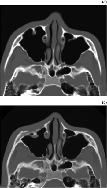
A 13-year-old male presented with a 10-year history of slowly progressive facial deformity in the right infraorbital region. There were no rhinological symptoms such as nasal obstruction, decreased facial sensation or visual problems, and no history of trauma or surgery. Computed tomography (CT) scans of the facial bone showed an abnormally aerated and enlarged right maxillary sinus with an outwardly displaced anterior bony wall. Neoplastic lesions were excluded and so surgical treatment was postponed until maxillary growth was complete.
Two years later, the patient returned complaining of progression of the right maxillary protrusion. Follow-up CT scans showed that the right maxillary sinus had expanded during the two-year interval. We performed a right middle meatal antrostomy by endoscopy to achieve permanent pressure equilibrium, as described in previous reports.Reference Wolfensberger and Herrmann2, Reference Mauri, de Oliveira and Franche3
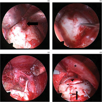
Two years post-operation, a follow-up examination showed that facial asymmetry remained (Figure 1). To correct the facial asymmetry, we performed a reduction osteoplasty via a sublabial approach under general anaesthesia. A conventional incision and dissection was performed at the superior gingivobuccal sulcus, followed by exposure of the protruding right maxillary sinus anterior wall. There was no evidence of bony destruction. Using a 1-mm diamond bur, we circumferentially resected a section of anterior bony wall measuring approximately 3 × 4 cm. We observed that the maxillary sinus was lined with normal mucosa, and that the ostium opened during previous endoscopic surgery was patent. The anterior wall of the maxillary sinus was then reconstructed to prevent the right anterior cheek from sinking. We manipulated the removed bone and divided it into three pieces using a 1-mm diamond bur. The inferior portion of the defect was reconstructed using those bones and fixed with wiring. However, there was insufficient bone to cover the entire defect. We therefore harvested left conchal cartilage to cover the remaining superior portion of the defect without wiring. The cartilage was not manipulated because its inherent curvature was suited to the anterior maxillary wall. The reconstructed anterior wall of the maxillary sinus was then covered with Tutoplast-processed fascia lata to achieve a natural contour and smooth surface (Figure 2).
Axial computed tomography images showing: (a) pre-operative abnormal dilatation of the right maxillary sinus (which contained only air, with no noticeable bony erosion or destruction); and (b) decreased volume and improved contour of the right maxillary sinus at four-months' post-operation.

Endoscopic photographs showing: (a) the protruding anterior wall of the right maxillary sinus with the infraorbital nerve (arrow) exposed; (b) a 3 × 4 cm piece of protruding anterior bony wall, which was circumferentially resected from the maxillary sinus; (c) the protruding bone removed; and (d) anterior maxillary defect reconstructed using the removed bone for the inferior portion (arrow) and harvested left conchal cartilage for the superior portion (arrowhead). The reconstructed wall was covered with Tutoplast-processed fascia lata (asterisk).

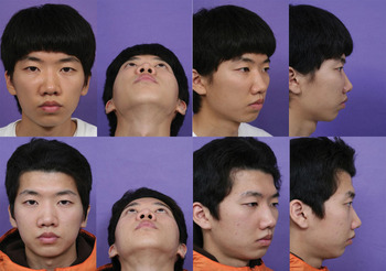
No significant complications such as bleeding or swelling occurred during or after the procedures. At 18-months' follow up, the patient was satisfied with the cosmetic results, and there was no evidence of recurrence (Figures 1 and 3).
Pre-operative (top) and 18-month post-operative views (bottom) showing correction of the facial deformity. Published with patient's permission.

Discussion
Maxillary pneumosinus dilatans usually causes cosmetic deformities such as cheek protrusion and exophthalmos. We used reduction osteoplasty and reconstruction with autologous bone, cartilage and Tutoplast-processed fascia lata via a sublabial approach to treat cosmetic deformities associated with maxillary pneumosinus dilatans. At the follow up, the patient showed a good cosmetic outcome and was satisfied with the results.
The pathogenesis of pneumosinus dilatans remains unclear. It is suggested that factors causing abnormal pneumatisation of the sinus may be involved, including spontaneous drainage of the mucoceles, gas-forming micro-organisms, congenital abnormalities and a one-way valve mechanism.Reference Trimarchi, Lombardi, Tomenzoli, Farina and Nicolai4 Local processes involving obstruction of the sinus ostium with a one-way valve mechanism are commonly accepted pathogenic factors.Reference Smith, Maran and von Haacke5, Reference Urken, Som, Lawson, Edelstein, McAvay and Biller6 Specifically, the long-term effect of air trapping produces positive intrasinus pressure resulting in sinus dilatation.
Although there is no established surgical treatment protocol for maxillary pneumosinus dilatans, a few surgical options have been proposed. Based on a presumed one-way valve mechanism, treatments have included direct sinus needle puncture, creation of a nasoantral window via Caldwell–Luc exploration, endoscopic middle meatal antrostomy to create a nasoantral window, and endoscopic maxillary sinus lateral wall resection.Reference Wolfensberger and Herrmann2, Reference Trimarchi, Lombardi, Tomenzoli, Farina and Nicolai4 The present patient underwent an endoscopic middle meatal antrostomy, but the facial deformity persisted. Additional corrective surgery is inevitable in such patients. Viehweg et al. described using an iliac bone graft and a bilateral sagittal split osteotomy for treating bilateral maxillary pneumosinus dilatans.Reference Viehweg and Hudson7 Breidahl et al. reported two maxillary pneumosinus dilatans cases treated with direct resection of the primary lesion and reconstruction using a reversed cystic wall and medial maxillary wall via an intraoral approach.Reference Breidahl, Szwajkun and Chen8
• Pneumosinus dilatans presents as an abnormal, air-filled dilatation of the paranasal sinuses with intact mucosa and bony wall
• Pneumosinus dilatans of the maxillary sinus is benign, but can lead to cosmetic deformity in the midface
• As the condition is rare, there is no established surgical treatment protocol
• In this case report, reduction osteoplasty resulted in a good cosmetic outcome
The approach used for our patient had some advantages over the procedures described above. First, our sublabial approach enabled wide exposure of the anterior maxillary contour and precise resection of the protruding lesion without a visible scar. Second, our technique was less invasive and has less associated morbidity. Third, we believe that exploiting the inherent curvature of the conchal cartilage and use of the manipulated bone assisted in achieving a natural anterior cheek contour. In addition, the Tutoplast-processed fascia lata covering provided a smooth surface and reduced irregularities. Fourth, the risk of complications such as post-operative infection was minimised by using autologous material for reconstruction. Although resorption of the autologous material and Tutoplast-processed fascia lata can occur, there were no signs of this in our patient at 18-months' follow up. However, such cases require continued follow up.
Conclusion
The present report describes a patient with maxillary pneumosinus dilatans with facial deformity treated by reduction osteoplasty, and reconstruction with autologous bone, cartilage and Tutoplast-processed fascia lata via a sublabial approach. This surgical technique was less invasive than other reported procedures and revealed good cosmetic results. We therefore recommend this reduction technique as a useful surgical option for treating maxillary pneumosinus dilatans patients with facial deformity.