Congenital anomalies of the coronary arteries are infrequent conditions with an incidence ranging from 0.17–2.2% in autopsy cases to 0.6–1.3% in cases with evaluated conventional coronary angiography.Reference Alexander and Grıffıth 1 – Reference Angelini, Velasco and Flamm 3 Although most of the coronary artery anomalies lack haemodynamic and clinical significance, some of them may cause sudden cardiac death or other symptoms of myocardial ischaemia, particularly in young adults and athletes after vigorous physical exertion.Reference Angelini, Velasco and Flamm 3 , Reference Eckart, Scoville and Campbell 4 Given the increase of interventional procedures, the detection of these anomalies is gaining major clinical importance. Anomalies of coronary arteries may be associated with major congenital heart diseases, such as bicuspid aortic valve, tetralogy of Fallot, or transposition of the great arteries.Reference Vastel-Amzallag, Le Bret and Paul 5 Furthermore, abnormal course of a coronary artery can complicate a cardiac surgery.
To date, diagnosis of the coronary artery anomalies was usually established during conventional coronary angiography. However, owing to the two-dimensional projectional nature of conventional coronary angiography, the visualisation of a complex three-dimensional vessel course and the clarification of the exact relationship to the surrounding anatomic structures – aorta and pulmonary artery – may be difficult, and misinterpretation is reported in up to 53% of the cases.Reference Shi, Aschoff, Brambs and Hoffmann 6 Establishing the proximal courses of the anomalous coronary arteries is of particular importance in determining whether surgical correction is needed. Multidetector computed tomographic angiography is currently considered the ideal tool for the three-dimensional visualisation of the complex and tortuous anatomy of the anomalous coronary arteries. It provides an excellent spatial resolution, which allows to determine the origin and course of the anomalous coronary arteries.Reference Vastel-Amzallag, Le Bret and Paul 5 – Reference Fujimoto, Kondo and Orihara 12
Most literature reports on coronary artery anomalies are based on findings obtained by conventional coronary angiography.Reference Yamanaka and Hobbs 2 – Reference Garg, Tewari, Kapoor, Gupta and Sinha 13 Previous studies with multidetector computed tomography demonstrated that coronary artery anomalies may be accurately detected and that their origins and courses can be clearly depicted.Reference Vastel-Amzallag, Le Bret and Paul 5 – Reference Koşar, Ergun, Oztürk and Koşar 10 , Reference Fujimoto, Kondo and Orihara 12 However, most of these studies were not aimed at evaluating the clinical impact and the additional value of multidetector computed tomography as compared with conventional coronary angiography. The purpose of this study is to define the prevalence of coronary artery anomalies in a large patient population who underwent multidetector computed tomographic angiography and to demonstrate in which cases multidetector computed tomographic angiograhy has an additional clinical value compared with conventional coronary angiography.
Materials and methods
Patient population
Between February, 2007 and September, 2011, a total of 2375 consecutive patients (1472 men and 903 women; mean age 61.6; age range 18–82 years) with known or suspected coronary artery disease due to chest pain or other reasons such as one or more cardiovascular risk factors or electrocardiogram abnormality underwent coronary multidetector computed tomographic angiography at our institution. In 23 of these patients, multidetector computed tomography was requested by cardiologists after conventional coronary angiography suggested the presence of coronary artery anomalies. In a retrospective analysis of all multidetector computed tomography data files, 42 patients (24 men and 18 women; mean age 55.9; age range 22–78 years) were found to have origin and/or course anomalies of the coronary artery and 10 patients (three men and seven women; mean age 55.6; age range 34–67 years) were found to have anomalies of termination of the coronary arteries. These two groups (n = 52) constitute the study population for further analysis. The patient cohort consisted of two subgroups. In all, 29 patients were referred to cardiac multidetector computed tomography to prove or rule out coronary artery disease. These patients revealed anomalies of coronary arteries as incidental findings. In 23 patients – 17 had origin and/or course anomalies and six had fistulas – multidetector computed tomographic angiography was indicated for complementary diagnostics following conventional coronary angiography to determine the exact course or to terminate the suspicion of anomalous coronary artery. Informed consent was obtained in all cases. An ethical committee approval was not required because of the retrospective character of this study.
Multidetector computed tomography scanning protocol
All of the studies were completed in our institution with either a 16-slice (GE Lightspeed Ultra 16; General Electrical Medical Systems, Milwaukee, Wisconsin, United States of America) or a 64-slice (Aquilion; Toshiba Medical Systems, Tokyo, Japan) scanner with retrospective electrocardiogram gating. In all, 928 examinations were performed on a 16-slice scanner and 1447 examinations on a 64-slice scanner. The following parameters were used for 16-slice multidetector computed tomography: the detector collimation of 16 × 0.625 mm, tube voltage of 120 kV, tube current of 320–460 mA s, tube rotation time of 500 ms, slice thickness of 0.625 mm, increment of 0.4 mm; for 64-slice multidetector computed tomography, the parameters used were as follows: the detector collimation of 64 × 0.5 mm, tube voltage of 120 kV, tube current of 400–500 mA s, tube rotation time of 400 ms, slice thickness of 0.5 mm, and increment 0.3 mm.
Patients with a pre-scan heart rate above 70 beats/min received a single dose of 50–100 mg metoprolol 1 h before the multidetector computed tomography scan, and if the heart rate was above 65 beats/min at the time of multidetector computed tomography scanning metoprolol (5–20 mg) was administered intravenously 5 min before the scan. A bolus of contrast agent (320–400 mg iodine/ml) was administered through an 18-gauge cannula positioned in an antecubital vein at a flow rate of 4–5 ml/s. The scanning delay was determined with a bolus tracking technique by placing the region of interest in the proximal descending aorta and setting the trigger threshold to 180 HU. The imaging data were acquired during an intravenous injection of 100–130 ml (16-slice multidetector computed tomography) or 80–90 ml (64-slice multidetector computed tomography) iodinated contrast agent at a rate of 4.5–5 ml/s, followed by 30 ml saline chaser with the same injection rate. The raw data sets were reconstructed during the diastolic phase at 75% of the R–R cycle. If image quality in this data set was not optimal, additional reconstructions were performed and the data sets with the optimal image were chosen for further evaluation.
Multidetector computed tomography image analysis
For three-dimensional image reconstruction, the raw multidetector computed tomography data were processed on a separate workstation (Advanced Workstation 4.2; GE Healthcare, Milwaukee, Wisconsin, United States of America or Vitrea 2 Workstation; Vital Images Incorporation, Plymouth, Minnesota, United States of America). For the evaluation of the coronary artery anomalies, all multidetector computed tomography images were reviewed first in axial projection, then with different post-processing tools such as multiplanar reconstruction, curved multiplanar reconstruction, thin-slab maximum-intensity projection, and volume-rendered technique. Curved multiplanar reconstruction allows visualisation of the entire course of a coronary artery on a single image. Three-dimensional volume-rendered reconstructions were used to ascertain the morphology and course of vessels. Segments were classified according to the American Heart Association scheme. Multidetector computed tomography images were reviewed by two investigators experienced in cardiovascular radiology and a consensus report was written.
Conventional coronary angiography
In all, 23 of the 52 patients (44.23%) identified with anomalies of the coronary arteries underwent catheter angiograhy with transfemoral or transbrachial approach before multidetector computed tomographic angiography scanning. Angiograms were evaluated by a blinded independent observer in multiple views. The results of catheter angiography were compared with multidetector computed tomographic angiography findings.
Classification of the coronary artery anomalies
Normal coronary anatomy is defined as follows: the left main coronary artery arises from the left coronary sinus and divides into left anterior descending and left circumflex arteries. The left anterior descending artery passes posterior to the main pulmonary artery into the anterior interventricular groove. The left circumflex artery passes posteriorly into the left atrioventricular groove. The right coronary artery arises from the right coronary sinus and passes into the anterior atrioventricular groove. Any deviation from the above definition was considered to be abnormal coronary artery anatomy.
First, dominance of the coronary artery system was determined according to the origin of the posterior descending artery. Coronary artery systems with posterior descending artery originating from the right coronary artery were defined as right dominant, and those with posterior descending artery arising from the left circumflex artery were defined as left dominant. Coronary artery systems where posterior descending artery was supplied by right coronary artery and a significant portion of the posterior wall of the left ventricle was supplied by posterolateral branches from the left circumflex artery were termed as co-dominant.
Coronary artery anomalies were classified according to the modified version of a classification system developed by Greenberg et al. The classification of coronary artery anomalies was performed according to anatomical criteria – origin, course, intrinsic anatomy, and termination – and pathophysiology–clinical relevance – “malignant” because of their clinical relevance, correlated with myocardial ischaemia or associated sudden death, or “benign” because of without functional and clinical relevance. The assessment of the coronary atherosclerotic disease was made, but not restricted to the purpose of study.
Statistical analysis
All statistical analyses were performed using the SPSS software package (SPSS 16.0 for Windows; SPSS, Chicago, Illinois, United States of America). Descriptive statistics was performed with regard to the incidence of the anomalous coronary arteries within total patient cohort, and relative frequencies of the anomalies were calculated. The additional clinical value of multidetector computed tomography versus conventional coronary angiography was defined as the percentage of the cases in which multidetector computed tomography images were essential in providing additional clinical information in comparison to conventional coronary angiography.
Results
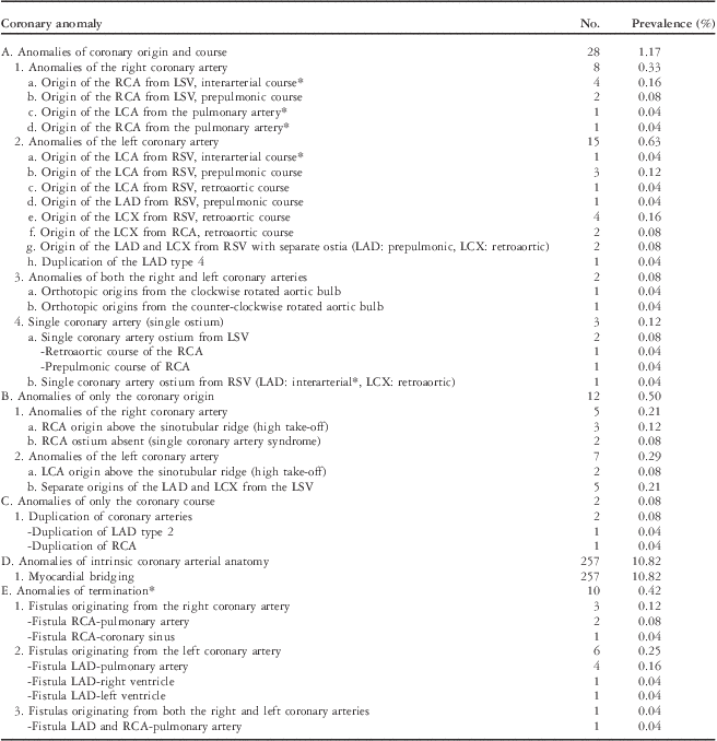
Multidetector computed tomography angiography was performed without complications in all patients. All examinations proved to be of diagnostic quality and appropriate for evaluation of coronary artery anomalies. Of the 2375 patients, 1971 (82.99%) had right dominance, 190 (8.0%) had left dominance, and 214 (9.01%) had balanced, co-dominant distribution. The incidence of the origin and/or course anomalies of the central coronary artery segments was 1.76% (n = 42) and that of fistulas was 0.42% (n = 10). The most common abnormality was abnormal origin of the left main coronary artery or branch arising from the right sinus of Valsalva or right coronary artery. Among these, the most common type was an ectopic left circumflex artery arising from the right sinus of Valsalva or proximal right coronary artery with retroaortic course. Most of (80.95%, n = 34) the origin and/or course anomalies of coronary arteries were benign and eight cases (19.05%) were malignant. In all, 10 cases with fistulas were also judged as malignant. The incidence of coronary artery anomalies observed in this study is shown in Table 1.
Classification and prevalence of anomalous coronary arteries in the study (n = 2375).

LAD = left anterior descending artery; LCA = left coronary artery; LCX = left circumflex artery; LSV = left sinus of Valsalva; RCA = right coronary artery; RSV = right sinus of Valsalva
*Malignant coronary artery anomalies
Origin and course anomalies of the coronary arteries (n = 28)
In all, 28 patients were found to have origin and course anomalies of the central coronary artery segments, and in this subgroup six were judged as “malignant” because of interarterial courses between the aortic root and the pulmonary trunk, either of the right coronary artery (n = 4) or the left main coronary artery/left anterior descending artery (n = 2). In addition, one case with the anomalous origin of the left main coronary artery from the pulmonary artery and another one case with anomalous origin of the right coronary artery from the pulmonary artery were judged as “malignant”.
Anomalies of the right coronary artery (n = 8)
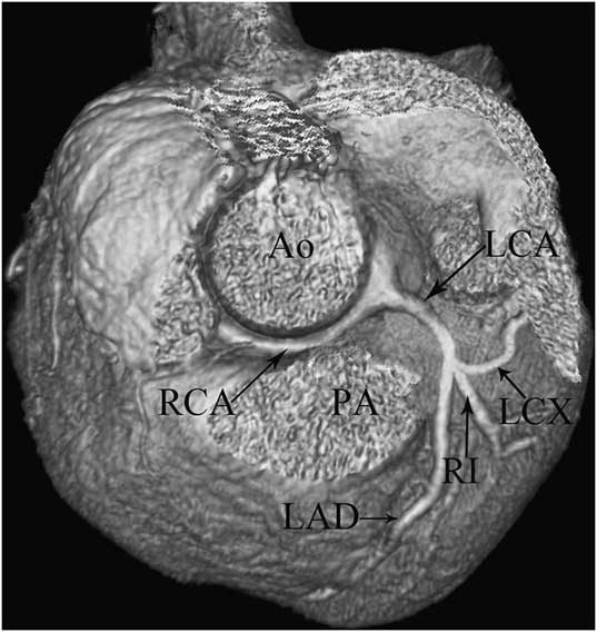
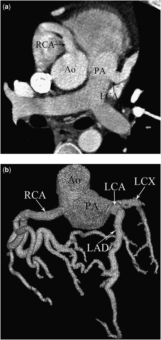
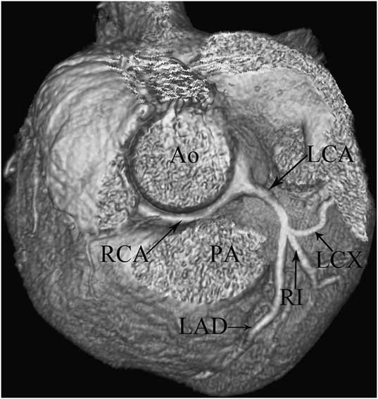
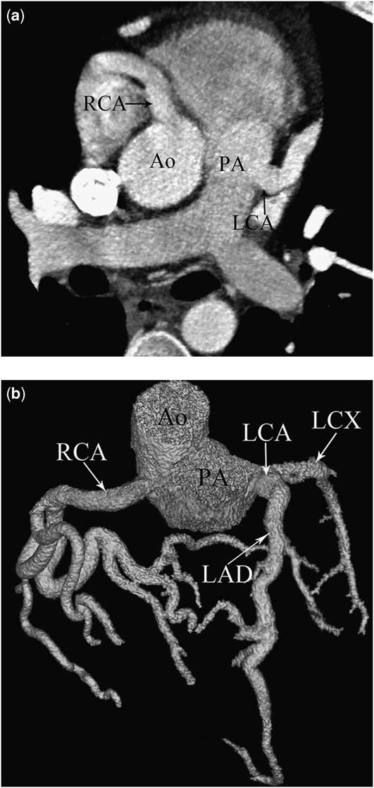
There were eight patients who showed anomalous origin and course of the right coronary artery. In six patients, the right coronary artery arose from the left sinus of Valsalva separately from the left main coronary artery (four with interarterial course and two with prepulmonic course; Fig 1). There was one patient who had anomalous origin of the left main coronary artery from the pulmonary artery (Fig 2) and another patient who had anomalous origin of the right coronary artery from the pulmonary artery.
Right coronary artery (RCA) arising from the left sinus of Valsalva with an interarterial course in a 44-year-old man. Volume-rendering multidetector computed tomography image ( a ) shows the RCA originating from the left sinus of Valsalva and coursing between the aorta (Ao) and the pulmonary artery (PA). LAD = left anterior descending artery; LCA = left coronary artery; LCX = left circumflex artery; RI = ramus intermedius.
Anomalous origin of the left coronary artery (LCA) from the pulmonary artery (PA) in a 22-year-old man. Axial ( a ) and volume-rendering ( b ) multidetector computed tomography images show the LCA arising from the PA and right coronary artery (RCA) arising from the aorta (Ao). Note dilatation of the LCA and RCA. LAD = left anterior descending artery; LCX = left circumflex artery.
Anomalies of the left coronary artery (n = 15)
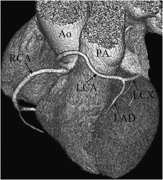
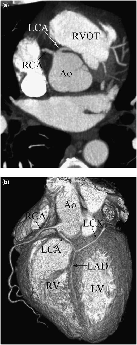
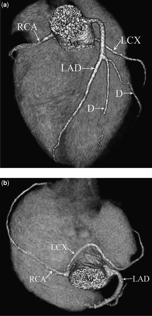
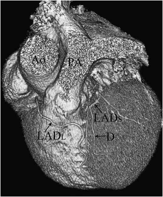
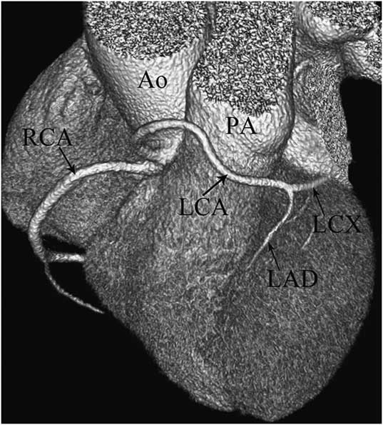
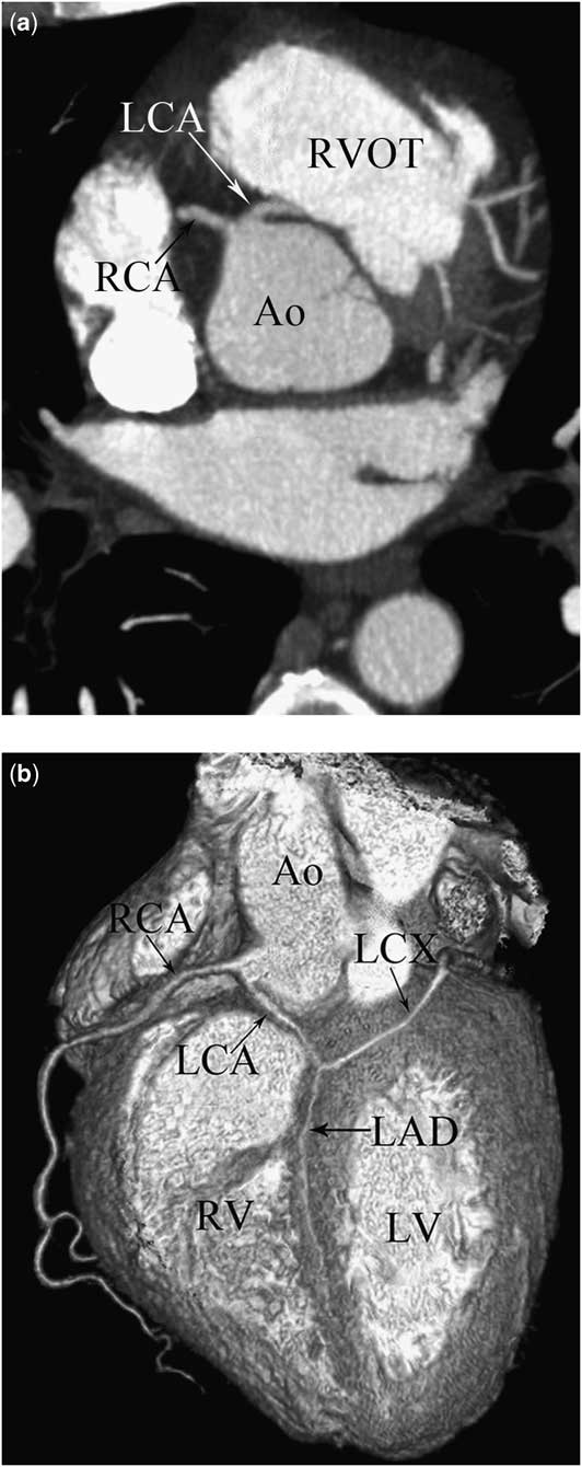
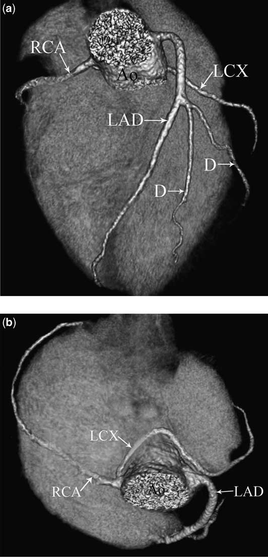
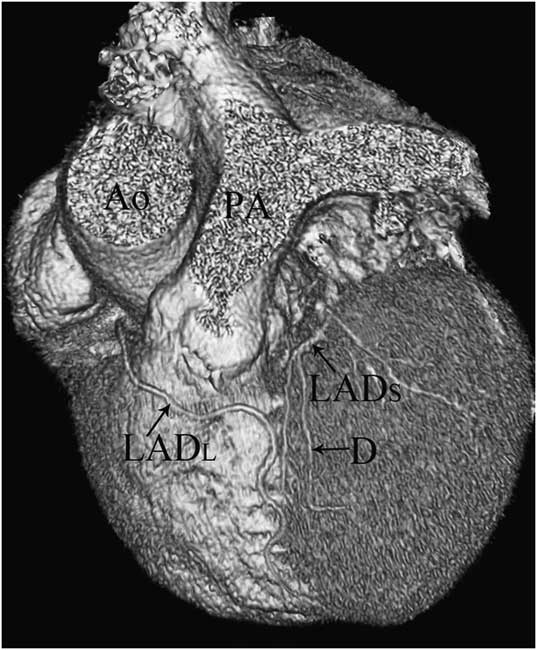
In all, 15 patients presented anomalous origin and course of the left main coronary artery. Right sinus of Valsalva origin of the left main coronary artery was seen in five patients (three with prepulmonic course, one with retroaortic course and one with interarterial course; Figs 3 and 4,) and right sinus of Valsalva origin of the left anterior descending artery with prepulmonic course was seen in one patient. In four patients, the left circumflex artery originated from the right sinus of Valsalva with its own orifice (Fig 5) and in two patients from the proximal right coronary artery segment. In all of these six patients, the proximal left circumflex artery segments coursed posterior to the aorta and the left anterior descending artery originated from a separate orifice of the left sinus of Valsalva with normal peripheral distribution. In two patients, separate origins of all three coronary arteries from the right sinus of Valsalva were presented. In these patients, the left anterior descending artery had a prepulmonic course and left circumflex artery had a retroaortic course. In one patient, type 4 duplication of the left anterior descending artery was detected. In this patient, the long left anterior descending artery originated from the right sinus of Valsalva, followed an anomalous course, and entered the anterior interventricular groove (Fig 6).
Left coronary artery (LCA) arising from the right sinus of Valsalva with a prepulmonic course in a 60-year-old woman. Volume-rendering image shows the LCA arising from the right sinus of Valsalva, coursing anterior to pulmonary artery (PA) and dividing to the left anterior descending artery (LAD) and left circumflex artery (LCX). Ao = aorta; RCA = right coronary artery.
Left coronary artery (LCA) arising from the right sinus of Valsalva with an interarterial course in a 43-year-old man. Axial oblique ( a ) and volume-rendering ( b ) multidetector computed tomography images show LCA arising from the right sinus of Valsalva, coursing between the aorta (Ao) and the right ventricular outflow tract (RVOT), dividing to left anterior descending (LAD) and left circumflex (LCX) arteries. The LAD courses intramyocardial in the interventricular septum. LV = left ventricle; RCA = right coronary artery; RV = right ventricle.
Left circumflex (LCX) artery arising from the right sinus of Valsalva with a retroaortic course in a 42-year-old man. Volume-rendering multidetector computed tomography images ( a , b ) show the LCX artery arising from the right sinus of Valsalva and coursing posterior to the aorta (Ao) and left anterior descending (LAD) artery originating from a separate orifice of the left sinus of Valsalva. D = diagonal branches; RCA = right coronary artery.
Type 4 duplication of the left anterior descending (LAD) artery in an 80-year-old man. Volume-rendering multidetector computed tomography image shows long LAD (LADL) originating from the right sinus of Valsalva, following an anomalous prepulmonic course, and entering the anterior interventricular groove. Short LAD (LADS) originated from the left sinus of Valsalva and terminated high in anterior interventricular groove. Ao = aorta; D = diagonal branch; PA = pulmonary artery.
Anomalies of both the right and left coronary arteries (n = 2)
In two patients, origin anomalies of both the right coronary artery and left main coronary artery were caused by rotation of the aortic root between 45° and 90° with normal coronary origin from the sinuses of Valsalva. The direction of the aortic root rotation was clockwise in one patient and counter-clockwise in the other one.
Single coronary artery (single ostium; n = 3)
In two patients, a common ostium of the right coronary artery and left main coronary artery from the left sinus of Valsalva was seen – one with a prepulmonic course of the right coronary artery and one with a retroaortic course of the right coronary artery. In the other patient, a single ostium from the right sinus of Valsalva was detected – the left anterior descending artery had an interarterial course and the left circumflex artery had a retroaortic course.
Anomalies of only the coronary origin (n = 12)
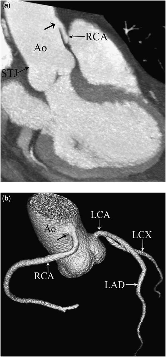
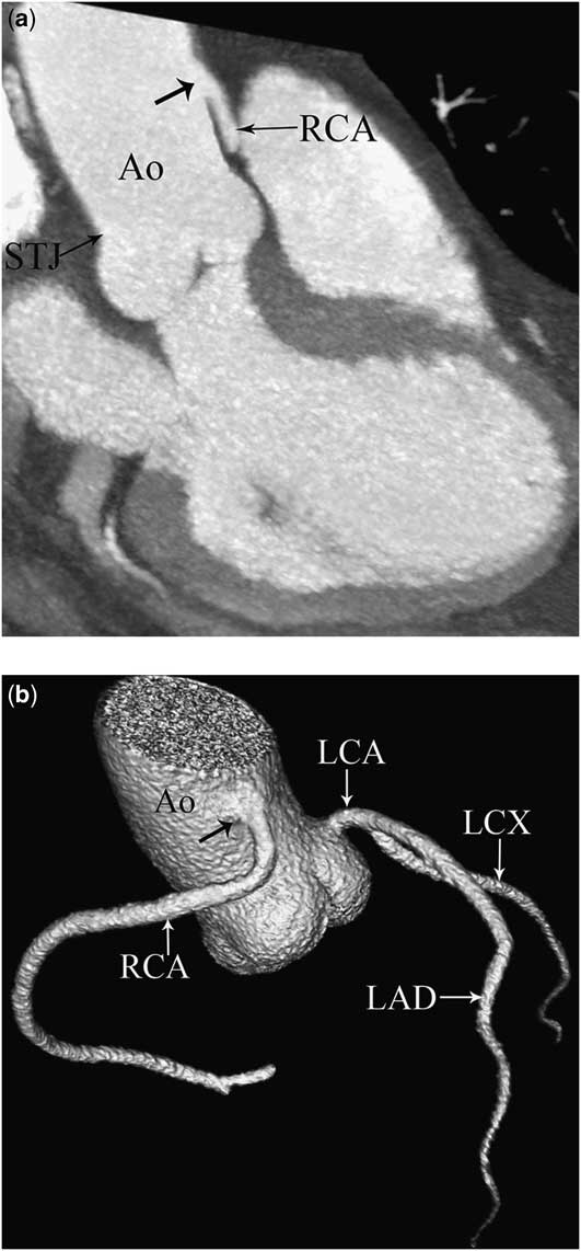
In all, 12 patients were found to have only the coronary artery origin anomalies. In three patients, the right coronary artery originated above the sinotubular ridge – high take-off position – and in two patients (Fig 7) the left main coronary artery originated above the sinotubular ridge. In two patients, the ostium and the proximal segment of the right coronary artery were absent and the left circumflex artery flowed in the posterior aspect of the right atrioventricular groove through the track of the right coronary artery – absence of the proximal right coronary artery. There were five patients who had separate origins of the left anterior descending artery and left circumflex artery – absent left main coronary artery – from the left sinus of Valsalva.
High take-off of the right coronary artery (RCA) in a 74-year-old man. Coronal oblique thin-slab maximum intensity projection ( a ) and volume-rendering ( b ) multidetector computed tomography images show the RCA demonstrating a high take-off (black arrow) above the sinotubular junction (STJ). Ao = aorta; LAD = left anterior descending artery; LCA = left coronary artery; LCX = left circumflex artery.
Anomalies of only the coronary course (n = 2)
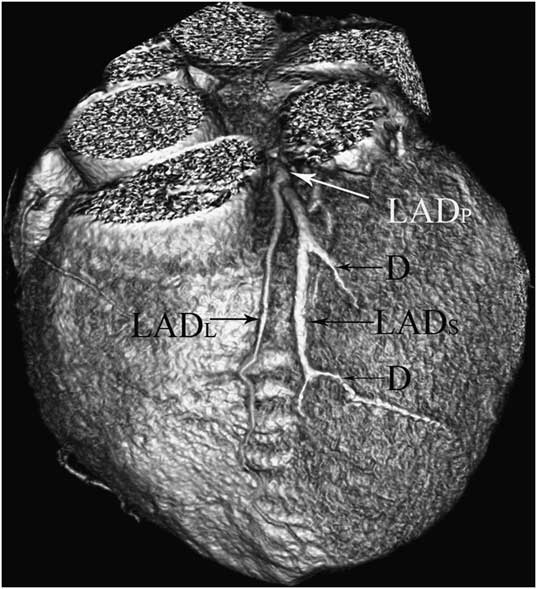
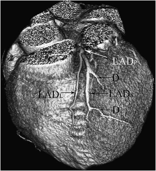
There were two patients who were found to have only the coronary artery course anomalies and one patient had type 2 duplication of the left anterior descending artery. In this patient, the left anterior descending artery proper gave rise to a short left anterior descending artery that terminated in the midportion of the interventricular groove, and the long left anterior descending artery originated from left anterior descending artery proper, descended on the right ventricular side of the anterior interventricular groove, coursed parallel to the short left anterior descending artery, and reentered the distal anterior interventricular groove (Fig 8). In the other patient, the double right coronary artery arising from two separate ostia in the right sinus of Valsalva – termed duplication of the right coronary artery – was present.
Type 2 duplication of the left anterior descending artery (LAD) in a 72-year-old woman. Volume-rendering multidetector computed tomography image shows short LAD (LADS) terminating high in the anterior interventricular groove and giving rise to diagonal branches (D). Long LAD (LADL) originates from LAD proper (LADP), descends on the right ventricular side of the anterior interventricular groove, courses parallel to the short LAD, and reenters the distal anterior interventricular groove.
Anomalies of intrinsic coronary arterial anatomy (n = 257)
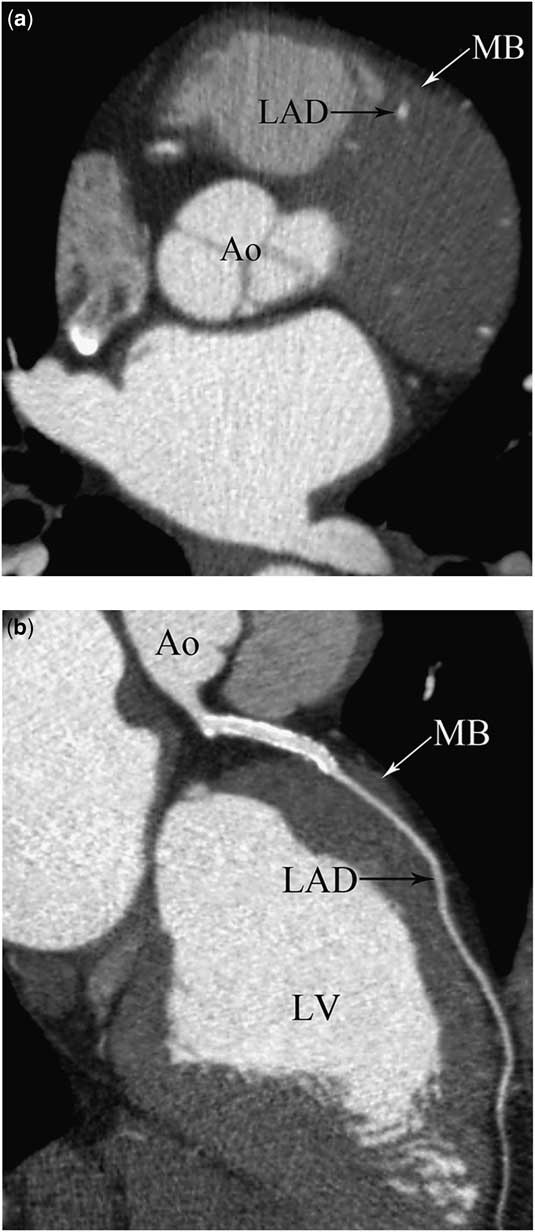
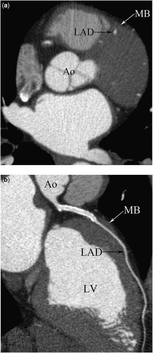
Myocardial bridging was detected in 257 patients (10.82%; Fig 9). The coronary arteries involved in this type of anomaly were as follows: mid left anterior descending artery (n = 221, 85.99%), distal left anterior descending artery (n = 13, 5.05%), proximal left anterior descending artery (n = 1, 0.38%), first diagonal (n = 3, 1.16%), second diagonal (n = 2, 0.77%), ramus intermedius (n = 3, 1.16%), first obtuse marginal (n = 3, 1.16%), second obtuse marginal (n = 2, 0,77%), mid left anterior descending artery and first diagonal (n = 3, 1.16%), mid left anterior descending artery and first obtuse marginal (n = 4, 1.55%), and mid left anterior descending artery and second obtuse marginal (n = 2, 0.77%).
Myocardial bridging of the left anterior descending (LAD) artery in a 45-year-old man. Axial ( a ) and curved planar reformatted ( b ) multidetector computed tomography images show a band of myocardial muscle overlying the mid segment of the LAD artery corresponding to myocardial bridge (MB) and a stent in the proximal LAD artery, immediately before the bridge. Ao = aorta; LV = left ventricle.
Anomalies of termination (n = 10)
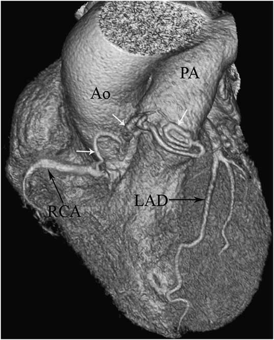
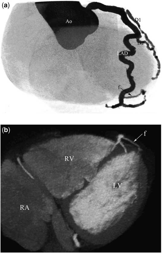
In all, 10 patients presented with anomalies of termination. In three patients, fistulas originating from the right coronary artery were seen (two had a fistula from the right coronary artery to pulmonary artery and one had a fistula from the right coronary artery to the coronary sinus; Fig 10). In six patients, fistulas originating from the left main coronary artery were detected (four had a fistula from the left anterior descending artery to pulmonary artery, one had a fistula from the left anterior descending artery to right ventricle, and one had a fistula from the left anterior descending artery to the left ventricle; Fig 11).There was one patient who had a fistula originating from both the right coronary artery and the left main coronary artery to main pulmonary artery.
Coronary artery fistula in a 65-year-old woman. Volume-rendering multidetector computed tomography image shows a dilated and tortuous fistula (white arrows) extending from the right coronary artery (RCA) to the main pulmonary artery (PA). Ao = aorta; LAD = left anterior descending artery.

Coronary artery fistula in a 55-year-old woman. Volume-rendering ( a ) and coronal thin-slab maximum intensity projection ( b ) multidetector computed tomography images show a fistula (f) originating from the distal left anterior descending (LAD) artery and draining into the left ventricle (LV). Ao = aorta; D1 = first diagonal; D2 = second diagonal; RA = right atrium; RV = right ventricle.

Comparison of multidetector computed tomography and conventional catheter angiography findings
In 23 of 52 (44.23%) patients with coronary artery anomalies – 17 cases with origin and/or course anomalies and six cases with fistulas – multidetector computed tomographic angiography was indicated for complementary diagnostics following conventional coronary angiography to determine the exact course or termination of a suspected anomalous coronary artery. In 17 patients with origin and/or course anomalies, conventional coronary angiography was unsuccessful and it did not allow the complete evaluation of the origin and/or course of the anomalous coronary artery. In these 17 patients (100%), multidetector computed tomography detected the anomalous vessels and provided additional information regarding its origin and course, as well as its relationship with the aortic root and the main pulmonary trunk. In six of these 17 cases, anomalous coronary artery originating from the opposite coronary sinus with interarterial course – malignant anomalies – was found. In one case with anomalous origin of the left coronary artery from the pulmonary artery and another case with anomalous origin of the right coronary artery from the pulmonary artery conventional coronary angiography failed to detect their origins, which was correctly visualised by multidetector computed tomography. In six patients with coronary artery fistulas, conventional coronary angiography failed to detect their terminations, which were clearly depicted by multidetector computed tomography.
Discussion
Classification criteria for coronary artery anomalies have been extensively discussed in the literature, but to date no general accepted classification scheme exists. In a recent publication, a comprehensive and systematic approach to anatomical patterns has been proposed by Angelini et al.Reference Angelini, Velasco and Flamm 3 As a general guide, anomalies can be divided into three general classifications, which include anomalies of origin and course, anomalies of intrinsic coronary arterial anatomy – myocardial bridging, and anomalies of coronary termination – fistulas. In this article, we have classified the coronary artery anomalies using a modified version of a classification system developed by Greenberg et al.Reference Greenberg, Fish and Spindola-Franco 14
The incidence of right and left coronary dominance has been reported to be 80–85% and 7–9%, respectively.Reference Yamanaka and Hobbs 2 , Reference Cademartiri, La Grutta and Malagò 9 , Reference Koşar, Ergun, Oztürk and Koşar 10 In the present study, the right coronary artery was dominant in 83% of the cases, and the left main coronary artery was dominant in 8% of the cases, in agreement with the main studies reported in the literature. The incidence of co-dominance was 9% in our study, which falls in the range of 5–10% as reported in the literature.Reference Yamanaka and Hobbs 2 , Reference Cademartiri, La Grutta and Malagò 9 , Reference Koşar, Ergun, Oztürk and Koşar 10
The prevalence of coronary artery anomalies ranges from 0.17–2.2% in autopsy cases to 0.6–1.3% in conventional coronary angiography studies.Reference Alexander and Grıffıth 1 – Reference Angelini, Velasco and Flamm 3 Generally, lower ratios are reported in autopsy studies. Alexander and GriffithReference Alexander and Grıffıth 1 detected only 54 coronary artery anomalies in 18,950 cases (0.3%). However, these autopsy studies are limited by entry bias and lack of clear diagnostic criteria, which are both prerequisites for defining the true prevalence in a general population. The study with the largest number of cases, performed on 126,595 patients who underwent conventional coronary angiography, reported an incidence of coronary artery anomalies of 1.3%.Reference Yamanaka and Hobbs 2 In another study performed by Angelini et al,Reference Angelini, Velasco and Flamm 3 the authors prospectively analysed 1950 consecutive cineangiograms and they reported an incidence for coronary artery anomalies of 5.6%, which is higher than the usually cited prevalence derived from angiographic reports.Reference Yamanaka and Hobbs 2 In one study conducted using 4- and 16-slice multidetector computed tomography, the authors reported an incidence of coronary artery anomalies, including fistulas, of 2.5%.Reference Schmitt, Froehner and Brunn 7 Fujimoto et alReference Fujimoto, Kondo and Orihara 15 studied 5869 patients using 64-slice multidetector computed tomography, and they reported the prevalence of coronary artery anomalies, not including coronary fistulas and myocardial bridging, to be 1.52%. de Jonge et alReference de Jonge, van Ooijen and Piers 16 described an incidence of 7% of coronary artery anomalies including coronary fistulas in their patient population. Cademartiri et alReference Cademartiri, La Grutta and Malagò 9 demonstrated a much higher incidence of coronary anomalies including fistulas and myocardial bridging, equal to 18.4% in their study. These discrepancies in reported incidence may be caused by referral bias. Some of these patients with coronary artery anomalies might have been or were referred because of known presence of coronary artery anomalies and not because of unrelated factors as in the general population. In the present study, a total of 42 patients (1.76%) with coronary artery anomalies – not including coronary fistulas and myocardial bridging – were detected. A total of 40.47% (n = 17) of these patients were specifically referred for multidetector computed tomography imaging after coronary artery anomalies had been verified or suggested in conventional coronary angiography. The remaining 25 patients had incidental findings, representing 1.05% of all cases examined with multidetector computed tomographic angiography. This result is quite similar to that observed in a large angiographic series, as well as large previously published multidetector computed tomography studies.Reference Yamanaka and Hobbs 2 , Reference Fujimoto, Kondo and Orihara 15 , Reference von Ziegler, Pilla and McMullan 17 However, even such large studies do not reflect general population, as only symptomatic patients with indications for either conventional coronary angiography or multidetector computed tomography were considered.
Most common coronary artery anomalies are vessels arising from the contralateral side: anomalous origins of the right coronary artery from the left sinus of Valsalva or the proximal left main coronary artery, or anomalous origins of the left main coronary artery from the right sinus of Valsalva or the proximal right coronary artery. The anomalous coronary artery originating from contralateral side passes to the opposite side, thereby following four possible pathways: anterior to the pulmonary artery (prepulmonic course), posterior to the aortic root (retroaortic course), between the aortic root and the pulmonary artery (interarterial course), and within the interventricular septum (septal/subpulmonic course).Reference Ropers, Gehling and Pohle 18 Anomalous right coronary artery arising from the left coronary sinus of Valsalva generally takes an interarterial course.Reference Yamanaka and Hobbs 2 In the present study, an interarterial course of the right coronary artery was observed in 66.7% of cases with this anomaly. An interarterial course of the left main coronary artery arising from the right sinus of Valsalva is rare but more dangerous.Reference Bunce, Lorenz and Keegan 19 In the present study, five cases with the left main coronary artery arising from the right sinus of Valsalva were detected: the left main coronary artery followed a prepulmonic course in three (60%), interarterial course in one (20%), and retroaortic course in another one (20%).
Coronary artery anomalies may also be classified as haemodynamically either insignificant or significant. Most coronary artery anomalies are benign and clinically insignificant and are usually detected as incidental findings. Haemodynamically significant anomalies of the coronary arteries, sometimes termed “malignant” or “potentially lethal”, are characterised by abnormalities of myocardial perfusion, which lead to an increased risk of myocardial ischaemia or sudden death because their position or orientation may lead to coronary flow compromise.Reference Bunce, Lorenz and Keegan 19 – Reference Click, Holmes, Vlietstra, Kosinski and Kronmal 21 These anomalies include an anomalous course between the pulmonary artery and the aorta of either the right coronary artery arising from the left sinus of Valsalva or the left main coronary artery arising from the right sinus of Valsalva, an anomalous origin of either the left main coronary artery or the right coronary artery from the pulmonary artery, occasional myocardial bridging, and congenital coronary artery fistulas. The present study revealed six patients with anomalous coronary artery segments coursing between the aortic root and pulmonary trunk (four cases of the right coronary artery, one case of left main coronary artery, and one case of left anterior descending artery). These interarterial – “malignant” – segments are associated with a high risk of exercise-induced ischaemia, myocardial infarction, or sudden death. The risk is estimated to be up to 30% in children and young adults, especially in competitive athletes.Reference Click, Holmes, Vlietstra, Kosinski and Kronmal 21 Mechanisms are explained that the ostium of the anomalous coronary artery bifurcates sharply at an acute angle from the aorta, becoming slit shaped, and at exertion the aorta and the pulmonary artery expand as pressure of both vessels increases, compressing the coronary artery that runs between the two vessels.Reference Bunce, Lorenz and Keegan 19 It is important to determine whether or not the anomalous coronary artery follows an interarterial course when a coronary artery anomaly originating from the opposite sinus is detected. Owing to the fact that conventional coronary angiography is a two-dimensional projectional imaging modality, it may sometimes be insufficient in determining the course of the abnormal coronary artery and evaluating its relationship with the aorta and the pulmonary artery. Therefore, it is more appropriate to evaluate such cases using multidetector computed tomography angiography. In our study, it has been possible to correctly evaluate the interarterial course using multidetector computed tomography in all of the six cases (100%) for which conventional coronary angiography failed to give a clear evaluation of the interarterial course.
Anomalous origin of the left main coronary artery from the pulmonary artery is an uncommon and very serious anomaly, reported in one of every 300,000 live births.Reference Kragel and Roberts 20 – Reference Dodge-Khatami, Mavroudis and Backer 22 It usually results in death during infancy, and only a few patients survive to adulthood.Reference Dodge-Khatami, Mavroudis and Backer 22 Anomalous origin of the right coronary artery from the pulmonary artery is unusual, but when it does occur it is a relatively benign anomaly and may rarely lead to sudden death in case of right dominant coronary circulation. If the collateral circulation between the right coronary artery and the left main coronary artery remains adequate, the individual may survive into adult life and develop a left-to-right shunt from the right coronary artery to the pulmonary artery.Reference Williams, Gersony and Hellenbrand 23 The imaging features include tortuosity of the coronary arteries and multiple dilated intercoronary collaterals. In our series, we encountered one patient, 22-year-old, with anomalous origin of the left main coronary artery from the pulmonary artery and another patient, 57-year-old, with anomalous origin of the right coronary artery from the pulmonary artery. In these two patients, multidetector computed tomography demonstrated ostial origin and proximal course of the anomalous arteries better than conventional coronary angiography.
The most common anomaly (35.71%) in the present study was anomalous origin of the left main coronary artery or branch arising from the right coronary sinus or right coronary artery. Among these, the most common type was an ectopic left circumflex artery arising from the right sinus of Valsalva or proximal right coronary artery. The incidence of this anomaly is found to be 0.25%, which is slightly lower than the reported ranges of 0.32–0.67%.Reference El-Menyar, Das and Al-Suwaidi 24 In 66.7% of these cases, the left circumflex artery arose from a separate ostium in the right coronary sinus of Valsalva, and in 33.3% of the cases it originated as a branch of the right coronary artery. In all of the cases, the left circumflex artery coursed posterior to the aortic root (retroaortic course). The clinical significance of detecting the ectopic left circumflex artery arising from the opposite sinus with retroaortic course is not very clear. Although it may predispose to atherosclerosis, the retroaortic left circumflex artery is considered benign. Moreover, knowledge of an anomalous left circumflex artery is important before aortic valve surgery because of the anomalous artery circles behind the aortic root just beneath the aortic valve.
In rare instances, all the three coronary arteries arise from the right or left coronary sinus of Valsalva with separate ostia. The independent origin of both the left anterior descending artery and left circumflex artery from two separate ostia in the right sinus of Valsalva is an exceedingly rare anomaly.Reference El-Menyar, Das and Al-Suwaidi 24 The jeopardy of this coronary anomaly depends on the left anterior descending artery course. A left anterior descending artery course between the aorta and the pulmonary artery is potentially responsible for myocardial ischaemia and sudden death. The present study revealed two patients with origin of the three coronary arteries from the left sinus of Valsalva. In both these patients, the left anterior descending artery had a prepulmonic course and the left circumflex artery had a retroaortic course.
Single coronary artery – single ostium – is a relatively rare coronary artery anomaly and the prevalence is reported to be between 0.024% and 0.044%.Reference Desmet, Vanhaecke and Vrolix 25 The incidence in the present study was 2.10%, higher than the previous reports. In our study, we observed a single ostium from left sinus of Valsalva in two patients and right sinus of Valsalva in one patient. In another two patients, the ostium and the proximal segment of the right coronary artery were absent, and the left circumflex artery was a dominant vessel, which flowed in the posterior aspect of the right atrioventricular groove through the track of the right coronary artery. Increased risk of sudden death is reported in patients with a single coronary artery in whom the left main coronary artery lies between the aorta and the pulmonary artery. The clinical course of the case in which a coronary artery originates as a terminal extension of another is relatively benign. In addition, proximal stenosis of a single coronary artery may be devastating if there is an inability to develop collateral channels.
‘High take-off’ refers to an unusually high origin of either the right or left coronary artery at a point above the junctional zone between its sinus and the tubular part of the ascending aorta.Reference Kim, Seo and Do 26 The present study revealed three patients with high take-off of the right coronary artery and two patients with high take-off the left main coronary artery. The incidence of this anomaly was found to 0.21% in our study. The occurrence of separate orifices of the left anterior descending and left circumflex arteries from the left sinus of Valsalva – namely absent left main coronary artery – is a rare anomaly. The incidence of this anomaly was 0.21% in the present study, which is slightly lower than the reported ranges of 0.41–0.52%.Reference Montaudon, Latrabe, Iriart, Caix and Laurent 27 Separate orifices of the left anterior descending artery and left circumflex artery from the left sinus of Valsalva and high take-off positions of right coronary artery do not any haemodynamic significance, but they may lead to unexpected angiographic problems while localising and engaging the orifices.Reference Greenberg, Fish and Spindola-Franco 14
Duplication of the left anterior descending artery is a haemodynamically benign condition, but to be aware of this anomaly may be important for diagnosis and therapeutic planning. Owing to the inability to visualise the additional vessels, especially when the long left anterior descending artery arises from the right coronary sinus, some risk exists for mistaking the variant anatomic features at conventional coronary angiography for mid left anterior descending artery occlusion. Furthermore, this anomaly represents a potential cause of error for the cardiac surgeon to treat a healthy vessel instead of the diseased one when creating anastomosis. In all, four subtypes of this anomaly have been described. In types 1 and 2, a long left anterior descending artery originates as a branch from the left anterior descending artery proper, takes a course parallel to the short left anterior descending artery in its proximal course on either the left ventricle (type 1) or the right ventricle (type 2), and reenters the anterior interventricular groove in the distal aspect. Type 3 duplication of the left anterior descending artery is characterised by a proximal intramyocardial course of the long left anterior descending artery. Type 4 is a distinct type in which the long left anterior descending artery arising from the right coronary sinus takes an anomalous course and enters the anterior interventricular groove.Reference Agarwal and Kazerooni 28 In our series, we encountered type 2 and type 4 duplications of left anterior descending artery.
Intramyocardial course of a coronary artery, which normally has an epicardial course, is referred to as myocardial bridging. The incidence of this anomaly varies greatly and is reported to be 0.5–4.9% in conventional coronary angiography studies,Reference Kim, Hur and Kim 29 , Reference Leschka, Koepfli and Husmann 30 15–85% in autopsy studies,Reference Loukas, Curry and Bowers 31 and 3.5–38.5% in multidetector computed tomography studies.Reference Kim, Hur and Kim 29 , Reference Leschka, Koepfli and Husmann 30 , Reference Lazoura, Kanavou, Vassiou, Gkiokas and Fezoulidis 32 The actual incidence of myocardial bridges is probably overestimated by postmortem studies because of the preparation and selection of the hearts undergoing autopsy. However, conventional coronary angiography underestimates the true incidence of myocardial bridges, as it is only able to detect deep myocardial bridges in which the lumen is reduced during systole.Reference Kim, Hur and Kim 29 , Reference Leschka, Koepfli and Husmann 30 Multidetector computed tomographic angiography clearly depicts the intramyocardial path of the involved coronary arterial segment. Comparison of the images obtained during the systolic and diastolic phases allows the assessment of the luminal narrowing during the systolic phase. Myocardial bridging typically involves the middle portion of the left anterior descending artery, but the left circumflex artery, right coronary artery, and diagonals are rarely involved. It is considered a benign finding, and in the majority of patients it does not cause myocardial ischaemia. Occasionally, it causes acute myocardial infarction, fatal arrhythmia, and sudden cardiac death.Reference Hostiuc, Curca, Dermengiu, Dermengiu, Hostiuc and Rusu 33 Therefore, this abnormality should be ruled out in young patients with chest pain or arrhythmia. In the present study, the incidence of this anomaly was 10.82%. Similar to the results of the other studies, the most common coronary artery involved by myocardial bridging was the midportion of the left anterior descending artery.
The present study revealed the presence of 10 coronary artery fistulas in our study population. The incidence of this anomaly is found to be 0.42%, which falls in the range of 0.1–0.67%, as reported in the literature.Reference Song, Park, Kang and Chun 34 Patients with small coronary fistulas remain asymptomatic, whereas those with high-flow fistulas and enlarged vessels may develop myocardial ischaemia and congestive cardiac failure. Fistulas more commonly involve the right coronary artery (60%) than the left main coronary artery.Reference Zenooz, Habibi, Mammen, Finn and Gilkeson 35 In <5% of the cases, fistulas originate from both the left main coronary artery and the right coronary artery. The right ventricle is the most common (45%) site of drainage, followed by the right atrium (25%) and the pulmonary artery (15%).Reference Zenooz, Habibi, Mammen, Finn and Gilkeson 35 The drainage site of the fistula has a greater clinical and physiologic importance than does the artery origin. The drainage sites may be poorly visible in conventional coronary angiography because of significant dilution of the contrast medium. Multidetector computed tomography has proven to be very useful in the detection and characterisation of coronary artery fistulas. In the present study, for the six patients with coronary artery fistulas, conventional coronary angiography failed to detect their terminations, which were clearly depicted by multidetector computed tomography.
The principal findings of our study are that multidetector computed tomographic angiography is not only an optimum non-invasive imaging modality for determining coronary artery anomalies and depicting three-dimensionally their anomalous course, but also has an additional clinical role in those patients for whom conventional coronary angiography fails to diagnose coronary artery anomalies or, being inconclusive, is not able to differentiate between benign and malignant anomalies. In our study population, all of (100%) the 17 patients with origin and/or course anomalies who had inconclusive conventional coronary angiography were better defined by multidetector computed tomographic angiography. In these patients, multidetector computed tomography detected the anomalous vessels and provided additional information regarding their origin and proximal course, as well as their relationship with the aortic root and the main pulmonary trunk; eight malignant cases were found. In addition, in all of (100%) the six cases with coronary artery fistulas, conventional coronary angiography failed to detect their terminations, which were clearly depicted by multidetector computed tomography.
In conclusion, multidetector computed tomographic angiography is an accurate and a reliable non-invasive imaging modality to detect and define anomalous coronary arteries and to delineate their course. It should be used as the first-rank diagnostic tool in patients with a suspected a coronary artery anomaly. Multidetector computed tomography is superior to conventional coronary angiography in delineating the ostial origin and the proximal course of anomalous coronary arteries. Furthermore, it reveals the exact relationship of anomalous coronary arteries with the aortic root and the pulmonary artery trunk. Anomalies of intrinsic anatomy and termination of coronary arteries are also better visualised with multidetector computed tomographic angiography.