Report
An asymptomatic 6-year-old child patient was admitted to the Outpatient Cardiology Department for a cardiovascular check-up. There was no history of congenital heart disease in the family or any history of recurrent pregnancy loss. There was neither a history of maternal infections or alcohol and smoke consumption during the pregnancy.
The physical exam revealed a soft systolic heart murmur heard at the left sternal border (1/6 Levine’s scale), disappearing in the sitting position; its characteristics suggested an innocent heart murmur. Percutaneous peripheral oxygen saturation was 99% on air; arterial blood pressure was 90/60 mmHg. Blood exams were in range. The 12-lead ECG showed sinus rhythm with heart rate 88/minute, QRS axis at 45° on the frontal plane, PQ of 0.12 seconds, QRS of 0.08 seconds, and corrected QT of 0.36 seconds.
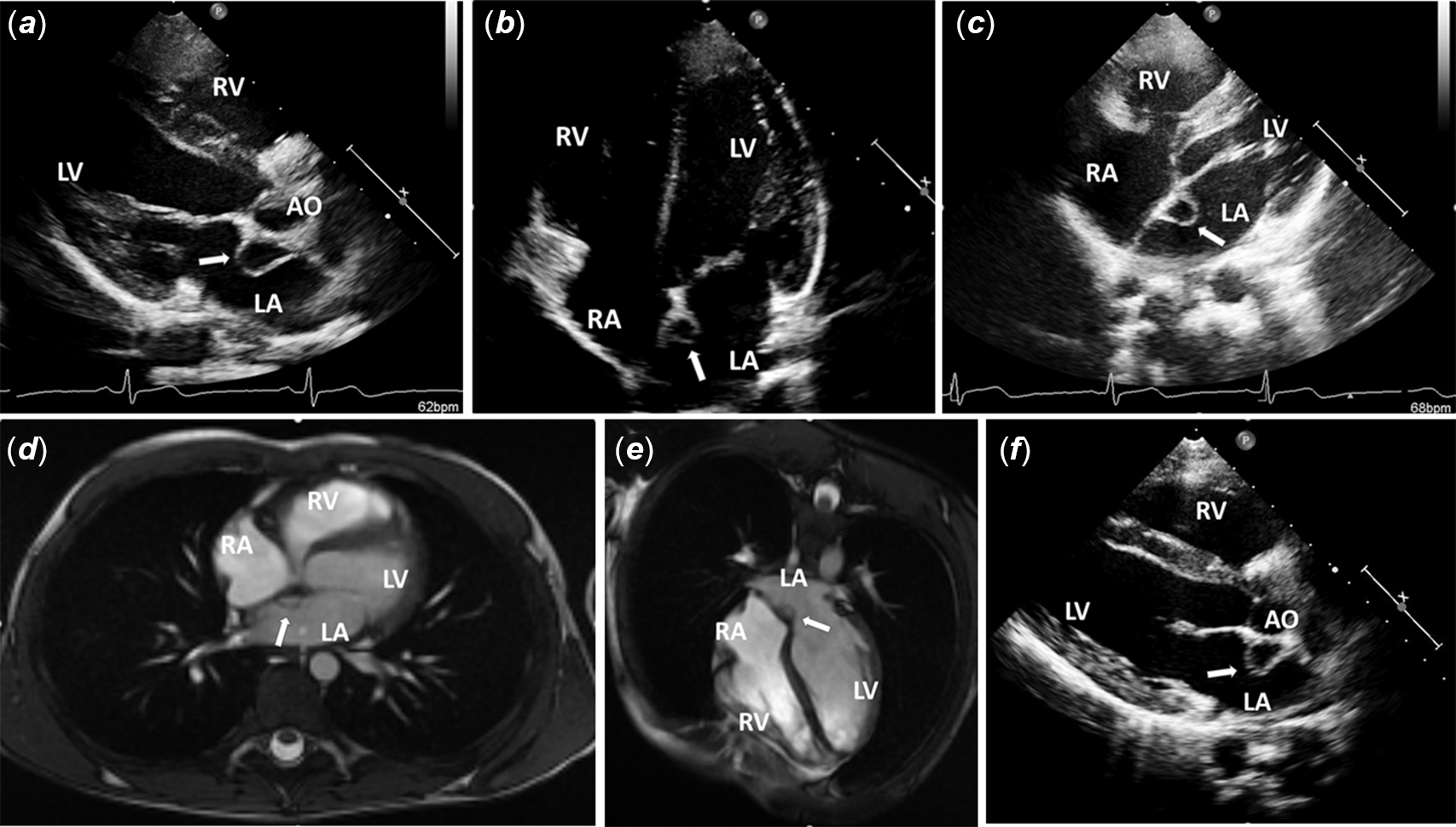
Transthoracic colour Doppler echocardiography was normal, but a saccular and semi-circular structure with anechogenic content, measuring about 10 mm, was visible in the left atrium, near the fossa ovalis (Fig 1a–c). The structure appeared mostly semi-circular, with a free side floating during the different stages of the heart cycle (Clip 1, Clip 2, Clip 3).
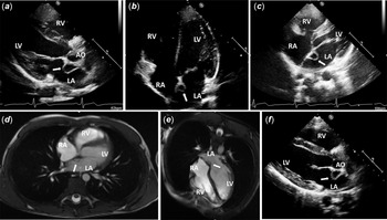
( a) Left parasternal long axis echocardiographic image. ( b) Apical four chamber echocardiographic image. ( c) Off axis echocardiographic image (modified apical four chamber view obtained by moving the probe of two intercostal spaces superiorly and approximately 3 cm medially, to better visualise the atrial septum). ( a – c) The left atrial septal pouch (arrow) was seen like a saccular structure, with anechogenic content, in the left atrium, near the fossa ovalis, posteriorly the posterior sinus of the aorta. ( d, e ) Cardiac magnetic resonance view of the left atrial septal pouch (arrow): its content had the same density as the blood into the left cardiac chambers. ( f ) Left parasternal long axis echocardiographic image. Five years later, the left atrial septal pouch (arrow) was unchanged. AO, aortic bulb; LA, left atrium; LV, left ventricle; RA, right atrium; RV, right ventricle.
The diagnosis of interatrial septal aneurysm was excluded because it usually appears like a localised, saccular deformity, generally at the level of the fossa ovalis, that bulges into the right or left atrium or in both sides; in contrast, in our patient the structure was roughly semi-circular (apparently closed on all sides), with a “rigid” base formed by a portion of the interatrial septum and a remaining free floating portion into the left atrium. In addition, the structure was located more caudal the oval pit than the interatrial septal aneurysm usually is.
No thrombus in the pouch and no interatrial communication were detected. Moreover, no valvular dysfunction or hemodynamic instability was detected in spite of its presence. However, the saccular structure was large and unusual; a cardiac magnetic resonance was performed to further evaluate the mass and rule out other possible differential diagnosis. The cardiac magnetic resonance showed that the content of the pouch had the same density of the blood into the left cardiac chambers, and it meant the pouch was opened into the left atrium (Fig 1d and e). No signs of atrial wall inflammation or infiltration were detected.
A transesophageal echocardiogram was not performed because the transthoracic image quality was very accurate; a transesophageal echocardiogram would not have added any additional information. An agitated saline solution was not used to rule out an interatrial right to left shunt because the child was not able to do a Valsalva manoeuvre. However, in our patient, the quality of the echocardiographic images was very good and there was no evidence of interatrial patency with colour Doppler. A serological echinococcus test was performed, and the result was negative. Moreover, the ultrasound homogeneous appearance of the cavity without septation was not in accordance with the typical polycystic aspect of a hydatid cyst. The diagnosis of cystic myxoma also was excluded because its content was completely anechogenic.
A diagnosis of the left atrial septal pouch was made.
The left atrial septal pouch is a kangaroo pouch-like structure on the left side of the interatrial septum, opened into the left atrial cavity without a connection between the left and right atria. It occurs when the foramen ovale is absent but the septum primum and septum secundum are only partially fused.
The left atrial septal pouch was previously described, Reference Krishnan and Salazar1 but actually all publications describe cases of the left atrial septal pouch in adulthood. Reference Krishnan and Salazar1,Reference Hołda, Koziej and Hołda2 Based on our knowledge, this is the first case of the left atrial septal pouch in childhood and our patient is the youngest left atrial septal pouch patient described.
A left atrial septal pouch may result from an incomplete fusion of the septum primum and the septum secundum. The interatrial septum develops to divide the primordial common atrium into right and left halves. It develops by formation and fusion of two septa: the septum primum and the septum secundum. At the beginning, the septum primum grows from the atrial roof toward the endocardial cushions, narrowing the foramen primum. As the septum primum grows, perforations develop to form a large central window (the foramen secundum). Then, the foramen primum gradually closes. During this time, remodelling occurs in the atrial roof and the septum secundum forms as an infolding of the atrial wall. It folds inward to the right of the septum primum and produces an incomplete septal partition because it leaves a small gap, an oval-shaped window known as foramen ovale. In particular, the septum secundum forms the anterosuperior, superior, and posterior margins of the muscular rim of the foramen ovale. The remaining portion of the thinner septum primum forms a flap-like valve over the foramen ovale. During the neonatal period, pulmonary artery resistance decreases, and the right atrial pressure becomes lower than the left atrial pressure; in this way, the septum primum is forced against the septum secundum, achieving physiological closure. Over time, the development of adhesions serves to close the foramen. The anatomical closure occurs in up to 75% of adults. Reference Hara, Virmani and Ladich3–Reference Fisher, Fisher and Budd5 When the fusion between septum primum and septum secundum does not occur along the entire overlap zone, pockets could be observed. Reference Krishnan and Salazar1,Reference Hołda, Koziej and Hołda2 Fusion limited to the caudal portion of the overlap zone leads to the left septal pouch. Reference Hołda, Koziej and Hołda2 Autopsy studies showed that the prevalence of the left atrial septal pouch is 47% of population. Reference Hołda, Koziej and Hołda2 However, in many cases, the pocket on the atrial septum is small and so it could not be visualised by transthoracic echocardiography. Our description is rare because of the atrial septal pouch is very large.
Redundancy of the septal pouch flap may mimic an atrial mass or an organised structure. Reference Foley, Irwin and Abidin6 In these patients, the transesophageal echocardiogram or cardiac magnetic resonance could help to clarify the diagnosis. In our patient, at the first step, we made the diagnosis by transthoracic echocardiography. In children, this method accurately identifies the anatomic and functional characteristics of heart and interatrial septum and it helps to choose the most appropriate management.
In paediatric age, finding a saccular structure with anechogenic content on the left side of the oval pit could be a left atrial septal pouch. In doubtful cases, the transesophageal echocardiogram, the cardiac magnetic resonance, and a cardiological check after few months should be done.
A saccular and mobile structure on the interatrial septum (with a variable intra-atrial bulging, influenced by the different stages of the heart cycle) may cause intra-atrial haemodynamic alterations, and it may be a substrate for supraventricular arrhythmias, in common daily life or during exercise. Reference Yetkin, Ileri, Korkmaz and Ozturk7 Despite our patient was asymptomatic, he underwent an ECG Holter and cardiac stress testing; these exams were normal as initial check as well as at the follow up at 12, 36, and 60 months later.
In some patients, the left atrial septal pouch may solve spontaneously due to the natural history of patent foramen ovale closure that consists of a fusion between the septum primum and septum secundum progressing throughout the lifetime. After following up with our patient at 12, 36, and 60 months, the anatomical characteristics of the left atrial septal pouch and its measurements were unchanged (Fig 1f).
Due to its anatomical features (a pouch separating a space with low flow, partially opened into the left atrium), the left atrial septal pouch might serve as a site for thrombus formation and source of thromboembolism, especially in patients with conditions associated with stagnant blood (atrial fibrillation, mitral stenosis, heart failure with high left ventricular filling pressures). Reference Hołda, Koziej and Hołda2,Reference Hołda and Koziej8–Reference Gurudevan, Shah and Tolstrup10 Actually, no data have been published concerning primary thromboembolic prophylaxis in the left atrial septal pouch, especially in paediatric age. In our young patient, no thrombus was detected in the pouch; no antithrombotic therapy was administered because he did not have cardiac or haematological predisposing factors for thromboembolism. We scheduled periodic cardiology check-ups.
To the best of our knowledge, our patient has the left atrial septal pouch with the longest follow up described. The anatomical characteristics of the left atrial septal pouch, in our patient, remained unchanged over a period of 5 years; moreover, he did not have arrhythmias or limitations in his ability to exercise.
Supplementary material
To view supplementary material for this article, please visit https://doi.org/10.1017/S1047951121004236
Acknowledgements
None.
Financial support
This research received no specific grant from any funding agency, commercial or not-for-profit sectors.
Conflicts of interest
None.