INTRODUCTION
During sampling conducted to study invasive species in the Gulf of California in summer 2011, small colonies of an unknown tube-worm were found as part of the dock fouling community in La Paz, Baja California Sur, Mexico. While extracting worms from their tubes, many individuals were found undergoing fission, with several developing offspring posterior to the parent, forming chains. This taxon was identified as belonging to a new species of the genus Pseudobranchiomma Jones, Reference Jones1962.
The genus Pseudobranchiomma is a quite heterogeneous taxon composed of 15 mid-sized sabellid species, most of them with short, lateral serrations in paired radiolar flanges. This feature is quite different from the paired transversely orientated epithelial flaps (stylodes) present in Branchiomma Kölliker, Reference Kölliker1858, which lacks radiolar flanges (Nogueira & Knight-Jones, Reference Nogueira and Knight-Jones2002). However, flanges and serrations can be differently developed in the genus Pseudobranchiomma.
Species of Pseudobranchiomma were divided by Knight-Jones & Giangrande (Reference Knight-Jones and Giangrande2003) into three categories based on the presence and extension of serrations along the radiolar length. Thus, group A was characterized by having radiolar serrations extending for most of the radiolar length. This group contains six known species: P. grandis (Baird), P. serratibranchis (Grube), P. orientalis (McIntosh), P. emersoni Jones, P. paraemersoni Nogueira, Silva & López and P. paulista Nogueira, Silva & López. Group B includes those species with flanges only in the distal parts of the radioles and includes P. tricolour (Grube), P. zebuensis (McIntosh), P. odhneri (Fauvel) and P. bocki (Johansson). Group C includes those species with flanges reduced or absent (without serrations) and includes P. longa (Kinberg), P. punctata (Treadwell), P. minima (Nogueira & Knight-Jones), P. perkinsi Knight-Jones & Giangrande, and P. tarantoensis Knight-Jones & Giangrande.
In this paper the new species is described belonging to group A, and compared with all species in that group. Information about live coloration, glandular pattern and reproduction is also provided.
MATERIALS AND METHODS
Sampling was carried out on docks from marinas in La Paz, Baja California Sur (southern Gulf of California) during the summer of 2011. The fouling community was sampled by hand and placed in a seawater-filled container for transportation to the laboratory. Organisms were scraped and maintained in aquaria to document their live coloration and reproductive mode. Worms were anaesthetized with menthol crystals and fixed in 10% formalin–seawater or ethanol 95%. Specimens fixed in formalin were washed in the laboratory with tap water for 24 h and transferred to 70% ethanol for long-term preservation.
A Leica MZ75 stereomicroscope and Olympus CH30 optical microscope were used for identification and digital photographs were taken with an attached Canon S5 digital camera. Some specimens were processed and examined after final dehydration in two changes of 100% ethanol at the Laboratorio de Microscopía Electrónica de Barrido (Centro de Ciencias Básicas, Universidad Autónoma de Aguascalientes, México). Specimens were critical point dried using CO2 before being mounted on stubs with platinum tape and coated with gold (200 Å thickness). They were then viewed under a scanning electron microscope. The thoracic glandular pattern was revealed by staining worms with methyl green.
Complete specimens were measured to record the width of the posterior thorax, body length (from peristomium to pygidium), branchial crown length, number of radiolar pairs, number of thoracic and abdominal segments, and the presence of gametes and offspring. Description is based on holotype, with variation of paratypes as indicated in parentheses. Type material and topotypes were deposited at the Museum of Comparative Zoology, Harvard Institution (MCZ); Universidad Autónoma de Aguascalientes (UAA); Colección de Referencia de Invertebrados (EMU–ICML) and Colección de Anélidos Poliquetos de México (CNP–ICML) and Instituto de Ciencias del Mar y Limnología, Universidad Nacional Autónoma de México.
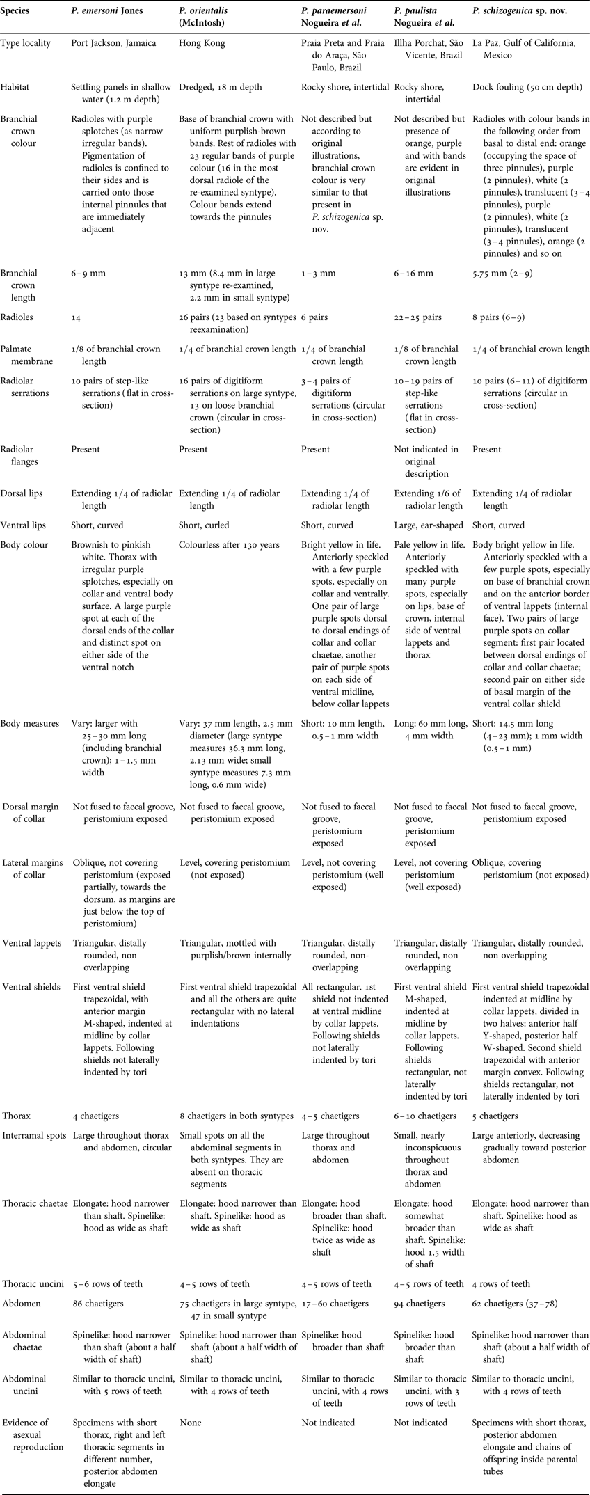
A comparative table of diagnostic characters for species of Pseudobranchiomma belonging to group A fide Knight-Jones & Giangrande (Reference Knight-Jones and Giangrande2003) and the new species described here is included. Pseudobranchiomma grandis (Baird) and P. serratibranchis (Grube) were not included since both descriptions are brief and lack figures. According to Knight-Jones & Giangrande (Reference Knight-Jones and Giangrande2003) and Nogueira et al. (Reference Nogueira, Rossi and López2006) these two species have compound radiolar eyes. Thus, the comparison included here only considers species without compound radiolar eyes. Features of P. orientalis (McIntosh) and P. emersoni Jones were based on original descriptions and also in the examination of type materials from the Natural History Museum (London), the American Museum of Natural History (New York) and National Museum of Natural History (Washington), respectively. Characters of P. paraemersoni Nogueira et al., and P. paulista Nogueira et al., were taken from original descriptions.
SYSTEMATICS
Order SABELLIDA Latreille, Reference Latreille1825
Family SABELLIDAE Latreille, Reference Latreille1825
Genus Pseudobranchiomma Jones, Reference Jones1962
Pseudobranchiomma schizogenica sp. nov.
(Figures 1–5)
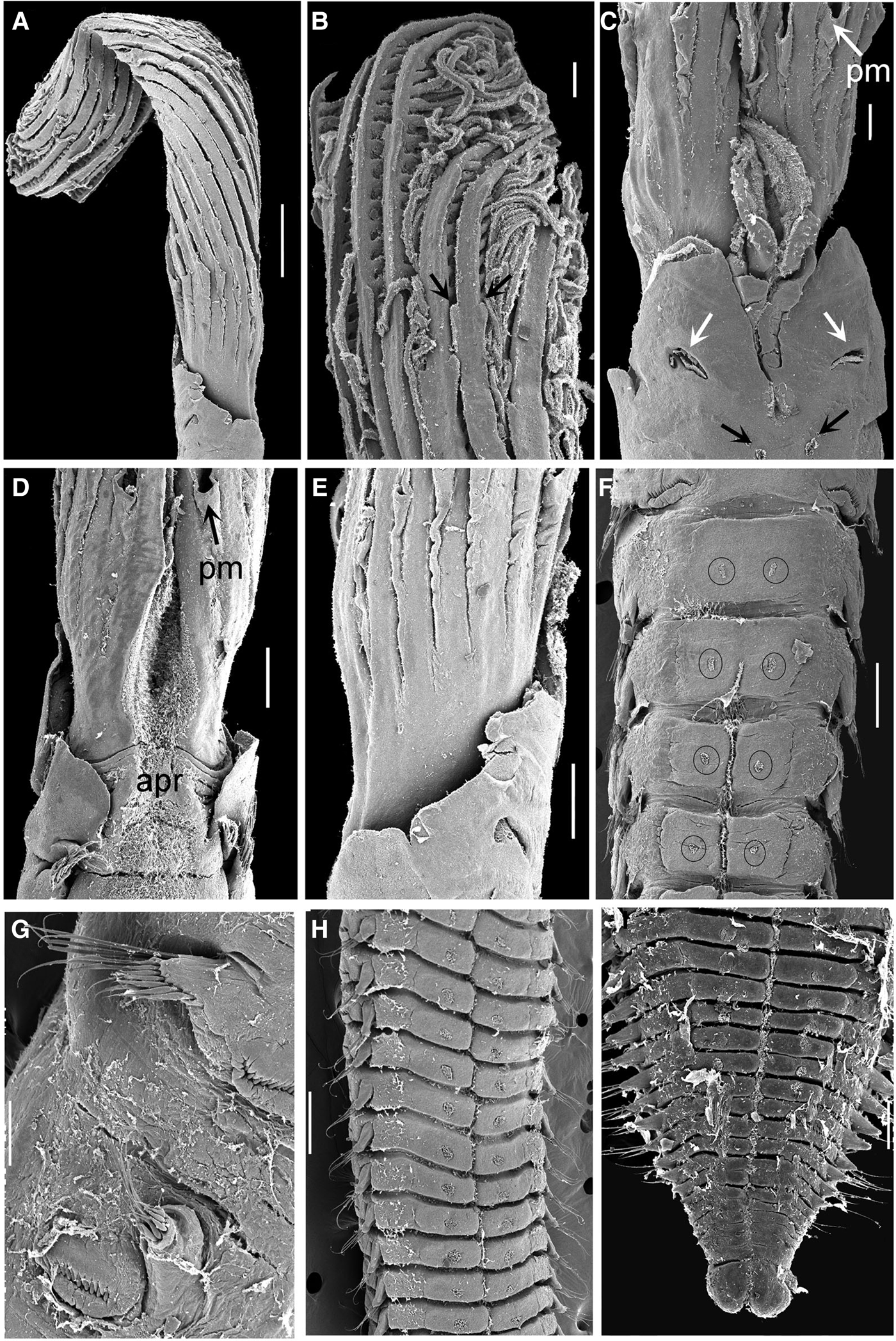
Body structures of Pseudobranchiomma schizogenica sp. nov., under scanning electron microscopy: (A) collar segment and branchial crown; (B) radiolar serrations as indicated with arrows; (C) ventral lappets and base of branchial crown, patches of cilia as indicated with arrows; (D) collar segment and base of branchial crown, dorsal view; (E) same, lateral view; (F) last thoracic and anterior abdominal ventral shields, patches if cilia in circles; (G) chaetal inversion; (H) abdominal patches of cilia encircled; (I) posterior abdomen and pygidium. Abbreviations: apr, anterior peristomial ring; pm, palmate membrane. Scale bars: A, 500 µm; B, 400 µm; D, F, H, 200 µm; C, G, I, 100 µm.

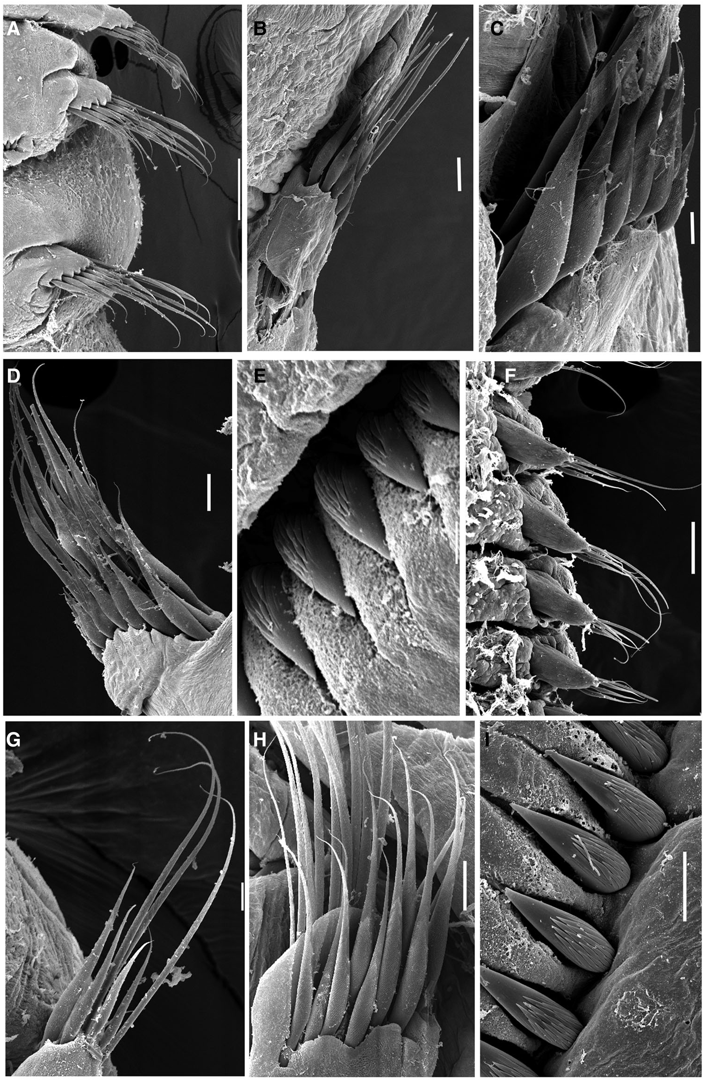
Uncini and chaetae of Pseudobranchiomma schizogenica sp. nov., under scanning electron microscopy: (A, B, D) thoracic notopodial lobes; (C) thoracic spinelike chaetae; (E) thoracic uncini; (F–H) abdominal neuropodial lobes; (I) abdominal uncini. Scale bars: A, 100 µm; B, D, H, 20 µm; C, G, I, 10 µm; E, 5 µm; F, 50 µm.
Life colour of Pseudobranchiomma schizogenica sp. nov.: (A) dorsal view; (B) ventrolateral view; (C) branchial crown; (D) colony attached to a floating dock. Scale bars: A, B, 500 µm; C, 300 µm; D, not scaled.
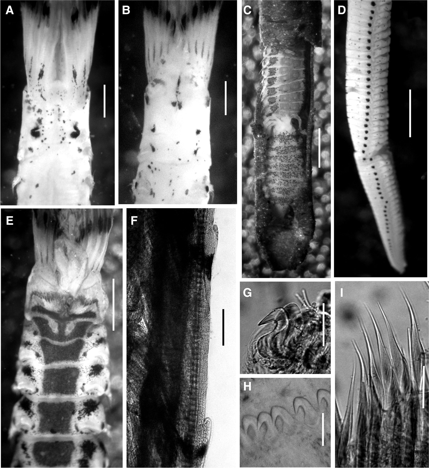
Post-fixation colour of Pseudobranchiomma schizogenica sp. nov., and methyl green staining pattern: (A) anterior thorax, dorsal view; (B) same, ventral view; (C) offspring in posterior end of an adult; (D) interramal spots; (E) ventral shields revealed with methyl green; (F) radiolar serrations; (G) thoracic uncini; (H) abdominal uncini; (I) thoracic chaetae. Scale bars: A, B, 250 µm; C, 500 µm; D, E, 700 µm; F, 100 µm; G, H, 45 µm; I, 25 µm.
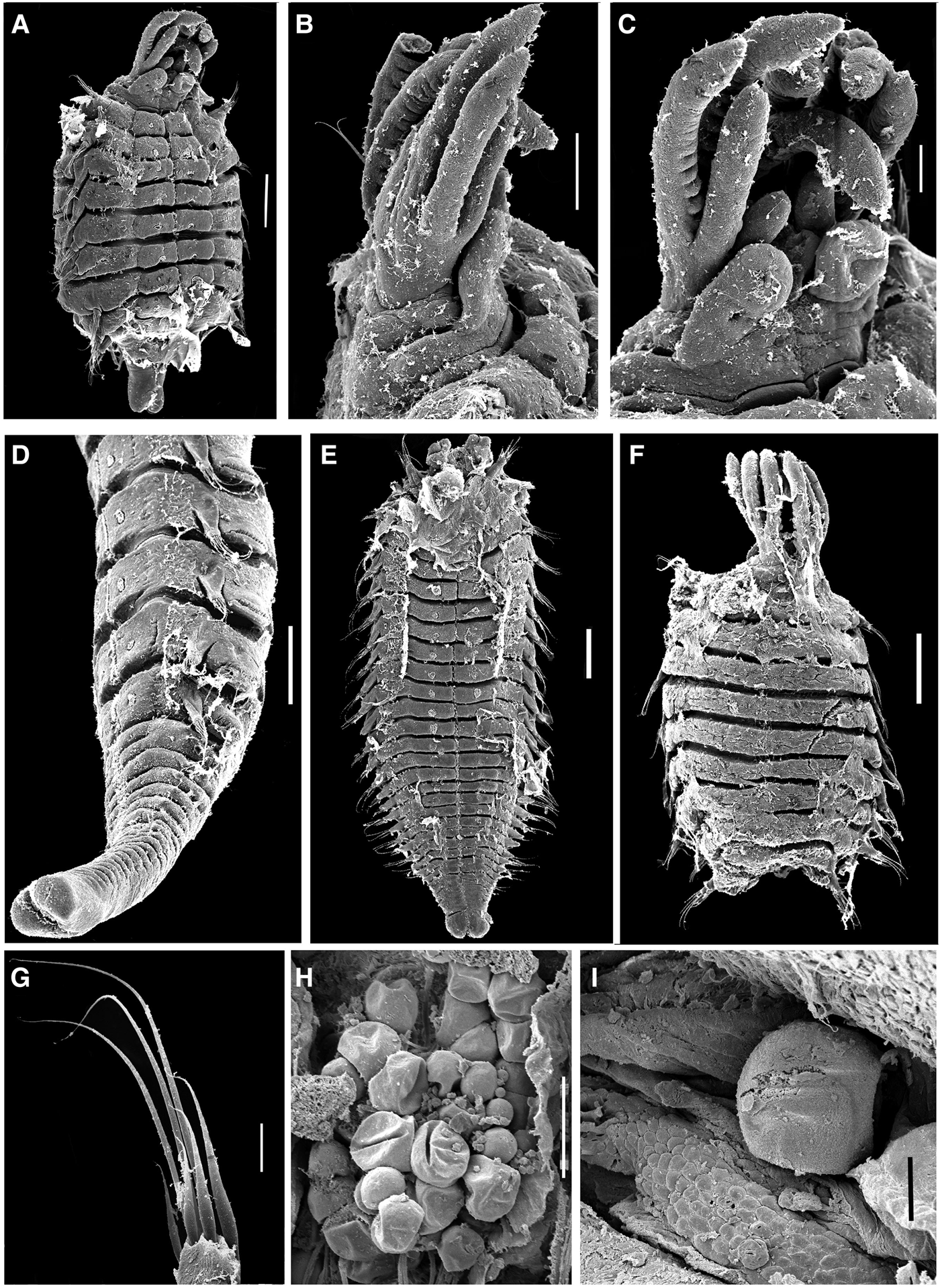
Offprings and oocytes of Pseudobranchiomma schizogenica sp. nov., under scanning electron microscopy: (A) offspring with branchial crown, collar and pygidium restored; (B) same, detail of branchial crown, lateral view; (C) same, detail of ventral lappets and branchial crown; (D) offspring with posterior end restored; (E) offspring with rudimentary radioles; (F) offspring with crown and collar restored; (G) abdominal chaetae; (H) oocytes; (I) oocyte and germinal cells. Scale bars: A, D, E, F, 200 µm; B, 200 µm; C, 50 µm; G, 20 µm; H, 100 µm; I, 20 µm.
TYPE MATERIAL
Holotype: MCZ-27848, 10 paratypes MCZ-27818, 27821–22, 27825–26, 27828, 27832, 27843, 27846–47: Club de Yates Palmira, La Paz, Baja California Sur, southern Gulf of California, México, 24°10.992′N 110°18.185′W, dock 3, replicate 1, 50 cm depth, 15 August 2011, Colls. TFVG & JMAC.
Paratypes: 25 paratypes EMU–ICML–10036–37, 25 paratypes CNP–ICML–POP–72–002, 4 paratypes UAA–M142C, M143B, 144A, 146E (mounted in gold for scanning electron microscopy (SEM)): Club de Yates Palmira, La Paz, Baja California Sur, southern Gulf of California, México, 24°10.992′N 110°18.185′W, dock 3, replicate 1, 50 cm depth, 15 August 2011, Colls. TFVG & JMAC.
Topotype material: 382 specimens GEOMARE: Club de Yates Palmira, La Paz, Baja California Sur, southern Gulf of California, México, 24°10.992′N 110°18.185′W, dock 3, replicate 1, 50 cm depth, 15 August 2011, Colls. TFVG & JMAC.
COMPARATIVE MATERIAL EXAMINED
Dasychone orientalis McIntosh, Reference McIntosh1885: Types NHML 85.12.1.393, dredged off Hong Kong, 10 fms, Challenger.
Pseudobranchiomma emersoni Jones, Reference Jones1962: Holotype AMNH 3612, Port Royal, Jamaica, West Indies 1 July 1961. Fourteen specimens USNM 3613 (same information as holotype). Three specimens USNM 3632 (labelled as non-type, same information as holotype).
DIAGNOSIS
Radioles with short, digitiform serrations along the entire radiolar length (Figures 1A, B, 4F). Lateral margins of collar oblique, covering anterior peristomial ring (Figure 1E). Ventral shield of collar trapezoidal; divided in two halves: anterior half Y-shaped, posterior half W-shaped (Figure 4E). Thoracic superior chaetae with hoods narrower than shafts (Figure 2A, D). Thoracic inferior chaetae spinelike with hoods as width as shafts (Figures 2C, 4I). Abdominal neurochaetae spinelike with hoods narrower than shafts (about a half width of shaft) (Figure 2H). Paired patches of cilia in ventral lappets of collar, thoracic and abdominal shields (Figure 1C, F, H).
DESCRIPTION
Gregarious sabellin with short body (Figure 3A, B, D): 23 mm length (4–23 mm), 1 mm width (0.5–1 mm). Branchial crown long (Figures 1A, 3C): 9 mm length (2–9 mm) with eight pairs of radioles (6–9 pairs) arranged in two semicircles, not involuted midventrally, and basally joined by low palmate membrane (1 mm length, occupying 1/4 of the branchial crown length) (Figure 1C–E). Outer margins of radioles flat with thin narrow flanges along the entire length (Figure 1A, B). Ten pairs of short, digitiform serrations (6–11 pairs) along the entire length of radioles (Figure 1A, B), circular in cross-section, each pair associated with pigmentation units; compound radiolar eyes absent. Dorsal lips with radiolar appendages (purple mid-rib), extending 1/4 of radiolar length, joined by a membrane to dorsalmost pair of radioles, ventral margins of dorsal lips fused to each other by transverse lamellar process (Figure 1C). Ventral lips short (1/2 of the dorsal lips length), curved. Ventral sacs (sand sacs) originating from dorsal end of parallel lamellae, projecting above ventral lappets (Figure 1C). Anterior (peristomium) and posterior peristomial rings (collar) present. Dorsal margins of collar not fused to faecal groove, peristomium exposed dorsally (Figures 1D). Lateral margins of collar oblique, covering anterior peristomial ring (peristomium not exposed) (Figure 1E). Ventral lappets triangular, distally rounded, not overlapping, with a pair of long, rounded, oval or half-moon-shaped patches of cilia in the middle area (100 µm long each) (Figures 1C, 4B, E). Thorax with five segments; notopodial lobe forming bundles (Figures 1G, 2A, B, D). Ventral shield of collar trapezoidal and indented at midline by collar lappets; divided in two halves: anterior half Y-shaped, posterior half W-shaped (Figure 4E). Between both halves there is a pair of small patches of cilia (half-moon, oval or rounded shaped). Second shield trapezoidal, with anterior margin convex (Figure 4E). Following ventral shields rectangular, not indented by tori, with a pair of patches of cilia per shield (Figures 1F, H, 4E). Collar chaetae elongate, narrowly hooded, arranged in an arc, with spinelike chaetae inside this arc. Thoracic notopodial lobe with superior row of chaetae as elongate, narrowly hooded chaetae, arranged in arc, hood narrower than shaft (Figure 2A, B, D). Inferior thoracic notochaeta spinelike, hood as width as shaft (Figures 2C, 4I). Thoracic uncini with about four rows of equal-sized teeth above main fang, covering around 1/2 of extension of main fang (Figure 2E), breast swollen and handle extending for 1/2 of distance between breast and crest (Figure 4G). Abdomen with 68 segments (37–78 segments) and conical neuropodial lobes (Figures 1G, 2F, G). All abdominal shields with small patches of cilia (Figure 1F, H, I). Anterior row of abdominal neurochaetae as modified, elongate, narrowly hooded chaetae, arranged in a fascicle inside arc formed by posterior chaetae (Figure 2F, G); posterior row of abdominal neurochaetae as spinelike chaetae (Figure 2H), hood narrower than shaft (about a half width of shaft). Abdominal uncini similar to thoracic uncini, with four rows of teeth above main fang (Figures 2I, 4H). Bilobed pygidium (Figure 1I). Tubes leathery-looking covered with a thin coat of mud (Figure 3D).
LIVE COLOUR
Body bright yellow in life (Figure 3A, B), anteriorly speckled with a few purple spots, especially on base of branchial crown (Figure 3C) and on anterior border of ventral lappets (internal face). Two pairs of large purple spots on collar segment: first pair dorsal, located between dorsal endings of collar and collar chaetae; second pair ventral on either side of basal margin of the collar shield. Palmate membrane purple (Figure 3C). Radioles with colour bands in the following order from basal to distal end: orange (occupying the space of three pinnules), purple (2 pinnules), white (2 pinnules), translucent (3–4 pinnules), purple (2 pinnules), white (2 pinnules), translucent (3–4 pinnules), orange (2 pinnules) and so on (Figure 3C). Interramal eyespots purple, large anteriorly, decrease gradually towards posterior end.
POST-FIXED COLOUR
Body pale yellow (Figure 4A, B, D). Purple spots remain along the entire body length, including the dorsal and ventral pairs of spots on collar segment (Figure 4A, B), and the interramal spots (Figure 4D). Branchial crowns fixed inside tubes maintain colour; those fixed outside tubes only have brown palmate membranes and some brown bands. Ventral shields whitish.
GLANDULAR PATTERN
Ventral shields stained deep blue except for the white ventral, paired patches of cilia. First shield (collar segment) divided in two halves: anterior half Y-shaped, posterior half W-shaped (Figure 4E). The largest pair of cilia patches is located in the first shield (always present). Second thoracic shield without cilia patches. Inconspicuous patches of cilia in third thoracic shield, except in one specimen in which they were not found. Remaining shields with conspicuous patches of cilia until last abdominal segments. Diffuse blue areas stain above thoracic tori (Figure 4E).
REPRODUCTION
The 82% of the population in the southern Gulf of California was found to be undergoing architomy, thus P. schizogenica sp. nov., reproduces mainly via asexual reproduction. Only females were found within the population, containing asynchronous oocytes (25–70 µm) and clusters of germ cells (Figures 5H, I). Sperm were not observed.
Many individuals were found with the posterior end enlarged and composed of about 26 segments with a protruding pygidium and 3–4 developing fragments immediately posterior to the parent (Figure 4C). Other individuals were found with the abdomen abruptly fragmented but, in these cases, 3–4 developing fragments were also found posterior to the parental body. All these offspring formed chains inside the parental tubes (Figures 4C), but morphologically these fragments were different.
Some fragments regenerated the branchial crown and the collar (Figure 5F), whereas other fragments restore both the branchial crown and collar in the anterior end, and pygidium posteriorly (Figure 5A–E). In the first case, offspring had nine abdominal segments and a restored branchial crown and collar but the pygidium was not present (Figure 5F). In the second case, offspring were found bearing nine abdominal segments with a restored branchial crown, collar and pygidium (Figure 5A, E). The branchial crown had four pairs of radioles with inconspicuous pinnules (Figure 5B, C). The peristomium segment was well differentiated in the anterior and posterior peristomial rings (Figure 5B, C). Ventral lappets (Figure 5C) and ventral shields with paired patches of cilia (Figure 5A). Abdominal neuropodial lobes with 4–6 spinelike and 4–6 elongate, narrowly hooded chaetae (Figure 5G) and abdominal tori with 5–6 uncini with four rows of teeth above the main fang. The parental tubes are unbranched except for occasional short branches near the proximal end. Presumably the clonal offspring leave the parental tube at one end or the other before they secrete their own tube attached to a neighbouring tube in the base of the aggregation.
HABITAT
Colonies of P. schizogenica sp. nov., were part of the dock fouling community in La Paz, southern Gulf of California (Mexico) and occurred at a density of 487 ind m−2 in association with the invasive sabellid polychaete Branchiomma bairdi (McIntosh) (Figure 3D).
ETYMOLOGY
Named after the process of modification and separation of the posterior abdomen into new individuals.
REMARKS
The species here documented here as Pseudobranchiomma schizogenica sp. nov., belongs to group A of Knight-Jones & Giangrande (Reference Knight-Jones and Giangrande2003) which is characterized by having radiolar serrations extending for most of the extension of radioles. Pseudobranchiomma schizogenica sp. nov. is distinguished from all known species in group A by the presence of a unique set of features related to the covering of the peristomium by the lateral collar margins, the shape of the first ventral shield, the size of the interramal eyespots, and the width of the hood of thoracic and abdominal spinelike chaetae (Table 1).
Comparison of closely-related species of Pseudobranchiomma.
Among species in group A, only Pseudobranchiomma grandis and P. serratibranchis have compound radiolar eyes. Radiolar serrations are reduced and step-like (flat in cross-section) in P. emersoni and P. paulista, whereas in P. orientalis, P. paraemersoni and P. schizogenica sp. nov., serrations are conspicuously digitiform (circular in cross-section). The palmate membrane is very low in the species of Pseudobranchiomma reviewed here, occupying 1/4 (P. orientalis, P. paraemersoni and P. schizogenica sp. nov.) to 1/8 of the total branchial crown length (P. emersoni and P. paulista). The large ear-shaped ventral lips of P. paulista are remarkable as all other species have short, curved ventral lips.
An important character to separating species in Pseudobranchiomma is the shape of lateral collar margins and the degree of exposition of the anterior peristomial ring. In P. orientalis and P. paraemersoni lateral margins of the collar are level, but in the former, margins cover the anterior peristomial ring while in the latter the anterior peristomial ring is well exposed above lateral collar margins. In P. schizogenica sp. nov., lateral collar margins are oblique, covering the anterior peristomial ring. An intermediate state is found in P. emersoni, in which lateral collar margins are oblique, but the peristomium is partially exposed.
The shape of the ventral shield of the collar shows differences among species of Pseudobranchiomma. This structure is easy seen using methyl green staining. It is rectangular in P. paraemersoni, M-shaped in P. paulista, and trapezoidal in P. emersoni, P. orientalis and P. schizogenica sp. nov. The collar shield of P. schizogenica sp. nov., is differentiated from that of P. emersoni and P. orientalis by having two halves: an anterior, Y-shaped half and a posterior W-shaped half (entire, just one piece in P. emersoni and P. orientalis).
Interramal spots are large anteriorly, decreasing gradually toward the posterior abdomen in P. schizogenica sp. nov. In P. emersoni and P. paraemersoni spots are large throughout the thorax and abdomen. In P. paulista and P. orientalis spots are small, nearly inconspicuous (present along all body segments in P. paulista and only in abdominal segments in P. orientalis). In fact, P. orientalis is remarkable since interramal spots are present only in the abdomen as McIntosh (Reference McIntosh1885) described and corroborated by examination of syntypes.
The hood of superior and inferior thoracic chaetae varies among species and, as a consequence, is a useful character. In P. emersoni, P. orientalis and P. schizogenica sp. nov. the hood of the superior group of elongate chaetae is narrower than the shaft while it is broader than shaft in P. paraemersoni and P. paulista. The hood of the inferior group of spinelike chaetae is as wide as the shaft in all species except for P. paulista where the hood is 1.5× the width of shaft. Referring to the hood of the abdominal spinelike chaetae, it is broader than shafts in P. paraemersoni and P. paulista and narrower than the shafts in P. emersoni, P. orientalis and P. schizogenica sp. nov.).
Pseudobranchiomma schizogenica sp. nov., and P. paraemersoni are bright yellow in life with the anterior thorax speckled with a few purple spots (brownish to pinkish white in P. emersoni; pale yellow in P. paulista). Life colour was not documented by McIntosh (Reference McIntosh1885) in the case of P. orientalis and syntypes are colourless after approximately 130 years.
Lastly, the branchial crown length and the number of radioles seem to be features dependent on age. Several juveniles were observed with a short branchial crown and few radioles, whereas large specimens usually have long crowns with numerous radioles, although correlations have not been assessed. On the other hand, the ventral lappets are similar in all species except for the irregular purple spots that vary among species. Thus, these features are not considered here for comparative purposes.
On asexual reproduction
Peculiar features of the here described species P. schizogenica sp. nov., is the gregariouness and the presence of asexual reproduction. Asexual reproduction has been reported frequently in the sabellids, taking the form of architomy (spontaneous fission followed by subsequent regeneration). Thus, natural fission from the posterior end, as opposed to regeneration following damage or autotomy, has been recorded in several species: Sabella discifera (Rioja, Reference Rioja1929; Knight-Jones & Bowden, Reference Knight-Jones and Bowden1984), Perkinsiana rubra, Myxicola aesthetica (Knight-Jones & Bowden, Reference Knight-Jones and Bowden1984), Megalomma cinctum (as Megalomma sp. in Yuan, Reference Yuan1992), Perkinsiana milae (Gambi et al., Reference Gambi, Giangrande and Patti2000), Pseudobranchiomma punctata, P. minima (Nogueira & Knight-Jones, Reference Nogueira and Knight-Jones2002), P. emersoni and P. perkinsi (Knight-Jones & Giangrande, Reference Knight-Jones and Giangrande2003), Branchiomma curtum (Tovar-Hernández & Knight-Jones, Reference Tovar-Hernández and Knight-Jones2006), Bispira brunnea (Tovar-Hernández & Pineda-Vera, Reference Tovar-Hernández and Pineda-Vera2008), Branchiomma bairdi (Tovar-Hernández et al., Reference Tovar-Hernández, Méndez and Salgado-Barragán2009a, Reference Tovar-Hernández, Méndez and Villalobos-Guerrerob, Reference Tovar-Hernández, Yáñez-Rivera and Bortolini-Rosales2011; Arias et al., Reference Arias, Giangrande, Gambi and Anadón2013), Pseudopotamilla reniformis (Kolbalsova et al., Reference Kolbasova, Tzetlin and Kupriyanova2013) and also in the new species of Pseudobranchiomma here described. Among these species, S. discifera, B. bairdi, B. brunnea, P. reniformis and species of Pseudobranchiomma form dense aggregations and natural fission seems a process that leads to the formation of colonial forms as occurs in some serpulids such Salmacina and Filograna (Kupriyanova et al., Reference Kupriyanova, Nishi, Ten Hove and Rzhavsky2001; Pernet, Reference Pernet2001).
Asexual reproduction in species of Pseudobranchiomma has been poorly described due mainly to the presence of few, miscellaneous scissiparous offspring among samples, which are easily overlooked when parental worms are extracted from their tubes. Nogueira & Knight-Jones (Reference Nogueira and Knight-Jones2002) reported only one scissiparous offspring among the paratypes of Pseudobranchiomma punctata. It consisted of a small portion of the posterior abdomen with very short, narrow segments thus indicating recent posterior regeneration. The same authors also recorded the presence of only one scissiparous specimen of P. minima with budding radioles among a series of type specimens and several specimens with a short thorax typical of imperfect regeneration. For P. perkinsi, Knight-Jones & Giangrande (Reference Knight-Jones and Giangrande2003) mentioned the presence of scissiparity and illustrated a newly regenerated posterior abdomen with a bilobed pygidium, and they mentioned one offspring with rudimentary radioles, separated from the parental worm, inside the original tube. Based on re-examination of the type material of P. emersoni, Knight-Jones & Giangrande (Reference Knight-Jones and Giangrande2003) reported the presence of a single offspring with six distal buds which were the elements of the first crown radioles. The clonal offspring of P. schizogenica sp. nov., were found regenerating the crown and the collar, whereas others were found restoring both the crown and collar anteriorly and the pygidium posteriorly. A similar developmental pattern was also documented in Pseudopotamilla reniformis by Kolbalsova et al. (Reference Kolbasova, Tzetlin and Kupriyanova2013), although in this species a well-developed thorax was restored (in P. schizogenica sp. nov., only offsprings with collar were found).
It is common for this method of reproduction to be followed by imperfect regeneration, usually resulting in fewer than the eight thoracic segments found in most sabellid species that do not reproduce by fission. Thus, the low number of thoracic segments found in P. schizogenica sp. nov. can be explained by means of an incomplete regeneration, as showed in Branchiomma nigromaculatum (Berrill, Reference Berrill1978; Tovar-Hernández & Knight-Jones, Reference Tovar-Hernández and Knight-Jones2006), Pseudobranchiomma emersoni (Knight-Jones & Giangrande, Reference Knight-Jones and Giangrande2003), P. minima (Nogueira & Knight-Jones, Reference Nogueira and Knight-Jones2002) and Branchiomma bairdi (Tovar-Hernández et al., Reference Tovar-Hernández, Méndez and Villalobos-Guerrero2009b, Reference Tovar-Hernández, Yáñez-Rivera and Bortolini-Rosales2011). The number of thoracic segments in most of the species of Pseudobranchiomma reviewed here is low when compared with other sabellin genera. In P. emersoni, P. paraemersoni and P. schizogenica sp. nov., individuals were found with 4–5 thoracic segments. Both syntypes of P. orientalis presented the usual number of thoracic segments (8) whilst a wide range is present in P. paulista (6–10 segments).
On the segmental cilia
Another peculiar feature present in the new described taxon is the presence of paired patches of cilia in collar and body segments. Presence of ventral patches of cilia in collar has been recorded in the sabellids Laonome Malmgren, Reference Malmgren1866, Amphicorina Claparède, Reference Claparède1864, Paradialychone ecaudata (Moore) and Dialychone trilineata (Tovar-Hernández) (Tovar-Hernández, Reference Tovar-Hernández2008). In the first two genera, cilia are extensions of those present in the anterior peristomial ring; in P. ecaudata and D. trilineata the ventral shield of the collar is completely ciliated; in Pseudobranchiomma schizogenica sp. nov., the collar has two ventral patches of cilia. In the last three above mentioned species the cilia are not an extension of those present in the anterior peristomial ring.
The presence of paired patches of cilia in body segments in Pseudobranchiomma has not been described before. These contain typical cilia lacking heads (kinocilia-type). The presence of patches of cilia at least in the collar was corroborated in P. emersoni (revealed using methyl green in types), P. paraemersoni (SEM image contained in Nogueira et al., Reference Nogueira, Rossi and López2006: figure 2E) and P. paulista (SEM images contained in Nogueira et al., Reference Nogueira, Rossi and López2006: figure 5C, G). These ciliary patches may be of different kinds and not all of them are necessarily sensory, they could have different functions such as to generate water currents inside tubes, but further investigation is needed.
On the adhesion to tubes
McIntosh (Reference McIntosh1885) highlighted the tenacity with which the body of Pseudobranchiomma orientalis adheres to the tube and this also occurs in P. schizogenica sp. nov., where the tubes are strongly fixed to the body-wall and its removal often ruptures the epithelia. This mechanism might be used by worms to avoid predation. The mucus produced by the ventral shields could be responsible for such adhesion, but the chemical components have not been studied in any species within the genus and deserve further study. In fact, properties of mucus have been studied only for Sabella spallanzanii in which its roles include determination of fertilization success and chemical defense (Stabili et al., Reference Stabili, Schirosi, Licciano and Giangrande2009).
ACKNOWLEDGEMENTS
Thanks are given to Beatriz Yáñez, Tulio Villalobos and José Maria Aguilar for their help in collecting material. Araceli Adabache Ortiz and Marcelo Silva Briano (Universidad Autónoma de Aguascalientes, México) processed SEM photographs and images included in Figure 1, which were kindly taken by Humberto Bahena (ECOSUR). Literature was kindly provided by Luis Fernando Carrera-Parra (ECOSUR). We are grateful to anonymous referees for their useful suggestions.
FINANCIAL SUPPORT
This work was supported by Instituto Nacional de Ecología (grant INE/ADE-013/2011).