Introduction
Bipolar disorder (BD) is one of the most severe psychiatric disorders with a significantly increased risk of suicide (Gonda et al., Reference Gonda, Pompili, Serafini, Montebovi, Campi, Dome and Rihmer2012; Pompili et al., Reference Pompili, Gonda, Serafini, Innamorati, Sher, Amore and Girardi2013; Schaffer et al., Reference Schaffer, Isometsa, Tondo, D, Turecki, Reis and Yatham2015), physical health burden (Kilbourne et al., Reference Kilbourne, Cornelius, Han, Pincus, Shad, Salloum and Haas2004), cognitive deficit (Bourne et al., Reference Bourne, Aydemir, Balanza-Martinez, Bora, Brissos, Cavanagh and Goodwin2013; Martinez-Aran & Vieta, Reference Martinez-Aran and Vieta2015), and functional impairment (Grande et al., Reference Grande, Magalhaes, Chendo, Stertz, Panizutti, Colpo and Vieta2014; Kapczinski et al., Reference Kapczinski, Dias, Kauer-Sant'Anna, Frey, Grassi-Oliveira, Colom and Berk2009; Kapczinski et al., Reference Kapczinski, Magalhaes, Balanza-Martinez, Dias, Frangou, Gama and Berk2014). According to the World Health Organization (WHO) Mental Health Survey, BD has the second greatest impact on days out of role among common physical and mental disorders (Alonso et al., Reference Alonso, Petukhova, Vilagut, Chatterji, Heeringa, Ustun and Kessler2011). BD affects 1–3% of the world's population, irrespective of diverse nationality, ethnicity, or socioeconomic status (Merikangas et al., Reference Merikangas, Jin, He, Kessler, Lee, Sampson and Zarkov2011; Pedersen et al., Reference Pedersen, Mors, Bertelsen, Waltoft, Agerbo, McGrath and Eaton2014). Because depressive episodes are generally more frequent in BD and BD patients are much more likely to visit clinicians in outpatient setting when they are depressed (Angst et al., Reference Angst, Azorin, Bowden, Perugi, Vieta, Gamma and Group2011), many individuals with BD are initially misdiagnosed as unipolar major depressive disorder (MDD) (Hirschfeld, Cass, Holt, & Carlson, Reference Hirschfeld, Cass, Holt and Carlson2005; Hirschfeld, Lewis, & Vornik, Reference Hirschfeld, Lewis and Vornik2003), and they may receive inappropriate medications before presenting hypomanic or manic symptoms (Bowden, Reference Bowden2005; Ghaemi, Sachs, Chiou, Pandurangi, & Goodwin, Reference Ghaemi, Sachs, Chiou, Pandurangi and Goodwin1999; Lish, Dime-Meenan, Whybrow, Price, & Hirschfeld, Reference Lish, Dime-Meenan, Whybrow, Price and Hirschfeld1994; Morselli, Elgie, & Europe, Reference Morselli, Elgie and Europe2003). For accurate diagnosis and treatment, it is imperative to identify valid and reliable biomarkers for this disorder.
Structural brain imaging studies have increased the neurobiological understanding of BD. Patients with BD showed more alterations in cortical thickness, surface area, and overall gray matter volumes compared to healthy controls (HCs) (Hartberg et al., Reference Hartberg, Sundet, Rimol, Haukvik, Lange, Nesvag and Agartz2011; Padmanabhan et al., Reference Padmanabhan, Tandon, Haller, Mathew, Eack, Clementz and Keshavan2015). A large-scale coordinated and multicenter study showed that adult patients with BD had widespread bilateral patterns of reduced cortical thickness in the frontal, temporal, and parietal regions, specifically in the left pars opercularis, left fusiform gyrus, and left rostral middle frontal cortex (Hibar et al., Reference Hibar, Westlye, Doan, Jahanshad, Cheung, Ching and Andreassen2018). Although there were common volumetric reductions in the dorsomedial and ventromedial prefrontal cortex, the anterior cingulate cortex, and bilateral insula in both MDD and BD, patients with BD had relatively larger volumes in the left parahippocampal gyrus and right dorsolateral prefrontal cortex compared to patients with MDD (Wise et al., Reference Wise, Radua, Via, Cardoner, Abe, Adams and Arnone2017). However, to date, there have been no clear disease-specific brain imaging biomarkers for BD, which could differentiate from other diseases and help to treat this disorder.
Neurodevelopmental imaging biomarkers could be considered more stable and accurate indicators of cortical pathology reflecting early perinatal or childhood developmental disturbances (Zilles, Palomero-Gallagher, & Amunts, Reference Zilles, Palomero-Gallagher and Amunts2013), since most brain morphometric parameters are prone to various related factors such as state-dependent effects and medication status (Nenadic et al., Reference Nenadic, Maitra, Dietzek, Langbein, Smesny, Sauer and Gaser2015). Because early neurodevelopmental deviations are suspected to contribute to BD pathophysiology (Bortolato et al., Reference Bortolato, Kohler, Evangelou, Leon-Caballero, Solmi, Stubbs and Carvalho2017; O'Shea & McInnis, Reference O'Shea and McInnis2016), abnormal cortical folding, such as gyrification and sulcation, has been considered as a candidate endophenotype or a biomarker for BD. Gyrification and sulcation are developmental processes occurring in the fetal brain between the 10th and 44th gestational week (Dubois et al., Reference Dubois, Benders, Cachia, Lazeyras, Ha-Vinh Leuchter, Sizonenko and Huppi2008) and contribute to the early development of neural connectivity (Hilgetag & Barbas, Reference Hilgetag and Barbas2005; Rakic, Reference Rakic1988; Van Essen, Reference Van Essen1997; White, Su, Schmidt, Kao, & Sapiro, Reference White, Su, Schmidt, Kao and Sapiro2010).
Previous studies investigated distinct cortical folding patterns in patients with BD compared to those with schizophrenia, MDD, or HCs (Janssen et al., Reference Janssen, Aleman-Gomez, Schnack, Balaban, Pina-Camacho, Alfaro-Almagro and Desco2014; McIntosh et al., Reference McIntosh, Moorhead, McKirdy, Hall, Sussmann, Stanfield and Lawrie2009; Nanda et al., Reference Nanda, Tandon, Mathew, Giakoumatos, Abhishekh, Clementz and Keshavan2014; Nenadic et al., Reference Nenadic, Maitra, Dietzek, Langbein, Smesny, Sauer and Gaser2015; Penttila et al., Reference Penttila, Cachia, Martinot, Ringuenet, Wessa, Houenou and Paillere-Martinot2009a; Penttila et al., Reference Penttila, Paillere-Martinot, Martinot, Ringuenet, Wessa, Houenou and Cachia2009b; Sarrazin et al., Reference Sarrazin, Cachia, Hozer, McDonald, Emsell, Cannon and Houenou2018). Research on cortical folding patterns in patients with BD has usually focused on the prefrontal and limbic regions and has shown mixed results. Previously, Penttila et al. (Reference Penttila, Cachia, Martinot, Ringuenet, Wessa, Houenou and Paillere-Martinot2009a) found lower local sulcal index in the right dorsolateral prefrontal cortex in patients with intermediate-onset (onset age, 25–45 years) BD compared to HCs. Pentilla et al. also showed a significantly decreased right-sided global sulcal index in patients with bipolar or unipolar depression compared to patients with HC (Penttila et al., Reference Penttila, Paillere-Martinot, Martinot, Ringuenet, Wessa, Houenou and Cachia2009b). McIntosh et al., showed that patients with BD type I (BD-I) and schizophrenia had lower ventral and dorsal prefrontal gyrification compared to HCs (McIntosh et al., Reference McIntosh, Moorhead, McKirdy, Hall, Sussmann, Stanfield and Lawrie2009). Similarly, Janssen et al., found that patients with adolescent BD or schizophrenia had lower cortical thickness and higher sulcal width in the frontal cortex compared to HCs (Janssen et al., Reference Janssen, Aleman-Gomez, Schnack, Balaban, Pina-Camacho, Alfaro-Almagro and Desco2014). More recently, Nenandic et al. showed a significantly higher gyrification of the dorsal anterior and infragenual cingulate in patients with psychotic BD-I compared to HCs, whereas Nanda et al. showed no significant differences of local gyrification between patients with psychotic BD and HCs (Nanda et al., Reference Nanda, Tandon, Mathew, Giakoumatos, Abhishekh, Clementz and Keshavan2014; Nenadic et al., Reference Nenadic, Maitra, Dietzek, Langbein, Smesny, Sauer and Gaser2015). A large-sized multicenter study comprising 263 patients with BD-I and 320 HCs reported that among patients with BD, those with an early onset showed a higher local sulcal index in the right prefrontal dorsolateral region, whereas those with psychotic features showed a decreased local sulcal index in the left superior parietal cortex; however, their study only showed a trend toward association since they did not correct the statistical analyses for multiple comparisons (Sarrazin et al., Reference Sarrazin, Cachia, Hozer, McDonald, Emsell, Cannon and Houenou2018). Furthermore, the abovementioned studies have differences in methodology, including different sample sizes, diagnosis, symptom severity, illness duration, and used psychotropic medication. There has also been a paucity of studies that investigated the difference in the gyrification between BD and HCs, with controlling related factors such as BD subtype, illness duration, depressive symptom severity, or lithium treatment.
We hypothesized that patients with BD would show significantly less cortical gyrification than HCs. To explore disease-specific cortical folding patterns in the patients with BD, we set out to investigate the differences in the local gyrification index (LGI) in each cortical region (66 areas) at the whole-brain level between patients with BD and HCs. To explore the influence of clinically related factors on cortical folding patterns, we additionally analyzed the differences in LGI depending on the BD subtype (BD-I or BD-II), and investigated the association between LGI and illness duration, depressive symptom severity, and lithium treatment in patients with BD.
Methods and materials
Participants
In the present study, a total of 244 subjects were recruited between January 2015 and November 2018: 61 patients with BD from the outpatient psychiatric clinic of Korea University Anam Hospital (Seoul, Republic of Korea) and 183 HCs from the community using an advertisement. The inclusion criteria for patients with BD were as follows: (1) age between 18 and 64 years and (2) current euthymic or depressive state. The diagnosis of BD was determined by two board-certified psychiatrists (K.-M. Han and B.-J. Ham) using the Structured Clinical Interview for the DSM-IV-TR (Diagnostic and Statistical Manual of Mental Disorders, Fourth Edition, Text Revision) Axis I disorders (SCID). The exclusion criteria for patients with BD were as follows: (1) comorbidity of any other major psychiatric disorders (including personality and substance use disorders), (2) current psychotic symptoms, (3) high suicidal risk requiring immediate treatment, (4) history of a serious or unstable medical illness, (5) primary neurological illness (e.g. Parkinson's disease, cerebrovascular disease, or epilepsy), (6) recent abnormal results on physical examination or laboratory tests, (7) pregnant or nursing women, and (8) any contraindication for magnetic resonance imaging (MRI). Subjects were included in the HC group if two board-certified psychiatrists (K.-M. Han and B.-J. Ham) confirmed that they had no ongoing or past history of Axis I or II disorders. The same exclusion criteria of the BD group were applied for the HC group. The study protocol was approved by the Institutional Review Board (IRB) of Korea University Anam Hospital (IRB No: 2015AN0009). All participants provided written informed consent for their participation. The study methodology was in accordance with the approved guidelines and Declaration of Helsinki.
Clinical assessments
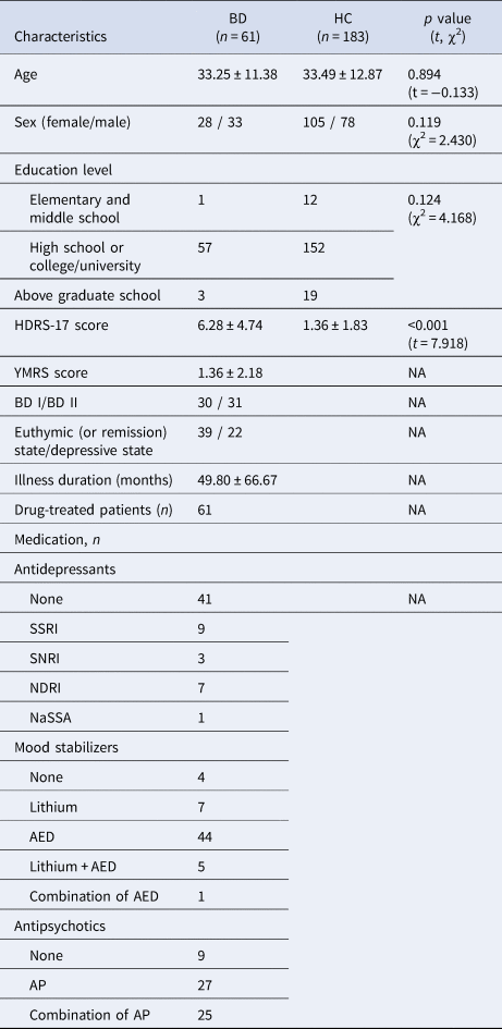
Demographic and clinical characteristics were collected from the two groups. We assessed the severity of depressive symptoms of all participants using the 17-item Hamilton Depression Rating Scale (HDRS) at the time of the MRI scan (Hamilton, Reference Hamilton1960). For the BD group, the 11-item Young Mania Rating Scale (YMRS) was applied to evaluate mania symptoms (Young, Biggs, Ziegler, & Meyer, Reference Young, Biggs, Ziegler and Meyer1978). The illness duration of patients with BD was assessed using the Life Chart Methodology. All patients with BD were under a psychopharmacological treatment during the study enrollment. Detailed information about psychotropic medications is presented in Table 1.
Demographic and clinical characteristics of patients with bipolar disorder and healthy controls

Data are mean ± standard deviation for age, HDRS-17 scores, illness duration.
p values for distribution of sex and education level were obtained using a chi-squared test.
p values for comparisons of age and HDRS scores were obtained using an independent t test.
HCs, healthy controls; BD, bipolar disorder; HDRS-17, 17-item Hamilton Depression Rating Scale; YMRS, Young Mania Rating Scale; BD I, bipolar I disorder; BD II, bipolar II disorder; SSRI, selective serotonin reuptake inhibitor; SNRI, serotonin and norepinephrine reuptake inhibitor; NDRI, norepinephrine-dopamine reuptake inhibitor; NaSSA, noradrenergic and specific serotonergic antidepressant; Combination of AD, combinations of two or more types of antidepressant; AEDs, anti-epileptic drugs; APs, antipsychotics; ADs, antidepressants.
MRI data acquisition
MRI scans were obtained parallel to the anterior commissure–posterior commissure line using the 3.0-T Siemens Trio whole-body imaging system (Siemens Healthineers, Munich, Germany), using T1-weighted magnetization-prepared rapid gradient-echo with the following parameters: repetition time, 1900 ms; echo time, 2.6 ms; field of view, 220 mm; matrix size, 256 × 256; slice thickness, 1 mm; coronal slices without gap, 176; voxels, 0.86 × 0.86 × 1 mm3; flip angle, 16°; and number of excitations, 1.
Imaging processing for LGI
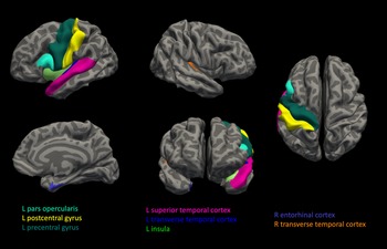
LGI values in each cortical parcellation of whole-brain were extracted using automated reconstruction procedures implemented in the FreeSurfer 5.3 development version (Laboratory for Computational Neuroimaging, Athinoula A. Martinos Center for Biomedical Imaging, Charlestown, MA, USA; http://surfer.nmr.mgh.harvard.edu). A three-dimensional model of the cortical surface reconstructions was computed from T1-weighted images that underwent pre-processing in FreeSurfer. Detailed procedures regarding cortical reconstruction performed were described in our previous studies (Han et al., Reference Han, Choi, Jung, Na, Yoon, Lee and Ham2014; Han et al., Reference Han, Won, Sim, Kang, Han, Kim and Ham2017b). We defined the average LGI as the ratio of the buried cortex surface area to the outer convex (hull) surface area in each parcellated cortical region (i.e. buried cortical surface area (mm2)/outer convex surface area (mm2)). We performed the processing extracting LGI from cortical parcellations according to a previously reported standard protocol (Nanda et al., Reference Nanda, Tandon, Mathew, Giakoumatos, Abhishekh, Clementz and Keshavan2014). Detailed information about the processes is described in our previous study on LGI (Han et al., Reference Han, Won, Kang, Kim, Yoon, Chang and Ham2017a). For the extraction of LGI, each hemisphere was parcellated into 33 cortical regions according to the Desikan–Killiany atlas, as shown in Fig. 1 (Desikan et al., Reference Desikan, Segonne, Fischl, Quinn, Dickerson, Blacker and Killiany2006). Then, we used the 66 LGIs of cortical regions in the bilateral hemispheres in the analyses. The list of cortical regions is shown in Table 2.
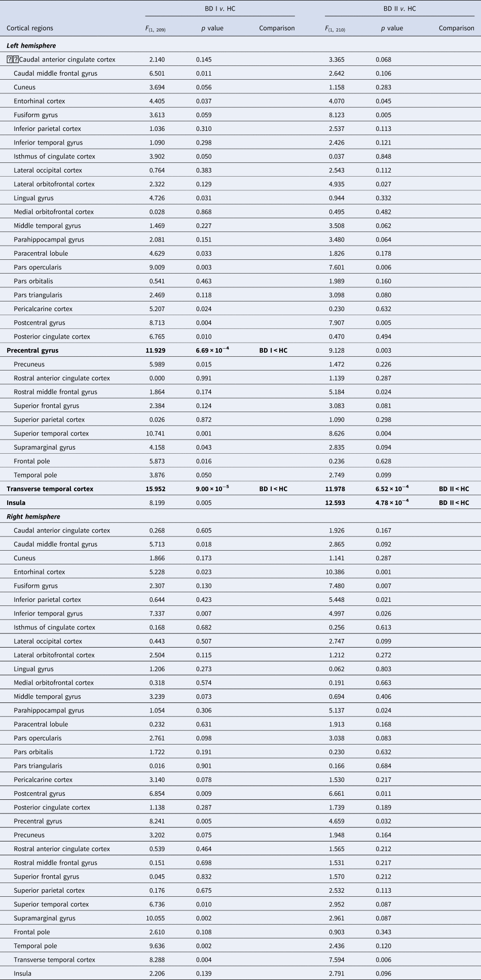
Schematic maps on the cortical regions with significant hypogyria in patients with Bipolar disorder (BD). For the extraction of lower local gyrification index (LGI), each hemisphere was parcellated into 33 cortical regions according to the Desikan-Killiany atlas. The patients with BD showed significantly lower LGI in left pars opercularis, left precentral gyrus, left postcentral gyrus, left superior temporal cortex, left insula, right entorhinal cortex, and both transverse temporal cortices (p < 0.05/66, colored regions).

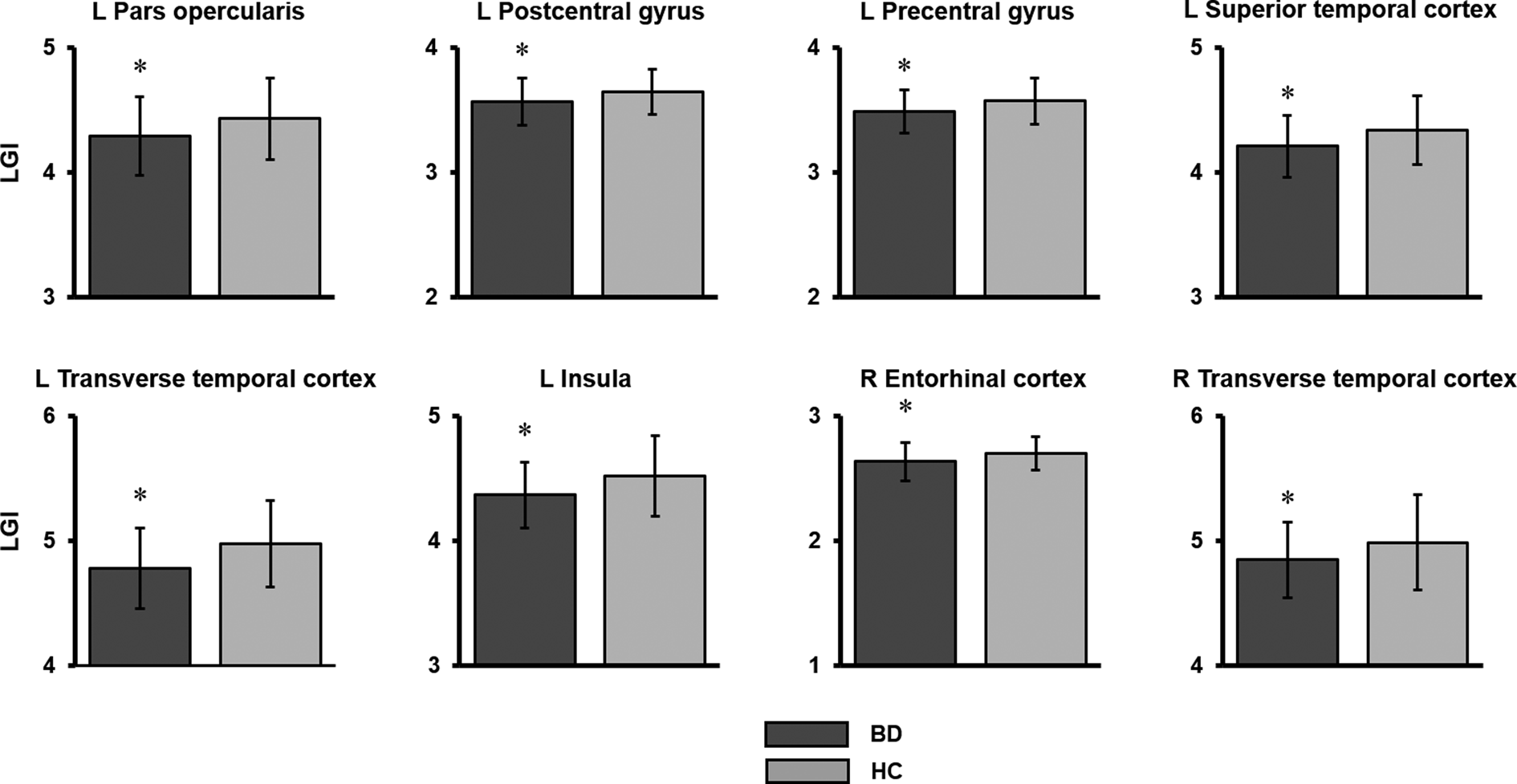
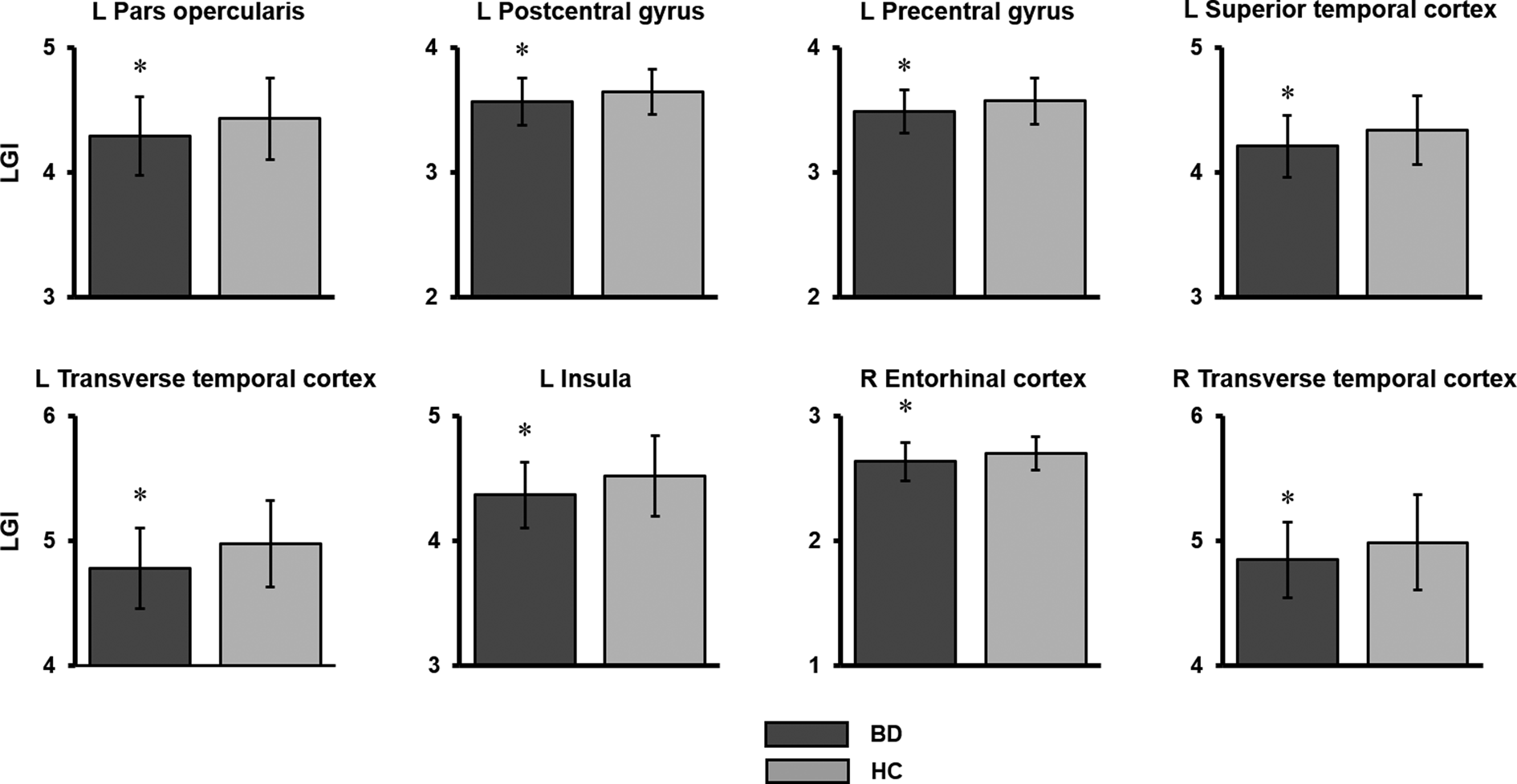
Comparisons of local gyrification index (LGI) between the BD and HC groups. The asterisk represents significantly different LGI in the comparison after Bonferroni correction. The error bar represents one standard deviation. BD, patients with bipolar disorder; HC, healthy control participants; L, left hemisphere; R, right hemisphere.
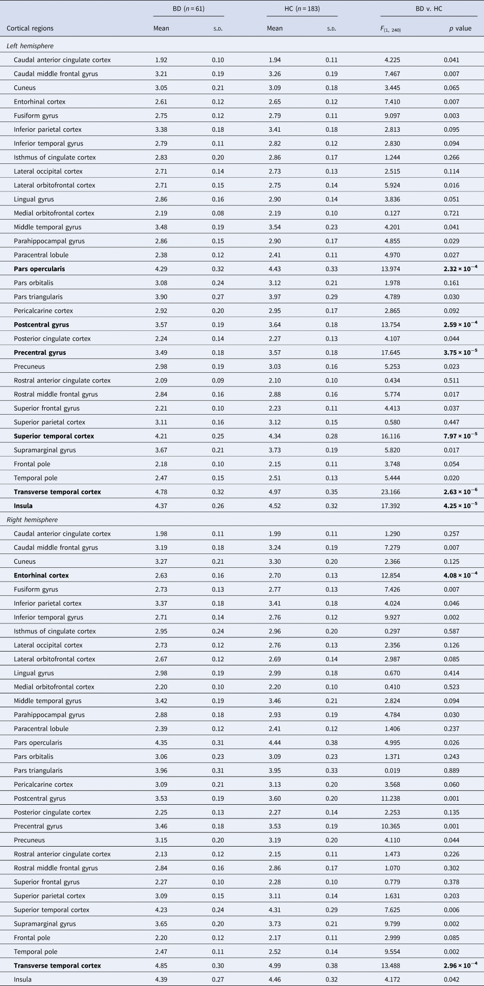
Comparison of local gyrification index between patients with bipolar disorders and healthy controls
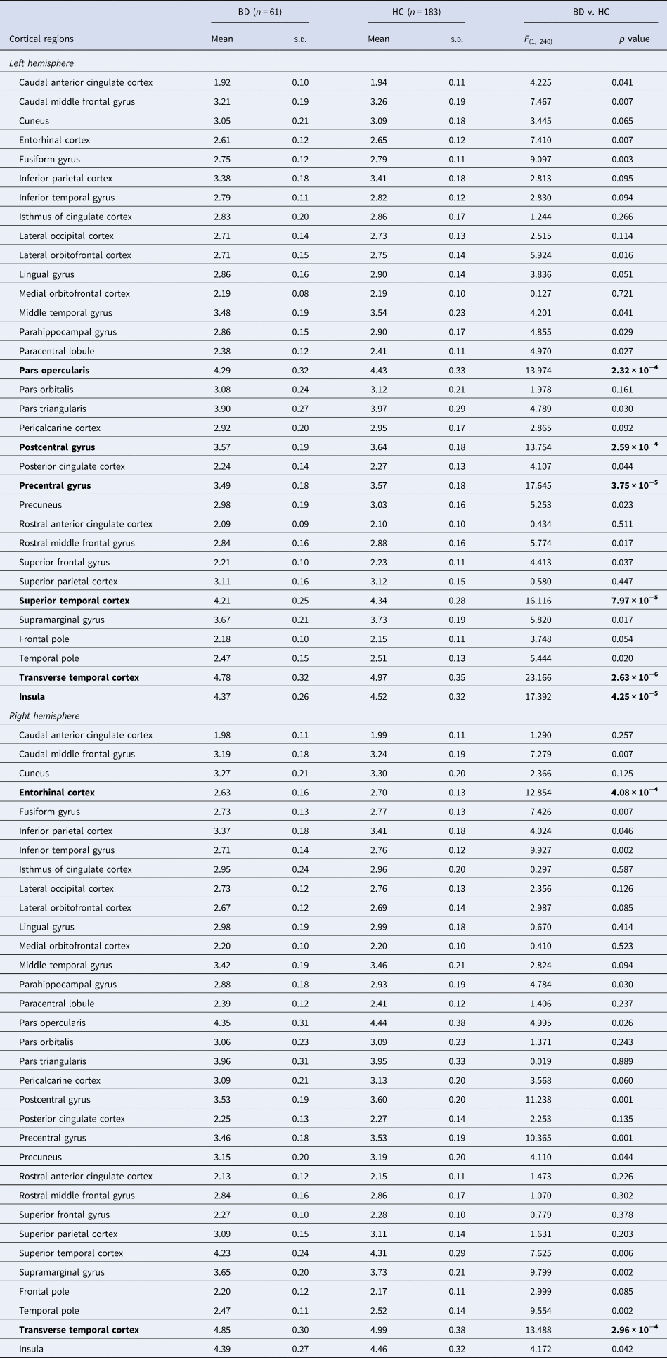
The F and p values were obtained using one-way analysis of covariance (ANCOVA) with the adjustment for age and sex as covariates.
Bonferroni correction was applied: p < 0.05/66 = 0.000758.
All the p values presented in the table are uncorrected values.
Significant group differences are presented in bold.
BD, patients with bipolar disorder; HC, healthy control participants; s.d., standard deviation.
Statistical analyses
As the main analysis, we performed a comparison of LGI between patients with BD and HCs using a one-way analysis of covariance (ANCOVA). The extracted 66 LGIs from each cortical parcellation were set as dependent variables and the groups (BD v. HC group) as independent variables. Age and sex were included as nuisance covariance in the analysis. For multiple comparisons, Bonferroni correction was applied to all the analyses (i.e. p < 0.05/66 = 0.000758 (66 cortical regions in the bilateral hemispheres)). In the sub-group comparison of LGI between BD type I and II, we used ANCOVA including age, sex, HDRS score, YMRS score, illness duration, and lithium treatment as covariates. To investigate the correlation of LGI with illness duration or HDRS score in the BD group, a two-tailed Pearson's partial correlation analysis was performed controlling for age, sex, illness duration (for HDRS score analysis), HDRS score (for illness duration analysis), YMRS score, BD type, and lithium treatment as covariates. To investigate demographic and clinical differences between the BD and HC groups, we used the independent t test to analyze age and HDRS scores and the chi-square test to analyze the difference of sex and education level distribution. All statistical analyses were performed using IBM SPSS Statistics for Windows, Version 24.0 (IBM Corp., Armonk, NY, USA).
Results
Sociodemographic characteristics
Demographics and clinical characteristics are shown in Table 1. Among the 61 patients with BD, 33 were male and 28 were female, having a mean age of 33.25 (standard deviation (s.d.) = 11.38). Among the 183 HCs, 105 were male and 78 were female, having a mean age of 33.49 (s.d. = 12.87). The BD group showed no statistical differences with regard to age, sex, and educational level compared to the HC group.
Clinical characteristics
Regarding their clinical characteristics, 30 and 31 patients were diagnosed with BD-I and BD-II, respectively, 39 were euthymic and 22 were in a depressive episode, and the mean illness duration was 49.80 (s.d. = 66.67) months. The BD group showed significantly higher HDRS scores than the HC group (t = 7.918, p < 0.001, Table 1). All patients were receiving psychotropic medications, and 12 patients were on lithium treatment at the time of enrollment. In the patient group, six patients were treated with a combination of two mood stabilizers (five patients, lithium with anti-epileptic drug (AED); one patient, combination of AEDs) and 25 patients were treated with a combination of two antipsychotics at the time of study enrollment. Detailed information about psychotropic medication use is presented in Table 1.
Differences in LGI values between patients with BD and HCs
Patients with BD showed a significantly lower LGI in the inferior frontal gyrus (pars opercularis, BD = 4.29 ± 0.32, HC = 4.43 ± 0.33, F (1, 240) = 13.974, p = 2.32 × 10⁻⁴), precentral gyrus (BD = 3.49 ± 0.18, HC = 3.57 ± 0.18, F (1, 240) = 17.645, p = 3.75 × 10⁻⁵), postcentral gyrus (BD = 3.57 ± 0.19, HC = 3.64 ± 0.18, F (1, 240) = 13.754, p = 2.59 × 10⁻⁴), superior temporal cortex (BD = 4.21 ± 0.25, HC = 4.34 ± 0.28, F (1, 240) = 16.116, p = 7.97 × 10⁻⁵), transverse temporal cortex (BD = 4.78 ± 0.32, HC = 4.97 ± 0.35, F (1, 240) = 23.166, p = 2.63 × 10⁻⁶), and insular cortex (BD = 4.37 ± 0.26, HC = 4.52 ± 0.32, F (1, 240) = 17.392, p = 4.25 × 10⁻⁵) in the left hemisphere and entorhinal cortex (BD = 2.63 ± 0.16, HC = 2.70 ± 0.13, F (1, 240) = 12.854, p = 4.08 × 10⁻⁴) and transverse temporal cortex (BD = 4.85 ± 0.30, HC = 4.99 ± 0.38, F (1, 240) = 13.488, p = 2.96 × 10⁻⁴) in the right hemisphere than HCs (Table 2 and Figure 2). These results were significant after the Bonferroni correction (p < 0.05/66). No cortical region of patients with BD showed a significantly greater LGI than that of HCs.
Differences in LGI values according to the BD subtype
There was no significant difference in LGI between patients with BD-I (n = 30) and BD-II (n = 31, Table S1 in the supplementary materials). Patients with BD-I showed a significantly lower LGI in the precentral gyrus (BD-I = 3.46 ± 0.19, HC = 3.57 ± 0.18, F (1209) = 11.929, p = 6.69 × 10⁻⁴) and transverse temporal cortex (BD-I = 4.74 ± 0.34, HC = 4.97 ± 0.35, F (1, 209) = 15.952, p = 9.00 × 10⁻⁵) and those with BD-II showed significantly smaller LGI in the transverse temporal cortex (BD-II = 4.83 ± 0.30, HC = 4.97 ± 0.35, F (1, 210) = 11.978, p = 6.52 × 10⁻⁴) and insular cortex (BD-II = 4.37 ± 0.27, HC = 4.52 ± 0.32, F (1, 210) = 12.593, p = 4.78 × 10⁻⁴) (Table 3).
Comparison of local gyrification index between patients with bipolar I and II disorders and healthy controls
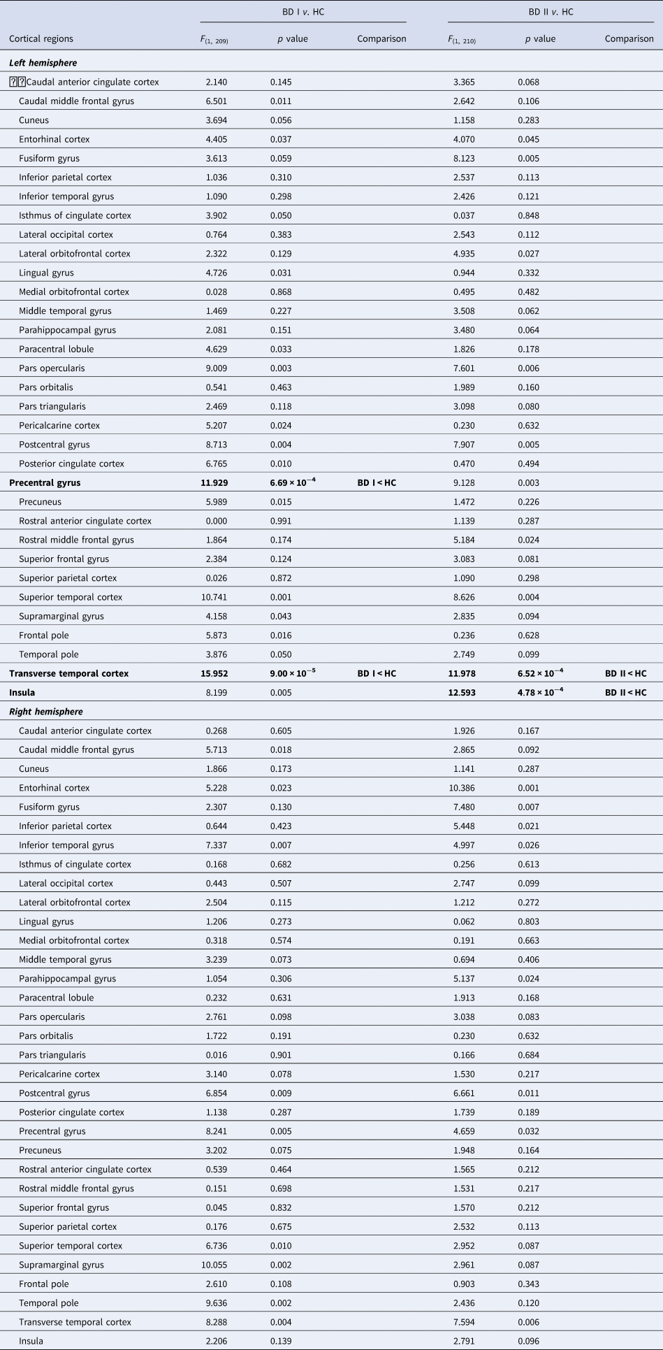
The F and p values were obtained using one-way analysis of covariance (ANCOVA) with the adjustment for age and sex as covariates.
Bonferroni correction was applied: p < 0.05/66 = 0.000758.
All the p values presented in the table are uncorrected values.
Significant group differences are presented in bold.
BD I, patients with bipolar I disorder; BD II, patients with bipolar II disorder; HCs, heathy controls; s.d., standard deviation.
Association between LGI and clinical characteristics in patients with BD
We could not find any significant correlation of LGI with illness duration or HDRS score in patients with BD after the Bonferroni correction (Table S2). Furthermore, we performed a comparison of LGI between patients with (n = 12) and without (n = 49) lithium treatment using ANCOVA including age, sex, illness duration, HDRS and YMRS scores, and BD subtype as covariates. There was no significant difference in LGI between patients with and without lithium treatment (Table S3). As a post-hoc analysis, we compared the LGIs between patients with and without antidepressant treatment using the same statistical method with the lithium treatment and observed no significant finding (Table S4). We also performed the post-hoc comparison of LGI between euthymic (n = 39) and depressive patients (n = 22) with BD using one-way ANCOVA with covariates of age, sex, BD subtype, illness duration, and lithium treatment, and no cortical regions showed significant difference of LGI between them after Bonferroni correction (Table S5).
Discussion
In the present study, patients with BD showed significant hypogyria in various cortical regions, including the left inferior frontal gyrus (pars opercularis), precentral gyrus, postcentral gyrus, superior temporal cortex, insula, right entorhinal cortex, and both transverse temporal cortices compared to HCs. BD-I patients had lower LGIs of the left precentral and transverse temporal gyrus and BD-II patients showed significant hypogyria of left transverse temporal gyrus and insula compared to HCs. However, there was no significant difference in LGI between patients with BD-I and BD-II. Furthermore, LGI was not significantly associated with clinical factors such as illness duration, depressive symptom severity, or lithium and antidepressant treatment in this study. Our findings are based on a relatively medium to large sample size compared with previous studies, along with conservative multiple comparison correction parameters and exclusion of covariates.
In this study, we found decreased gyrification of the subregion of the left ventrolateral prefrontal cortex (VLPFC, left pars opercularis or known as inferior frontal gyrus), superior temporal cortex, insula, and both transverse temporal cortices in patients with BD, which are consistent with those in prior reports of structural abnormalities in the VLPFC (Hanford, Nazarov, Hall, & Sassi, Reference Hanford, Nazarov, Hall and Sassi2016; Hibar et al., Reference Hibar, Westlye, Doan, Jahanshad, Cheung, Ching and Andreassen2018; Selvaraj et al., Reference Selvaraj, Arnone, Job, Stanfield, Farrow, Nugent and McIntosh2012), temporal cortex (Hanford et al., Reference Hanford, Nazarov, Hall and Sassi2016; Hibar et al., Reference Hibar, Westlye, Doan, Jahanshad, Cheung, Ching and Andreassen2018; Selvaraj et al., Reference Selvaraj, Arnone, Job, Stanfield, Farrow, Nugent and McIntosh2012), and insula (Selvaraj et al., Reference Selvaraj, Arnone, Job, Stanfield, Farrow, Nugent and McIntosh2012) in BD. Although there was right-sided grey matter volume reduction, which was different from our mostly left-sided positive results, the systematic review of VBM studies by Selvaraj et al., showed grey matter volume reduction in similar regions such as the inferior frontal gyrus, temporal cortex, insula, and claustrum (Selvaraj et al., Reference Selvaraj, Arnone, Job, Stanfield, Farrow, Nugent and McIntosh2012). Hanford et al., showed reduced cortical thickness in regions of the left cingulate, temporal, and bilateral frontal cortices compared to HCs in their review article (Hanford et al., Reference Hanford, Nazarov, Hall and Sassi2016). According to the study with high-powered sample from the ENIGMA Bipolar Disorder Working Group (whole sample, n = 6503, unrelated BD patients, n = 1837; unrelated HCs, n = 2582), researchers found that adults with BD had widespread reduced cortical thickness in the bilateral frontal, temporal, and parietal regions (Hibar et al., Reference Hibar, Westlye, Doan, Jahanshad, Cheung, Ching and Andreassen2018). Similar to our results, they found a significant and widespread pattern of reduced cortical thickness with the largest effects in the several regions including the left pars opercularis, left fusiform gyrus, and left rostral middle frontal gyrus (Hibar et al., Reference Hibar, Westlye, Doan, Jahanshad, Cheung, Ching and Andreassen2018).
Previous studies on LGI have shown significant differences among patients with BD, schizophrenia, schizoaffective disorder, and HCs. Nenandic et al., showed that psychotic BD-I patients had higher gyrification in the dorsal anterior and infragenual cingulate cortex compared to patients with schizophrenia or HCs, whereas patients with schizophrenia had higher gyrification in the anterior medial and orbitofrontal cortex than HCs, altogether indicating disease-specific changes in the prefrontal cortex (Nenadic et al., Reference Nenadic, Maitra, Dietzek, Langbein, Smesny, Sauer and Gaser2015). Nanda et al., also reported significant hypogyria in the regions such as the left pars opercularis, right inferior parietal, and right superior temporal gyrus in patients with schizoaffective disorder and the left rostral anterior cingulate in patients with schizophrenia compared to HCs, although they did not find any significant differences in the LGI between patients with psychotic BD and HCs (Nanda et al., Reference Nanda, Tandon, Mathew, Giakoumatos, Abhishekh, Clementz and Keshavan2014). McIntosh et al., investigated the differences of dorsal or ventral prefrontal gyrification between patients with BD or schizophrenia and HCs. According to their study, patients with BD showed reduced dorsal and ventral prefrontal gyrification compared with controls, but did not differ from patients with schizophrenia (McIntosh et al., Reference McIntosh, Moorhead, McKirdy, Hall, Sussmann, Stanfield and Lawrie2009). However, their study did not investigate the whole-brain level gyrification and only examined the dorsal and ventral prefrontal gyrification (McIntosh et al., Reference McIntosh, Moorhead, McKirdy, Hall, Sussmann, Stanfield and Lawrie2009). According to these studies, more prefrontal or orbitofrontal alterations in the gyrification were found in patients with schizophrenia or schizoaffective disorder than those in patients with BD. However, further studies will be required to investigate the differences in the gyrification between BD and schizophrenia, since there has been a paucity of studies to compare gyrification indices in patients with BD to those in patients with schizophrenia directly. Comparing the differences of LGI between BD and MDD, studies that compare the gyrification indices in patients with BD to those in patients with MDD directly are insufficient. Several studies on cortical folding differences between MDD and HCs have been reported (Han et al., Reference Han, Won, Kang, Kim, Yoon, Chang and Ham2017a; Peng et al., Reference Peng, Shi, Li, Fralick, Shen, Qiu and Fang2015; Schmitgen et al., Reference Schmitgen, Depping, Bach, Wolf, Kubera, Vasic and Wolf2019; Xiong et al., Reference Xiong, Dong, Cheng, Jiang, Sun, He and Yao2019; Zhang et al., Reference Zhang, Yu, Zhou, Li, Li and Jiang2009). Frontal and cingulate alterations are the most replicable findings in LGI studies on MDD (Han et al., Reference Han, Won, Kang, Kim, Yoon, Chang and Ham2017a; Peng et al., Reference Peng, Shi, Li, Fralick, Shen, Qiu and Fang2015; Schmitgen et al., Reference Schmitgen, Depping, Bach, Wolf, Kubera, Vasic and Wolf2019). Different from our results, hypergyrification in the cingulate regions might be a more specific finding for MDD compared to BD. However, since there have been insufficient studies that compare cortical folding patterns between BD and MDD directly, further studies will be required to differentiate BD from MDD and to determine specific brain imaging-based biomarkers for BD.
Consistent with most of the previous studies, we found hypogyria in the VLPFC in patients with BD compared to HCs (Janssen et al., Reference Janssen, Aleman-Gomez, Schnack, Balaban, Pina-Camacho, Alfaro-Almagro and Desco2014; McIntosh et al., Reference McIntosh, Moorhead, McKirdy, Hall, Sussmann, Stanfield and Lawrie2009; Nanda et al., Reference Nanda, Tandon, Mathew, Giakoumatos, Abhishekh, Clementz and Keshavan2014; Nenadic et al., Reference Nenadic, Maitra, Dietzek, Langbein, Smesny, Sauer and Gaser2015; Penttila et al., Reference Penttila, Cachia, Martinot, Ringuenet, Wessa, Houenou and Paillere-Martinot2009a). Additionally, several functional MRI studies also showed decreased activity of the ventral prefrontal cortex in patients with BD (Chen, Suckling, Lennox, Ooi, & Bullmore, Reference Chen, Suckling, Lennox, Ooi and Bullmore2011; Phillips, Ladouceur, & Drevets, Reference Phillips, Ladouceur and Drevets2008; Strakowski, Delbello, & Adler, Reference Strakowski, Delbello and Adler2005; Townsend et al., Reference Townsend, Torrisi, Lieberman, Sugar, Bookheimer and Altshuler2013). Also, decreased activation of the inferior frontal gyrus has been found in BD irrespective of the mood state, which is similar to our findings (Blumberg et al., Reference Blumberg, Leung, Skudlarski, Lacadie, Fredericks, Harris and Peterson2003; Chen et al., Reference Chen, Suckling, Lennox, Ooi and Bullmore2011). VLPFC has been widely studied in BD, and has been known to be associated with processing emotional salient external cues (Phillips et al., Reference Phillips, Ladouceur and Drevets2008; Strakowski et al., Reference Strakowski, Delbello and Adler2005; Townsend & Altshuler, Reference Townsend and Altshuler2012; Yamasaki, LaBar, & McCarthy, Reference Yamasaki, LaBar and McCarthy2002). Consistent with previous structural findings, we found decreased LGI in the left superior temporal gyrus and both transverse temporal gyri (Heschl's area) in patients with BD (Elvsashagen et al., Reference Elvsashagen, Westlye, Boen, Hol, Andreassen, Boye and Malt2013; Ratnanather et al., Reference Ratnanather, Cebron, Ceyhan, Postell, Pisano, Poynton and Barta2014). The left superior temporal gyrus includes the Wernicke's area, a region for processing language and speech, and bilaterally contains the Heschl's area, an area important for auditory processing (Griffiths & Warren, Reference Griffiths and Warren2002; Olson, Plotzker, & Ezzyat, Reference Olson, Plotzker and Ezzyat2007; Price, Reference Price2000; Price & Drevets, Reference Price and Drevets2010). Impairments in the superior temporal gyrus have been associated with auditory hallucinations (Barta, Pearlson, Powers, Richards, & Tune, Reference Barta, Pearlson, Powers, Richards and Tune1990; Kwon et al., Reference Kwon, McCarley, Hirayasu, Anderson, Fischer, Kikinis and Shenton1999) and errors in facial recognition (Adolphs, Reference Adolphs2003). Similarly to our findings, Nanda et al., showed decreased gyrification in the right transverse temporal gyrus, bilateral posterior cingulate, and bilateral caudal ACC in adult patients with psychotic symptoms, including those with BD, but did not find any significantly decreased gyrification in adult patients with BD without psychotic symptoms (Nanda et al., Reference Nanda, Tandon, Mathew, Giakoumatos, Abhishekh, Clementz and Keshavan2014). Our findings might be in line with the trait characteristics of the impairment in important frontotemporal connections, which are involved in processing emotion, auditory perception, and facial recognition in patients with BD (Adolphs, Reference Adolphs2003; Olson et al., Reference Olson, Plotzker and Ezzyat2007; Price & Drevets, Reference Price and Drevets2010). Contrary to the previous findings, we did not find any significant alterations in gyrification of the ACC in patients with BD (Hanford et al., Reference Hanford, Nazarov, Hall and Sassi2016; Nanda et al., Reference Nanda, Tandon, Mathew, Giakoumatos, Abhishekh, Clementz and Keshavan2014; Nenadic et al., Reference Nenadic, Maitra, Dietzek, Langbein, Smesny, Sauer and Gaser2015). Several studies reported cortical thinning in the ACC or paracingulate regions in patients with BD (Elvsashagen et al., Reference Elvsashagen, Westlye, Boen, Hol, Andreassen, Boye and Malt2013; Foland-Ross et al., Reference Foland-Ross, Thompson, Sugar, Madsen, Shen, Penfold and Altshuler2011; Fornito et al., Reference Fornito, Malhi, Lagopoulos, Ivanovski, Wood, Saling and Yucel2008; Rimol et al., Reference Rimol, Hartberg, Nesvag, Fennema-Notestine, Hagler, Pung and Agartz2010). Nenadic et al. also reported that increased gyrification in dorsal and inferior ACC was found in patients with BD with psychotic symptoms (Nenadic et al., Reference Nenadic, Maitra, Dietzek, Langbein, Smesny, Sauer and Gaser2015). However, their sample size was relatively small (patients with BD, n = 17) to evaluate the statistical significance of LGI between patients with BD and HCs. Structural and functional studies implicated that cortical thinning of the ACC or hyperactivity of this region could be associated with dysregulation of emotion processing (Altshuler et al., Reference Altshuler, Bookheimer, Townsend, Proenza, Eisenberger, Sabb and Cohen2005; Blumberg et al., Reference Blumberg, Stern, Martinez, Ricketts, de Asis, White and Silbersweig2000; Bush, Luu, & Posner, Reference Bush, Luu and Posner2000; Deckersbach et al., Reference Deckersbach, Rauch, Buhlmann, Ostacher, Beucke, Nierenberg and Dougherty2008; Strakowski et al., Reference Strakowski, Adler, Almeida, Altshuler, Blumberg, Chang and Townsend2012; Yamasaki et al., Reference Yamasaki, LaBar and McCarthy2002). To clarify the cortical folding pattern alteration of the ACC in BD, future studies with a large sample size and longitudinal design will be required to investigate the disease specific changes of ACC in patients with BD.
Previous researchers focused only on patients with BD-I (Janssen et al., Reference Janssen, Aleman-Gomez, Schnack, Balaban, Pina-Camacho, Alfaro-Almagro and Desco2014; McIntosh et al., Reference McIntosh, Moorhead, McKirdy, Hall, Sussmann, Stanfield and Lawrie2009; Nenadic et al., Reference Nenadic, Maitra, Dietzek, Langbein, Smesny, Sauer and Gaser2015; Sarrazin et al., Reference Sarrazin, Cachia, Hozer, McDonald, Emsell, Cannon and Houenou2018) or on those with BD with psychotic symptoms (Nanda et al., Reference Nanda, Tandon, Mathew, Giakoumatos, Abhishekh, Clementz and Keshavan2014), whereas we evaluated LGI of both BD-I and BD-II patients with and without psychotic symptoms. Consistent with previous high-powered studies on structural brain differences in the cortical and subcortical regions, we did not find any significant differences in LGI between BD-I and BD-II (Hibar et al., Reference Hibar, Westlye, van Erp, Rasmussen, Leonardo, Faskowitz and Andreassen2016; Hibar et al., Reference Hibar, Westlye, Doan, Jahanshad, Cheung, Ching and Andreassen2018).
We also checked that our results were not driven by differences in medication patterns, specifically lithium and antidepressant treatment (medication data are provided in Tables S3 and S4). Consistent with previous finding (Sarrazin et al., Reference Sarrazin, Cachia, Hozer, McDonald, Emsell, Cannon and Houenou2018), our findings did not show any significant differences in LGI between patients with lithium treatment and those without lithium treatment. However, the previous high-powered previous structural study showed that lithium was positively associated with the cortical thickness (Hibar et al., Reference Hibar, Westlye, Doan, Jahanshad, Cheung, Ching and Andreassen2018). This discrepancy in the results between our study and a previous structural study might be because our subjects with lithium use included subjects with lithium plus AED treatment and those with lithium monotherapy. AED treatment might have influenced our study findings. However, since there were only five patients with lithium plus AED subjects and only seven patients with lithium monotherapy, we could not evaluate the differences between the two groups. Regarding antidepressant use, we did not find any significant LGI differences between BD patients with antidepressant medication and those without antidepressant medication. Although lithium and antidepressants are known to alter grey matter volume and density, have neurotrophic properties, and had the possibility of acting as a confounding factor in our study (Bearden et al., Reference Bearden, Thompson, Dalwani, Hayashi, Lee, Nicoletti and Soares2007; Fuchs & Flugge, Reference Fuchs and Flugge2014; Moore, Bebchuk, Wilds, Chen, & Manji, Reference Moore, Bebchuk, Wilds, Chen and Manji2000; Rybakowski, Reference Rybakowski2012; Sassi et al., Reference Sassi, Nicoletti, Brambilla, Mallinger, Frank, Kupfer and Soares2002), we did not find any significant results regarding lithium or antidepressant medication effects on the LGI in patients with BD. Furthermore, we also found that factors that might reflect the clinical state of BD, including the severity of depression and duration of illness, did not influence the LGI values in the affected brain areas. In our speculative hypotheses, these findings may be interpreted in terms of neurodevelopmental deviations rather than a disease or state-dependent neurodegenerative processes (Palaniyappan & Liddle, Reference Palaniyappan and Liddle2012; Zilles et al., Reference Zilles, Palomero-Gallagher and Amunts2013). According to the tension-based morphogenetic hypotheses, the process of gyrification is deeply related to cortical connectivity, and regional variations in tension during brain development induce specific brain folding patterns (Zilles et al., Reference Zilles, Palomero-Gallagher and Amunts2013). Our findings may implicate that hypogyria in patients with BD reflects a trait factor associated with the pathophysiology of BD rather than the state of BD. To elucidate these hypotheses, large longitudinal studies will be needed to investigate the relationship between frontal or temporal gyrification and cognitive or emotion processing in young individuals at high risk of BD.
Despite the several strengths in our study, several limitations should be noted. First, even though gyrification or cortical folding pattern was known to be associated with the neurodevelopmental process (Han et al., Reference Han, Won, Kang, Kim, Yoon, Chang and Ham2017a; Zilles et al., Reference Zilles, Palomero-Gallagher and Amunts2013), we could not conclude whether alterations in gyrification preceded or followed BD due to the cross-sectional design of our study. Future prospective studies will be needed to investigate causal relationships between cortical folding patterns and the development of BD. Second, we did not include environmental factors, such as early-life adversity, which could affect the cortical folding pattern (Kelly et al., Reference Kelly, Viding, Wallace, Schaer, De Brito, Robustelli and McCrory2013). For that reason, we could not elucidate the interplay between early psychosocial environment factors and gyrification in patients with BD. Third, although we found no significant differences in cortical gyrification according to lithium or antidepressant treatment in patients with BD, we could not totally exclude the possibility that medications might influence the cortical folding patterns as a potential confounding factor. Fourth, since we included both euthymic and depressive BD patients in the study sample, we cannot exclude the possibility that the mood state of patients might have affected our results as a potential confounding factor. However, we additionally investigated the differences in LGIs between euthymic BD patients and depressive BD patients in our supplementary section and did not find any significant difference in cortical gyrification according to mood state. Further studies will be required to evaluate the LGI among BD patients in the same mood status (i.e. euthymic, depressive, hypomanic, or manic). Fifth, since we did not compare the LGI values between patients with BD and those with schizophrenia or other related disorders, we could not conclude that we found disease-specific biomarkers for BD using cortical folding patterns. For this reason, further studies will be required to compare cortical folding patterns between BD and other related disorders such as schizophrenia and MDD directly. Sixth, although we analyzed the medication effects on the LGI between BD patients with lithium or antidepressant use and those without lithium or antidepressant use, we could not address the polypharmacy effects on the LGI in patients with BD. To evaluate the specific medication and polypharmacy effects on the cortical folding pattern in BD, future studies that include a statistically appropriate sample size in the medication and combination of medication groups will be required. Seventh, we did not evaluate whether there is a significant association between LGI values and age in our main comparison. In this study, we compared the LGI between BD and HCs after adjusting age as nuisance covariance in the analysis to control the age effect. A recently published report concluded that the gyrification index in patients with BD-I and schizophrenia decreased faster than HCs during aging (Cao et al., Reference Cao, Mwangi, Passos, Wu, Keser, Zunta-Soares and Soares2017). To elucidate neurodevelopmental issues, further studies that evaluate the age group-specific LGI changes in BD with longitudinal study design will be required. Finally, we excluded psychiatric comorbidities on Axis I of DSM-IV-TR, such as anxiety disorders, schizophrenia, and substance use disorders, using SCID; however, we did not use a structured diagnostic interview tool for Axis II (i.e. personality) disorders. Although no psychiatrists diagnosed personality disorders in the patient or healthy control groups after the psychiatric interviews, we cannot be certain that participants with personality disorders have not been included in the sample as structured diagnostic interview tools for personality disorders were not used. There are overlaps in the symptomatology of BD and personality disorders, particularly for borderline personality disorder (Ghaemi and Dalley, Reference Ghaemi and Dalley2014; Paris and Black, Reference Paris and Black2015). Furthermore, we did not use an inter-rater reliability measurement during the diagnostic processes, and the inter-rater reliability was determined based on a consensus of two psychiatrists (K.-M. Han and B.-J. Ham). These points might affect the homogeneity of the sample and our results.
To summarize, our study revealed that patients with BD had significant hypogyria of the left VLPFC, left precentral and postcentral gyrus, left temporal area, left insula, bilateral transverse temporal cortices, and right entorhinal cortex compared to HCs. Since these regions have anatomical and functional connections with many other cortical and subcortical areas, our results could provide a better understanding of the neurodevelopmental aspects of BD.
Supplementary material
The supplementary material for this article can be found at https://doi.org/10.1017/S0033291720004079
Acknowledgements
We genuinely appreciate our participants and their families for taking part in this study. We also thank for the essential support from National Research Foundation of Korea, and Korea University. This research was supported by the Bio & Medical Technology Development Program of the National Research Foundation (NRF) funded by the Ministry of Science, ICT & Future Planning (NRF- 2016M3A9A7916996), by the National Research Foundation of Korea (NRF) grant funded by the Korea government (MSIT) (NRF-2019R1C1C1002748), and by the National Research Foundation of Korea (NRF) grant funded by the Korea government (MSIT) (NRF-2020R1C1C1003394). The NRF of Korea had no further role in study design; collection, analysis, and interpretation of data; writing of the report; or in the decision to submit the study for publication.
Conflict of interest
None
Ethical standards
The authors assert that all procedures contributing to this work comply with the ethical standards of the relevant national and institutional committees on human experimentation and with the Helsinki Declaration of 1975, as revised in 2008.