Case report
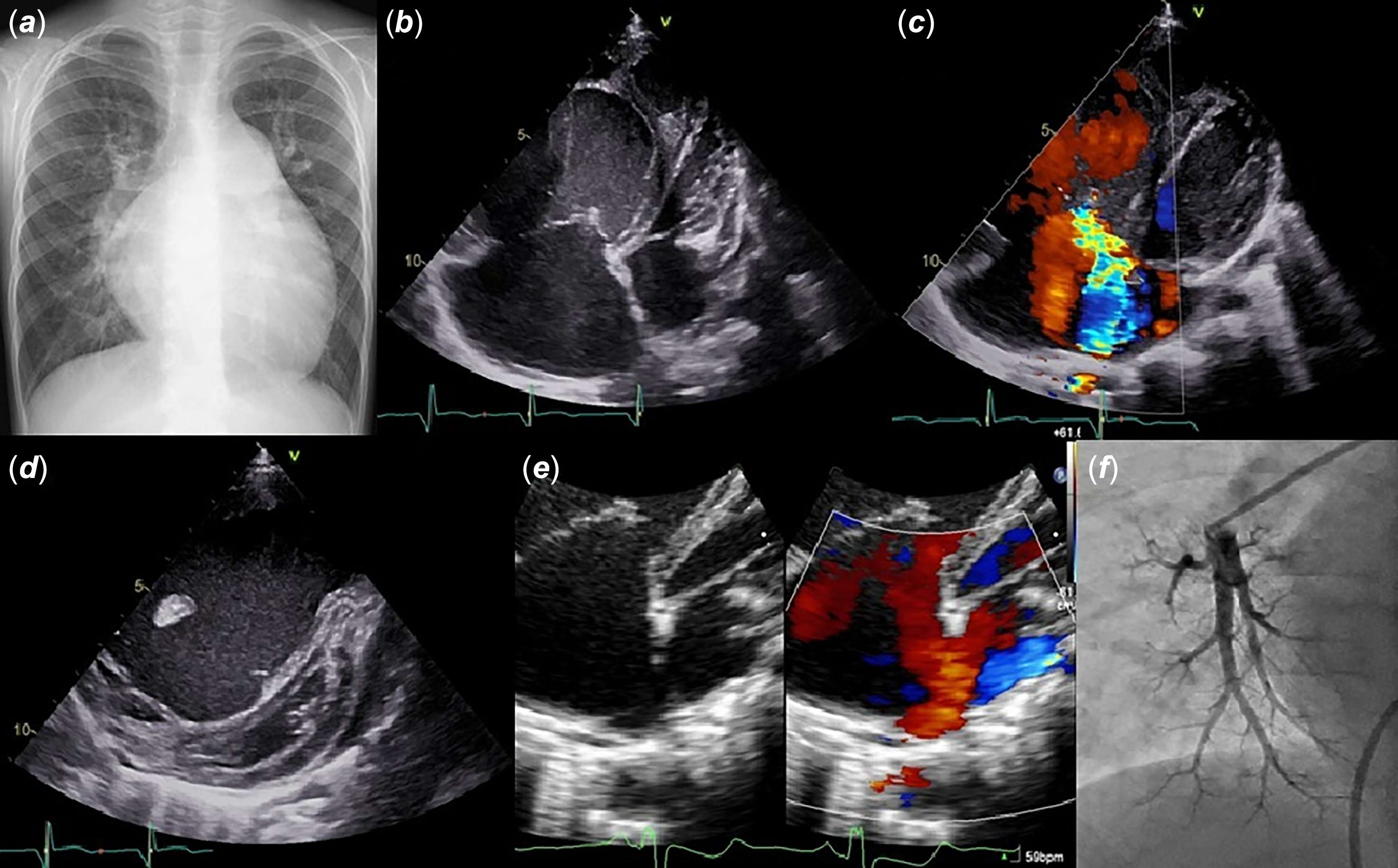
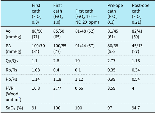
A 5-year-old boy, born at term with normal birth weight, required neonatal mechanical ventilation for respiratory distress but was discharged at 2 weeks old. A heart murmur was detected at 1 year old; atrial septal defect was identified and managed with observation. The patient remained asymptomatic until 1 year prior to presentation, when he developed exertional fatigue, followed by generalised oedema 9 months later. He was diagnosed with partial anomalous pulmonary venous connection and pulmonary arterial hypertension at a local clinic, where a diuretic and digoxin were initiated before referral to our hospital. Physical examination revealed no rales or signs indicating respiratory distress. No jugular venous distension, hepatomegaly, or peripheral oedema was observed. Laboratory tests revealed an elevated brain natriuretic peptide level (866 pg/mL), with no other significant abnormalities. Chest radiography revealed a cardiothoracic ratio of 60%, prominent left second arch, and pulmonary congestion (Figure 1(a)). Echocardiography revealed partial anomalous pulmonary venous connection of the right upper and lower pulmonary veins with a sinus venosus atrial septal defect (10 mm), severe tricuspid and pulmonary regurgitation, marked right heart enlargement, and severe pulmonary arterial hypertension (tricuspid regurgitation pressure gradient, 70 mmHg; Figure 1(b)–(e)). No significant CT or lung perfusion imaging findings were observed. Owing to the severe pulmonary hypertension with over-systemic pressure, immediate catheterisation was deemed high risk. A cautious treatment strategy of gradually introducing sildenafil with close monitoring for pulmonary congestion and heart failure was adopted. On day 37, catheterisation (fraction of inspired oxygen, 0.3) showed a pulmonary blood flow/systemic blood flow ratio, mean pulmonary artery pressure, and pulmonary vascular resistance index of 1.1, 84 mmHg, and 10.8 Wood units·m, Reference Hayashi, Hasegawa and Tanaka2 respectively. With oxygen loading (fraction of inspired oxygen, 1.0), the pulmonary blood flow/systemic blood flow ratio, mean pulmonary artery pressure, and pulmonary vascular resistance index (calculated using the Fick method with consideration of dissolved oxygen) were 2.8, 77 mmHg, and 2.77 Wood units·m, Reference Hayashi, Hasegawa and Tanaka2 respectively; with oxygen and nitric oxide (fraction of inspired oxygen 1.0 + nitric oxide 20 ppm), these values were 10, 67 mmHg, and 0.56 Wood units·m, Reference Hayashi, Hasegawa and Tanaka2 respectively (Table 1). Following sequential introduction of pulmonary vasodilators (sildenafil, 1.6 mg/kg; macitentan, 0.16 mg/kg; selexipag, 0.06 mg/kg), a calcium channel blocker was initiated based on acute vasoreactivity testing. On day 91, after introducing these four agents, catheterisation showed improved pulmonary vascular resistance (pulmonary blood flow/systemic blood flow ratio, 2.7; pulmonary artery pressure, 58 mmHg; pulmonary vascular resistance index, 3.5 Wood units·m), Reference Hayashi, Hasegawa and Tanaka2 prompting surgical planning. On day 102, fenestrated atrial septal defect closure and partial anomalous pulmonary venous connection repair were performed without pulmonary hypertensive crisis. On day 127, wedge angiography showed poor pulmonary vascular formation bilaterally (Figure 1(f)) (pulmonary blood flow/systemic blood flow ratio, 1.1; pulmonary artery pressure, 27 mmHg; pulmonary vascular resistance index, 4.0 Wood units·m). Reference Hayashi, Hasegawa and Tanaka2 The 6-min walk distance improved from 437 m at admission to 525 m postoperatively, with resolution of exertional dyspnoea. Genetic testing for heritable pulmonary hypertension, including known causative genes such as BMPR2 and ACVRL1, revealed no pathogenic mutations.
( a ) Chest radiography showed a cardiothoracic ratio of 60%, prominent left second arch, and pulmonary congestion. Echocardiography showed ( b ) marked right heart enlargement, ( c ) severe tricuspid regurgitation, ( d ) compressed interventricular septum at end-systole reflecting severe pulmonary arterial hypertension, and ( e ) partial anomalous pulmonary venous connection of the right upper pulmonary vein with a sinus venosus atrial septal defect of 10 mm. ( f ) Pulmonary artery wedge angiography showed a poor pulmonary vascular formation.

Cardiac catheterisation data

cath = catheterisation; FiO2 = fraction of inspired oxygen; ope = operation; Ao = aorta; PA = pulmonary artery; Qp/Qs = pulmonary blood flow/systemic blood flow ratio; Rp/Rs = pulmonary vascular resistance/systemic vascular resistance ratio; Pp/Ps = pulmonary pressure/aortic pressure ratio; PVRI = pulmonary vascular resistance index; SaO2 = saturation of arterial oxygen.
Discussion
Coexisting partial anomalous pulmonary venous connection and atrial septal defect can lead to early-onset severe pulmonary hypertension. In our case, sequential combination therapy—with multiple pulmonary vasodilators—enabled safe surgery, and the treat-and-repair strategy improved exercise tolerance.
Surgical intervention during infancy is uncommon in patients with isolated atrial septal defect. Symptoms arising from interatrial shunting or pulmonary hypertension typically emerge only with comorbidities, such as genetic syndromes, pulmonary lesions, chronic lung disease, or left ventricular diastolic dysfunction. Reference Charisopoulou, Bini and Riley1,Reference Hayashi, Hasegawa and Tanaka2 Partial anomalous pulmonary venous connection is often asymptomatic in children but should be suspected with unexplained right ventricular enlargements on echocardiography. Reference Jin, Wu and Wang3 Clinical symptoms are more likely with >50% anomalous pulmonary veins or coexisting conditions—such as atrial septal defect. Approximately 80% of cases are associated with sinus venosus atrial septal defect, Reference Fragata, Magalhães and Baquero4 especially right-sided and multiple partial anomalous pulmonary venous connections, whereas secundum septal defects are often associated with bilateral partial anomalous pulmonary venous connection. Reference Hatipoglu, Almogheer and Mahon5 MRI studies showed that a pulmonary blood flow/systemic blood flow ratio ≥2 is more common in right- than in left-upper partial anomalous pulmonary venous connections, possibly owing to lower pressure gradients in left-sided connections, which often drain into the innominate vein. Reference Seller, Yoo and Grant6 In right-sided multiple partial anomalous pulmonary venous connections and sinus venosus atrial septal defect cases, the pulmonary blood flow/systemic blood flow ratio is always ≥1.5 and averages 2.5. Reference Hatipoglu, Almogheer and Mahon5 In our case, prolonged exposure to high pulmonary blood flow likely led to pulmonary hypertension progression. Severe pulmonary hypertension due to partial anomalous pulmonary venous connection is rare (<5%) and associated with genetic predisposition, additional CHDs, anomalous veins, comorbidities including sickle cell disease, and pulmonary vascular bed involvement. Reference Pizzini, Sonnweber and Frank7 In this patient, extensive evaluation revealed no underlying causes other than CHD. Postoperative pulmonary artery wedge angiography showed poor bilateral pulmonary vascular formation, suggesting hereditary pulmonary hypertension. However, genetic testing failed to detect known pulmonary hypertension-associated mutations.
We initially deemed our case inoperable owing to severe pulmonary hypertension. Surgical indications for partial anomalous pulmonary venous connection include right ventricular enlargement, poor weight gain, respiratory symptoms, pulmonary blood flow/systemic blood flow ratio ≥1.5, pulmonary artery systolic pressure <50% of systemic pressure, and pulmonary vascular resistance <1/3 of systemic vascular resistance. Reference Jin, Wu and Wang3,Reference Choi, Greathouse and Darki8 To safely perform surgery, we initiated sequential combination therapy with multiple pulmonary vasodilators prior to surgery. Although epoprostenol was considered, the oral selective prostacyclin receptor agonist selexipag was selected because of its favourable administration route. In conjunction with three pulmonary vasodilators, a calcium channel blocker was initiated according to positive acute vasoreactivity testing. Reference Douwes, Loon and Hoendermis9 We considered titration of pulmonary vasodilators necessary to avoid worsening pulmonary congestion or heart failure, with careful monitoring of brain natriuretic peptide levels and pulmonary blood flow/systemic blood flow ratio by echocardiography and chest radiography findings. Although clinical and haemodynamic parameters have improved with combination therapy for severe pulmonary hypertension associated with partial anomalous pulmonary venous connection, Reference Pizzini, Sonnweber and Frank7,Reference El-Kersh, Homsy and Daniels10 no previous reports on severe pulmonary hypertension requiring a treat-and-repair strategy at a young age exist.
Conclusion
The treat-and-repair strategy involving sequential combination medical therapy, followed by definitive surgical repair, demonstrated successful outcomes in a patient with partial anomalous pulmonary venous connections and atrial septal defect complicated by severe pulmonary hypertension.
Acknowledgements
We would like to thank Editage (https://www.editage.jp/) for performing an English language review.
Financial support
This research received no specific grants from any funding agency, commercial, or not-for-profit sectors.
Competing interests
The authors declare none.
Ethical standard
This case report did not involve human and/or animal experimentation. The study protocol was approved by the Ethics Committee of Osaka Women’s and Children’s Hospital.