Introduction
Cognitive problems following breast cancer (BC) treatment received increasing attention over the past decade. Particularly after adjuvant chemotherapy (CT), BC patients frequently report cognitive problems (Poppelreuter et al., Reference Poppelreuter, Weis, Külz, Tucha, Lange and Bartsch2004; Pullens, De Vries, & Roukema, Reference Pullens, De Vries and Roukema2010). Cognitive decline relative to pre-treatment cognitive functioning (Ahles et al., Reference Ahles, Saykin, McDonald, Li, Furstenberg, Hanscom and Kaufman2010; Jansen, Cooper, Dodd, & Miaskowski, Reference Jansen, Cooper, Dodd and Miaskowski2011; Jenkins et al., Reference Jenkins, Shilling, Deutsch, Bloomfield, Morris, Allan and Winstanley2006) was observed in numerous prospective studies, and several studies report cognitive impairment up to 20 years after treatment (Collins, Mackenzie, Tasca, Scherling, & Smith, Reference Collins, Mackenzie, Tasca, Scherling and Smith2013; de Ruiter et al., Reference De Ruiter, Reneman, Boogerd, Veltman, van Dam, Nederveen and Schagen2011; Koppelmans, Breteler, et al., Reference Koppelmans, Breteler, Boogerd, Seynaeve, Gundy and Schagen2012; Vearncombe et al., Reference Vearncombe, Rolfe, Wright, Pachana, Andrew and Beadle2009; Wefel, Saleeba, Buzdar, & Meyers, Reference Wefel, Saleeba, Buzdar and Meyers2010). The incidence of cognitive problems following chemotherapy varies considerably with estimates ranging from 20 to 70% (Wefel & Schagen, Reference Wefel and Schagen2012). The most commonly affected domains include processing speed, memory, and executive function (Ahles, Root, & Ryan, Reference Ahles, Root and Ryan2012; Wefel & Schagen, Reference Wefel and Schagen2012).
Additionally, researchers have started to reveal potential neural substrates of this decline in cognitive functioning. Magnetic resonance imaging (MRI) studies reported reductions in brain gray matter volume and white matter microstructure in BC patients within a few months to many years post CT (de Ruiter et al., Reference De Ruiter, Reneman, Boogerd, Veltman, Caan, Douaud and Schagen2012; Koppelmans, de Ruiter, et al., Reference Koppelmans, de Ruiter, van der Lijn, Boogerd, Seynaeve, van der Lugt and Schagen2012). Functional MRI (fMRI) studies showed that brain activation during cognitive performance was altered in BC patients 1 month up to 10 years after completion of treatment (Conroy et al., Reference Conroy, McDonald, Smith, Moser, West, Kamendulis and Saykin2013; de Ruiter et al., Reference De Ruiter, Reneman, Boogerd, Veltman, van Dam, Nederveen and Schagen2011; Kesler, Bennett, Mahaffey, & Spiegel, Reference Kesler, Bennett, Mahaffey and Spiegel2009; Kesler, Kent, & O’Hara, Reference Kesler, Kent and O’Hara2011; McDonald, Conroy, Ahles, West, & Saykin, Reference McDonald, Conroy, Ahles, West and Saykin2012).
Given longer survival of BC patients, better insight into the long-term adverse effects of CT becomes increasingly relevant. The present study focuses on the long-term effects of cancer treatment on cognitive performance and brain function in BC patients, with a specific focus on dose and treatment strategies.
In a previous study by our group, task-specific hyporesponsiveness and concomitant reduction in task performance was found 10 years post-treatment in BC survivors treated with high-dose adjuvant chemotherapy (HI-CT) compared to patients treated with radiotherapy only (RT-only) (de Ruiter et al., Reference De Ruiter, Reneman, Boogerd, Veltman, van Dam, Nederveen and Schagen2011). Bilateral hypoactivation of posterior parietal cortex (PPC) was found both during an executive functioning task and a memory-encoding task. Furthermore, HI-CT showed task-specific hypoactivation of dorsolateral prefrontal cortex (DLPFC) and hippocampus, respectively. In addition, worse cognitive performance was found in the HI-CT group compared to the RT-only group. These findings suggest long-term adverse effects on cognition and neural function in brain areas that support executive function and episodic memory. However, whether these long-term adverse effects were specifically related to Hi-CT exposure remains unclear. To investigate this, we extended our measurements in the present study to BC survivors who received conventional dose (CON-) CT. All BC survivors who received CT previously participated in a trial that randomly assigned patients to either adjuvant HI-CT or CON-CT. Since these groups are highly homogeneous in terms of disease stage and disease grade, we were able to compare these cytotoxic regimens in a unique manner. Additionally, we compared these CT regimens with a cancer specific (RT-only) control group to examine the additional effect of CT. Finally, we included a healthy control group (HC). By comparing HC to RT-only we were able to investigate residual negative effects on cognition and altered brain function due to other aspects of BC (treatment) not related to CT, such as disease-specific factors (Phillips et al., Reference Phillips, Jim, Small, Laronga, Andrykowski and Jacobsen2012).
Methods
Participants
All BC survivors were recruited from the Netherlands Cancer Institute, VU University Medical Center, Leiden University Medical Center and the Erasmus University Medical Center-Daniel den Hoed Cancer Center. The review board of the Netherlands Cancer institute served as the central ethical committee for all participating hospitals and approved the study. This research was completed in accordance with the Helsinki Declaration.
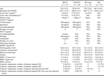
BC survivors who received adjuvant CT were diagnosed as high-risk patients and participated in a multicenter randomized trial comparing the efficacy of adjuvant HI-CT to CON-CT (Rodenhuis et al., Reference Rodenhuis, Bontenbal, Beex, Wagstaff, Richel, Nooij and de Vries2003) (details on disease stage and CT regimen are provided in Table 1). BC survivors who did not require CT had undergone locoregional surgery and RT. HCs were recruited among female friends and family of BC survivors. In an earlier neuropsychological study from our group, all study participants were evaluated within 2 years after treatment (Schagen, Muller, Boogerd, Mellenbergh, & van Dam, Reference Schagen, Muller, Boogerd, Mellenbergh and van Dam2006). The study inclusion/exclusion criteria are identical to our previous MRI study (de Ruiter et al., Reference De Ruiter, Reneman, Boogerd, Veltman, van Dam, Nederveen and Schagen2011). Written informed consent was obtained from all participants. The total sample consisted of 19 HI-CT, 24 CON-CT, 15 RT-only BC survivors and 27 HC. More detailed information on subject attrition can be found in our previous study (Stouten-Kemperman et al., Reference Stouten-Kemperman, de Ruiter, Koppelmans, Boogerd, Reneman and Schagen2014).
Characteristics of study population and results of cognitive assessment

Note. Values indicate mean (SD) unless specified otherwise. HI-CT=high-dose chemotherapy (CT); CON-CT=conventional-dose CT; RT-only=radiotherapy-only; HC=healthy controls. CON-CT and HI-CT were followed by radiotherapy and 2-5 years of tamoxifen treatment.
a HI-CT<CON-CT; HI-CT>RT-only; CON-CT>RT-only.
b HI-CT<CON-CT.
c RT-only<HC.
d One patient from the RT-only group was treated with tamoxifen for 5 years. M1: baseline measurement, up until 2 years after chemotherapy; M2: measurement ≥10 years after chemotherapy. EORTC QLQ-C30, European Organization for Research and Treatment of Cancer health-related Quality-of-Life Questionnaire (a higher score indicates better functioning, except for fatigue); HSCL-25, Hopkins Symptom Checklist (a higher score indicates worse functioning).
*p<.05.
Assessment Procedure
The experimental procedure lasted ~2.5 hr per participant. First, the neuropsychological test battery was administered, comprising the same battery of seven neuropsychological tests as was used in our earlier study (de Ruiter et al., Reference De Ruiter, Reneman, Boogerd, Veltman, van Dam, Nederveen and Schagen2011). The neuropsychological test battery consisted of the Trail Making Test A and B, Digit Symbol-Coding Test of the Wechsler Adult Intelligence Scale (WAIS)-III, number of correctly completed items, Stroop Color-Word Test, Dutch version of the California Verbal Learning Test (CVLT), Visual Reproduction Test of the Wechsler Memory Scale-Revised (WMS-R), immediate, Word Fluency Test, number of animals and number of professions and Fepsy Finger Tapping Test.
We investigated symptoms of anxiety and depression with the Hopkins Symptoms Checklist-25 (HSCL) (Hesbacher, Rickels, Morris, Newman, & Rosenfeld, Reference Hesbacher, Rickels, Morris, Newman and Rosenfeld1980) and health-related quality of life with the European Organization for Research and Treatment of Cancer (EORTC) Quality of Life Questionnaire-C30 (Aaronson et al., Reference Aaronson, Ahmedzai, Bergman, Bullinger, Cull, Duez and de Haes1993). After a short break, the MRI scanning session took place.
MR Imaging and Data Processing
Participants were scanned on a 3.0 Tesla Intera MRI scanner (Philips Medical Systems, Best, The Netherlands). The scan protocol, scanner configuration and scanner hardware were identical to that of our previous studies (de Ruiter et al., Reference De Ruiter, Reneman, Boogerd, Veltman, van Dam, Nederveen and Schagen2011, Reference De Ruiter, Reneman, Boogerd, Veltman, Caan, Douaud and Schagen2012). A sagittal T1 weighted spoiled gradient echo scan and T2*-weighted echo planar images (EPIs) were acquired. A sagittal T1-weighted spoiled gradient echo scan of 170 slices was acquired (repetition time/echo time [TR/TE]=9/3.53 ms, field of view [FOV] 232×256 mm, voxel size 1×1×1 mm3). For the fMRI runs, T2*-weighted echo planar images (EPIs) sensitive to the blood oxygen level dependent (BOLD) signal were obtained, containing 35 axial slices (TR/TE=2/25 ms, FOV 96×96 mm, voxel size 2.3×2.3×3 mm3). For consistency, the method of preprocessing and modeling of fMRI data was identical to our previous study (de Ruiter et al., Reference De Ruiter, Reneman, Boogerd, Veltman, van Dam, Nederveen and Schagen2011). Imaging data were processed and analyzed using SPM5 (Statistical Parametric Mapping; Wellcome Trust Centre for Neuroimaging, London, UK). After manual reorientation to the anterior commissure, EPIs were slice-timed, realigned and coregistered to the T1 image. Next, T1-images and the coregistered EPIs were normalized to the SPM T1 template. EPIs were spatially smoothed with an 8 mm full-width-half-maximum (FWHM) kernel.
From the fMRI time series contrast images were created for each participant, which were used in random effects analyses to identify voxels where brain activation differed between groups.
fMRI Paradigms
Tower of London
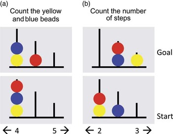
For the Tower of London (ToL) paradigm, an abbreviated (8 min) version of Van den Heuvel et al. (Reference Van den Heuvel, Groenewegen, Barkhof, Lazeron, van Dyck and Veltman2003) was used, identical to our previous study (de Ruiter et al., Reference De Ruiter, Reneman, Boogerd, Veltman, van Dam, Nederveen and Schagen2011). The ToL included two conditions. In both conditions, images of three colored beads (red, blue, yellow) placed on three vertical rods of decreasing height were presented (Figure 1). During the planning condition, a start configuration and a final target configuration were simultaneously displayed. Participants were instructed to count the minimum number of steps required to get from the start to the final target configuration, with the restriction that beads could only be moved one at a time. Two response options were displayed on the bottom of the screen (range: 1–5 moves). In the baseline condition, participants had to count to total number of yellow and blue beads. Again, two possible answers were presented. No feedback was provided regarding the correct answers Trials were self-paced, with a maximum response time of 60 s, and were presented in pseudorandom order. Before the scanning session the ToL was practiced outside the scanner. Participants were encouraged to focus on accuracy rather than speed.
Tower of London (ToL). (a) Baseline condition; (b) planning condition. In the baseline condition, participants had to count to total number of yellow and blue beads. (b) During the planning condition, participants were instructed to count the minimum number of steps required to get from the start to the final target configuration. In both conditions, two response options were displayed on the bottom of the screen (range: 1–5).

Paired Associates learning
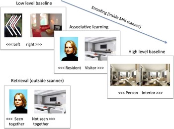
A Paired Associates (PA) memory encoding task was used that was identical to the task in our previous study (de Ruiter et al., Reference De Ruiter, Reneman, Boogerd, Veltman, van Dam, Nederveen and Schagen2011). This task consisted of three conditions (Figure 2). In the memory encoding condition, participants were asked to indicate whether they thought a person depicted on a portrait image was likely to live in a home of which the interior was depicted on a simultaneously presented photo, and to memorize this combination. They had to press the left or right button to indicate “resident” or “visitor.” In the high-level baseline condition participants were shown two identical portrait photos or interior design photos of which they had to indicate whether this image represented a person or interior design. In the low-level baseline condition participants were cued to press the left or right button according to the direction of three arrowheads presented on the screen. These were superimposed on a blurred portrait and interior design photo to match the visual input of the associative learning condition. The high level baseline was included to isolate memory encoding processes specifically related to associative learning. The low level baseline was included to capture additional memory encoding processes (such as novelty detection) that are not related to associative learning.
Paired Associates (PA) memory task. Participants had to press a button according to the direction of the arrows (low-level baseline) or indicate whether the picture represented a person or an interior design (high-level baseline). During associative learning participants indicated whether the depicted person was likely to live in the depicted interior. During the retrieval (outside the scanner) participants indicated whether they had seen the specific stimulus pair or not.

Each trial lasted 3 s (baseline conditions) or 7 s (memory encoding) and was followed by a white screen for 1 s. Six trials were presented per block and the sequence of three task blocks was repeated twice, so that overall task duration was 5 min 42 s. After the MRI scanning session, subjects performed a self-paced retrieval task during which they had to recognize specific combinations from the memory-encoding task (50% new combinations, 50% presented during memory encoding). Both the encoding and retrieval task were practiced outside the scanner.
Statistical Analysis
Demographic variables and patient-related outcomes were analyzed with IBM SPSS Statistics 20 (IBM, Armonk, NY), by means of analysis of variance (ANOVA). The reported neurocognitive data is collected longitudinally, while imaging data is collected cross-sectionally. Time point 1 (M1) neurocognitive data for all groups are collected from the neuropsychological assessment of our previous study that took place within 2 years after CT (Schagen et al., Reference Schagen, Muller, Boogerd, Mellenbergh and van Dam2006). For time point 2 (M2) neurocognitive data, HI-CT and RT-only data are collected from de Ruiter et al., Reference De Ruiter, Reneman, Boogerd, Veltman, van Dam, Nederveen and Schagen2011 as well as the imaging data for HI-CT and RT-only groups. CON-CT and HC M2 neurocognitive data and imaging data are collected as part of the present study.
Cognitive Impairment
We identified cognitive impairment based on frequently used cutoff scores. Therefore, each raw neuropsychological test score was converted into a standard score (Z-score) by using the mean test scores of the HC group as a reference. More information on raw neuropsychological scores can be found in Supplementary Table 1. Having a Z-score larger than −1.95 (2 SDs below the mean) was considered as being impaired on that specific test. An overall impairment score was calculated for each participant by taking the sum of tests on which they were impaired. Finally, to calculate the proportion of participants who were impaired, the fifth percentile of the overall impairment score (the total number of tests scored in the impaired range) of the HC group was taken as a cutoff score for neuropsychological impairment.
Furthermore, we investigated relative and absolute cognitive change over time between M1 and M2. To investigate relative cognitive change over time between M1 and M2, we subtracted the absolute decline (number of tests on which the participant deteriorated, defined as a decline of ≥1 SD) from the absolute improvement (number of tests on which the participant improved, defined as improvement of ≥1 SD). Our criteria for relative cognitive decline were stringent, since we considered a subject to have deteriorated only when having a relative decline in performance on least 3 tests (the 95th percentile of the HCs, including correction for practice effects which were based on the test–retest scores and standard deviations of the HC). Differences in proportion of impaired patients and differences between groups on decline and improvement were tested using logistic regression and χ2-tests.
Separate analyses per group were carried out on predictors of cognitive impairment. Potential predictors included were age, premorbid IQ, time since surgery, cognitive, emotional and physical functioning, and fatigue as measured with the EORTC QLQ-C30 questionnaire on M1, anxiety and depression as measured with the HSCL on M1 and the number of tests on which a participant scored in the impaired range on M1.
fMRI Analyses
fMRI preprocessing and modeling was identical to our previous study analyses (de Ruiter et al., Reference De Ruiter, Reneman, Boogerd, Veltman, van Dam, Nederveen and Schagen2011). fMRI performance data were analyzed by means of ANOVA. Analyses of reaction times were performed on correct answers. All fMRI time-series group-interactions were analyzed with two-sample t tests and were regarded significant at p<.001. Main task effects are reported at p<.05, whole-brain FDR corrected. Group interactions were masked with the appropriate main effect across groups at p<.05 to reduce the search volume to those voxels showing a main effect of task, and are reported at p<.001 with a cluster size threshold of 10 voxels. Region-of-interest (ROI) analyses of DLPFC and hippocampal area are reported at p<.005 with a cluster size threshold of 5. See de Ruiter et al. (Reference De Ruiter, Reneman, Boogerd, Veltman, van Dam, Nederveen and Schagen2011) for details on ROI masks. ROI analyses for the associations between performance and BOLD activation were performed separately for all groups. Age and estimated premorbid IQ were used as covariates in all our analyses.
Results
Demographic and Clinical Data
Characteristics of all participants and patient-related outcomes on cognition, fatigue, anxiety and depression are presented in Table 1. No significant differences were found between groups on age and estimated premorbid IQ. There was a significant overall difference between groups in time interval between surgery and assessment (F3,82=222.84; p<.001), reflecting earlier recruitment of the HI-CT and RT-only group in our previous study. The RT-only group scored significantly lower on self-reported cognitive functioning (as measured with the EORTC subscale “cognitive functioning”) than the HC group (F1,42=7.10; p=.011), indicating more problems with cognitive functioning in the RT-only group. No other significant differences were found between groups on measures of quality of life, depression, or anxiety.
Cognitive Test Performance
On the previous neuropsychological assessment (M1), 26.3% (n=5) of the HI-CT patients demonstrated impairments in cognitive functioning within 2 years after chemotherapy, compared to 16.7% (n=4) of the CON-CT, 0% of the RT patients, and 3.7% (n=1) of the HC group (Table 1). More than 10 years after chemotherapy (M2), the proportion of patients with late cognitive impairment was 26.3% (n=5), 12.5% (n=3), 0% and 3.7% (n=1), respectively. Group differences in the proportion of impaired participants did not reach statistical significance for both assessments. Sixty percent of the HI-CT (n=3) and 66.7% of the CON-CT patients (n=2) who demonstrated late cognitive impairment on M2 were also identified as cognitively impaired on M1, thereby showing stable cognitive dysfunction.
Comparing M1 and M2, we found that relative cognitive decline was present in 10.5% (n=2) of the HI-CT patients, 8.3% (n=2) of the CON-CT, and 6.7% (n=1) of the RT-only patients, whereas no cognitive decline was found in the HC group. Absolute cognitive decline (declining on more than three tests regardless of improvement) revealed identical [10.5% (n=2) of the HI-CT patients, 8.3% (n=2) of the CON-CT and 6.7% (n=1) of the RT-only patients, no decline in HC] percentages. Relative cognitive improvement was present in 5.3% (n=1) of the HI-CT patients, 4.2% (n=1) of the CON-CT, 14.3% (n=2) of the RT-only patients, and 3.7% (n=1) of the HC. Absolute cognitive improvement (improvement on more than 3 tests regardless of decline) was present in 10.5% (n=2) of the HI-CT patients, 4.2% (n=1) of the CON-CT, 20% (n=3) of the RT-only patients and 3.7% (n=1) in HC. Group differences were not significant.
For the HI-CT group, age (β=.61; t(8)=3.36; p=.01), time since surgery (β=.50; t(8)=2.61; p=.03) and physical functioning (β=.62; t(8)=2.70; p=.027) on M1 were predictors that were positively related to the number of tests on which a patient scored impaired on M2. Premorbid IQ was a predictor that was negatively related (β=−.54; t(8)=−2.77; p=.024) to the number of tests on which a patient scored impaired on M2 in this group.
For the CON-CT group, the number of tests on which a patient scored impaired on M1 was positively related to the number of impaired tests on M2 (β=−.56; t(13)=2.22; p=.045) meaning that impairment at M1 was predictive of impairment at M2. For the RT-only group and HC, no significant predictive variables were found.
Tower of London
Performance
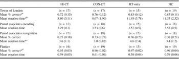
The Hi-CT group performed significantly worse on the planning condition of the ToL compared to the RT-only group (F 1,28=4.83; p=.036). Furthermore, both the Hi-CT (F 1,31=5.55; p=.026) and CON-CT (F 1,28=12.81; p<.001) group responded significantly faster than the RT-only group (Table 2).
fMRI task performance for Tower of London, Paired Associates encoding and recognition, and Flanker test

Values indicate mean (SD) unless specified otherwise. HI-CT=high-dose chemotherapy (CT); CON-CT=conventional-dose CT; RT-only=radiotherapy-only; HC=healthy controls.
a HI-CT<RT-only
b HI-CT<RT-only; CON-CT<RT-only
c HI-CT>CON-CT; RT-only>CON-CT; RT-only>HC
d HI-CT<RT-only
*p<.05.
fMRI
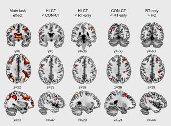
Imaging results for the TOL across groups showed significant BOLD activation in dorsolateral prefrontal cortex (DLPFC), ventrolateral prefrontal cortex (VLPFC), premotor cortex, precuneus and posterior parietal cortex (PPC) (Figure 3).
Tower of London. Main task effect and group comparisons (BOLD activations) for the Active>Baseline contrast (shown at p<.005). HI-CT: high-dose chemotherapy (CT); CON-CT: conventional-dose CT; RT-only: radiotherapy-only, HC: healthy controls.

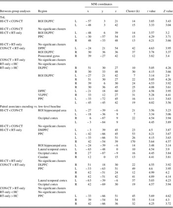
The HI-CT versus CON-CT group comparison showed hypoactivation of the HI-CT group in left DLPFC (ROI analysis). The HI-CT versus RT-only comparison demonstrated hypoactivation for the HI-CT group in bilateral PPC (whole-brain analyses) and right DLPFC (ROI analysis). The CON-CT versus RT-only comparison revealed hypoactivation for the CON-CT group in dorsal prefrontal cortex (DPFC), postcentral gyrus (whole-brain analysis), and right DLPFC (ROI analysis). Finally, the RT-only versus HC group comparison revealed hyperactivation in the RT-only group in DLPFC, DPFC, VLPFC, and PPC (whole-brain analysis) and DLPFC (ROI analysis) (Figure 3; Table 3).
fMRI between-group analyses for Tower of London and Paired Associates encoding

Note. HI-CT=high-dose chemotherapy (CT); CON-CT=conventional-dose CT; RT-only - radiotherapy-only; HC=healthy controls.
Paired Associates
Performance
Recognition memory performance was marginally worse for the HI-CT group versus the CON-CT group (F 1,30=3.64; p=.066) and the RT-only group (F 1,28=3.26; p=.081) (Table 2). The HI-CT group responded slower than the CON-CT group (F 1,31=12.29; p=.001). Furthermore, the RT-only group also responded slower than the CON-CT group (F 1,29=13.71; p=.001) and the HC group (F 1,29=5.44; p=.027). During memory encoding, the HI-CT group responded marginally faster than the RT-only group (F 1,28=3.74; p=.063).
fMRI
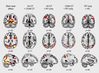
Across groups, the fMRI results showed a significant main task-effect for the encoding versus low-level baseline contrast in the ventral stream (occipital areas, fusiform gyrus) extending into parahippocampal gyrus and hippocampus proper (Figure 4). The HI-CT versus CON-CT comparison revealed hypoactivation in the HI-CT group in right occipital cortex (whole-brain analysis), and the left hippocampal area (ROI analysis) (Table 3; Figure 4). Compared to the RT-only group, the HI-CT group showed hypoactivation in the left dorsal medial prefrontal cortex (DMPFC), bilateral PPC, left lateral temporal cortex and right occipital cortex, caudate nucleus (whole-brain analysis), and the left hippocampal area (ROI analysis). Compared to RT-only, the CON-CT group showed hypoactivation in right DLPFC, bilateral PPC, left lateral temporal cortex, and right occipital cortex (whole-brain analysis). Significant hyperactivation for the RT-only group compared to the HC group was found in bilateral PPC (whole-brain analysis).
Main task effect and group comparisons for BOLD activations for the encoding>low-level baseline contrast (shown at p<.005). HI-CT: high-dose chemotherapy (CT); CON-CT: conventional-dose CT; RT-only: radiotherapy-only, HC: healthy controls.

Relation between Task Performance and BOLD
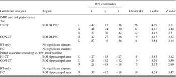
ROI regression analyses of the ToL with task-performance showed a positive association between task performance and brain activation in DLPFC in the HI-CT and CON-CT groups, but not in the RT-only and HC groups (Table 4).
fMRI ROI regression analyses for BOLD with task performance for Tower of London and Paired Associates encoding

Note. HI-CT=high-dose chemotherapy (CT); CON-CT=conventional-dose CT; RT-only=radiotherapy-only; HC=healthy controls.
For the paired associates task, ROI regression analyses showed a positive association between recognition memory performance outside the scanner and brain activation in the hippocampal area (Table 4) during encoding in the HI-CT, CON-CT, and HC groups.
Discussion
To our knowledge, this is the first study to show that very late side effects of adjuvant CT for BC on brain function depend on the specific cytotoxic regimen administered. In the present study, we compared HI-CT to CON-CT, HI-CT and CON-CT to RT-only, and RT-only to HC. These group comparisons were made based on the findings of our previous study, which showed treatment-dependent differences in white matter integrity and cognitive impairment (Stouten-Kemperman et al., Reference Stouten-Kemperman, de Ruiter, Koppelmans, Boogerd, Reneman and Schagen2014).
Worse cognitive functioning and worse performance on a planning task in both CT groups was observed, which could be explained by hypoactivation in task-related brain areas. The late effects were stronger after a more intense type of CT, containing an additional high dose of systemic compounds. Furthermore, this study adds to previous studies by showing the presence of stable cognitive dysfunction in patients treated with CT≥10 years earlier. Previous cross-sectional studies have shown late effects of CT on cognitive impairment (Koppelmans, Breteler, et al., Reference Koppelmans, Breteler, Boogerd, Seynaeve, Gundy and Schagen2012; Nguyen et al., Reference Nguyen, Yamada, Beglinger, Cavanaugh, Denburg and Schultz2013; Yamada, Denburg, Beglinger, & Schultz, Reference Yamada, Denburg, Beglinger and Schultz2010). However, studies with multiple measurements over a prolonged period of time, such as we present here, are scarce.
Task performance and fMRI data showed a coherent pattern of results for both executive function and episodic memory, as measured with the Tower of London and the Paired Associates Task (PAT). Task-related hypoactivation associated with worse performance was present in both tasks in cancer survivors who received CT, supporting the notion that treatment with cytotoxic regimens may be related to long-term failure of recruitment of relevant brain regions to perform well on such a task. Task-related hypoactivation in patients treated with CT was previously found (Conroy et al., Reference Conroy, McDonald, Smith, Moser, West, Kamendulis and Saykin2013; Kesler et al., Reference Kesler, Bennett, Mahaffey and Spiegel2009, Reference Kesler, Kent and O’Hara2011), 3 to 6 years post-treatment. Similar to the present study, hypoactive areas that were found included prefrontal and parietal areas during memory encoding, executive functioning and a working memory task. However, these studies are not fully comparable to the current study since different comparison groups were used. Nevertheless, the current study shows that task-related hypoactivation is found in patients late after cancer treatment/diagnosis, which is in line with the previous literature (de Ruiter & Schagen, Reference De Ruiter and Schagen2013).
The more intense hyporesponsiveness of task-specific brain regions after HI-CT compared to CON-CT may be driven by both substance-dependent and dose-dependent effects. While the CON-CT group received 5 cycles of FEC treatment, the fifth cycle was replaced by high-dose CTC in the HI-CT group. Since this cycle contains a 12-fold increase in cyclophosphamide, it may have caused an increase in neurotoxicity in the HI-CT versus CON-CT group. Indeed, preclinical studies have shown dose-dependent neurotoxicity of cyclophosphamide, for instance in the cortex and hippocampus (Dietrich, Monje, Wefel, & Meyers, Reference Dietrich, Monje, Wefel and Meyers2008), being in line with our findings of adverse effects in similar brain regions.
Furthermore, the (high) dosages of carboplatin and/or thiotepa that were incorporated in the CTC cycle may have more severe adverse effects, as shown in preclinical studies (Seigers, Schagen, Van Tellingen, & Dietrich, Reference Seigers, Schagen, Van Tellingen and Dietrich2013). Cyclophosphamide, 5-FU, and thiotepa affect neurogenesis and gliogenesis (Husain, Whitworth, Hazelrigg, & Rybak, Reference Husain, Whitworth, Hazelrigg and Rybak2003; Mignone & Weber, Reference Mignone and Weber2006). The cognitive deficits that were observed in preclinical studies also involved the hippocampal and frontal network systems, concordant with the results of the present study.
Our neuropsychological results concur with our fMRI data and suggest more severe adverse effects of HI-CT than CON-CT. This is demonstrated by the worse cognitive functioning in the HI-CT group and the higher percentage of cognitive decline (10.5% vs. 8.3%, 6.7% and 0% for the other groups), although this did not reach statistical significance at the 5% level. We found that different predictors are related to long-term cognitive impairment. For the HI-CT group, cognitive impairment on M2 was related to a lower premorbid IQ and older age at time of treatment and longer time since treatment and better physical functioning, whereas for the CON-CT group, only previous cognitive performance on M1 was related to performance on M2. Although these predictors are mentioned in the literature, no consistent predictors have been identified so far (Wefel & Schagen, Reference Wefel and Schagen2012). Furthermore, the relation between physical functioning and cognitive impairment is not straightforward to interpret and warrants further investigation.
Our study also shows differences between patients who only received RT-only and HC. These differences are mainly visible on fMRI, showing hyperactivation in brain areas involved in task-performance in the RT-only group, whereas task performance did not differ between groups. These combined findings suggest that this hyperactivation reflects a compensatory mechanism of the brain to perform at a similar level as HC (Manoach, Reference Manoach2003; Wagner et al., Reference Wagner, Sinsel, Sobanski, Köhler, Marinou, Mentzel and Schlösser2006). It seems that task performance of the RT-only group is less efficient, which could be associated with treatment-related factors but also with psychosocial aspects. Although we did not find significant differences between the RT-only and other groups on self-reported measures (except a significant difference between RT-only and HC on subjective cognitive functioning), it is of note that RT-only survivors report numerical lower global quality of life, lower self-reported cognitive functioning, and higher anxiety and depression than all other groups. A previous study in healthy postmenopausal midlife women showed that women with an increase in cognitive complaints showed increased brain activation during a working-memory task (Dumas et al., Reference Dumas, Kutz, McDonald, Naylor, Pfaff, Saykin and Newhouse2013).
Limitations of the present study include the cross-sectional design of the study regarding the fMRI data, which limits us to draw definite conclusions about the potential causal relationship between cancer treatment and brain dysfunction. Furthermore, BC survivors who received CT also received endocrine treatment. It has been shown that this can have additional negative effects on cognition and brain function (Collins, Mackenzie, Stewart, Bielajew, & Verma, Reference Collins, Mackenzie, Stewart, Bielajew and Verma2009; Eberling, Wu, Tong-Turnbeaugh, & Jagust, Reference Eberling, Wu, Tong-Turnbeaugh and Jagust2004; Schilder et al., Reference Schilder, Seynaeve, Beex, Boogerd, Linn, Gundy and Schagen2010), so it is difficult to completely rule out its confounding effects in the CT groups. However, because of differences in cognition and brain function between both CT groups it is not likely that endocrine treatment constitutes a large contributing factor to our findings. The failure of some statistical tests to reach conventional levels of statistical significance might be attributable to the relatively small sample size of this study. Since patient recruitment can be considered challenging in these type of studies, larger sample sizes, for example, by data pooling would be recommended for future studies. Consecutively, it would be interesting to additionally investigate CT and HC differences, for which adequate statistical power and therefore larger sample sizes are a prerequisite.
Some studies show that worse cognitive performance and lower brain activation are related to a longer time since treatment (Conroy et al., Reference Conroy, McDonald, Smith, Moser, West, Kamendulis and Saykin2013; Schilder et al., Reference Schilder, Eggens, Seynaeve, Linn, Boogerd, Gundy and Schagen2009). Although there are significant differences in this measure between groups, it was not possible to adequately adjust for this in the analyses because of non-overlapping ranges of this variable between groups. However, we showed that in the HI-CT group, higher cognitive impairment and time since treatment are positively related. Since the HI-CT group was measured shorter after treatment than the CON-CT group, this might indicate that our results in fact represent an underestimation of the impact of HI-CT on these measures.
The fact that BC survivors had been randomly assigned to different CT regimens is a major strength of the present study. This allowed us to directly compare cytotoxic regimens unconfounded by premorbid group differences. Furthermore, by including an RT-only group and HC group we were able to evaluate residual cognitive and fMRI effects in BC survivors who did not receive CT.
The extensive neuropsychological test battery and complete assessment of patient related outcomes are also important strengths of this study. The exact pattern of cognitive areas that are affected warrants further investigation. Apart from the known affected areas such as memory and processing speed, a recent study has shown that attentional dysfunction may also contribute to subjective and objective memory problems (Root et al., Reference Root, Ryan, Barnett, Andreotti, Bolutayo and Ahles2014). This would be an interesting additional focus for future studies.
In summary, our results show that, even more than 10 year post-treatment, adjuvant chemotherapy for breast cancer treatment is associated with cognitive problems and associated reductions in brain activation that are more severe with exposure to a more intense type of chemotherapy.
Acknowledgments
We thank all the participants and colleagues who contributed to our studies. All authors declare that they have no conflict of interest. This is a non–industry-sponsored study. This research was supported by the AMC Medical Research, grant 09.25.229 I 09.03.270.
Supplementary material
To view supplementary material for this article, please visit doi:10.1017/S1355617714001015