Introduction
Lymphangiomatosis is a rare disorder characterised by diffuse proliferation of lymphatic channels involving bones, parenchymal organs and soft tissue. It occurs mainly in children and young adults. Proliferating lymphatic channels are thin-walled vessels showing an absence of inflammation, and are associated with massive osteolysis with progressive, localised bone resorption.Reference Bickel and Broders1
We report a case of disseminated lymphangiomatosis affecting the temporal bone and the skull base.
Case report
A 17-year-old, male, right-handed student presented with a right earache with yellowish discharge. He was seen by a local ENT specialist and was given antibiotics. Three days later, the patient was noted to have altered consciousness followed by generalised tonic–clonic seizures. As a child, the patient had suffered osteomyelitis affecting the left upper arm. There was no history of blood transfusion, overseas travel, febrile convulsions or central nervous system trauma. There was no family history of seizures.
General medical examination revealed fever with a temperature 39.5°C. On otoscopy, there was a trace of yellowish fluid in the right external ear canal. Upon admission, the patient was unconscious. Tone and power were within the normal range. Plantar responses were equivocal bilaterally. There was no clonus. Local examination of the ear showed sagging of the postero-superior meatal wall with transmitted pulsations. The tympanic membrane was intact, dull and thickened, but there was no sign of inflammation.
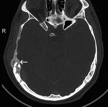
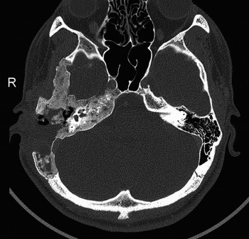
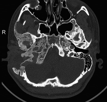
Computed tomography of the head showed heterogeneous, mixed density lesions occupying the right mastoid bone and the calvarium overlying the middle cranial fossa (Figures 1 to 3). Magnetic resonance imaging confirmed the presence of heterogeneous signal intensity in the right mastoid and temporal bones. Other bony lesions were noted over the left frontal and right parietal and occipital domes. There was no abnormal meningeal enhancement of the leptomeninges overlying the temporal lobe. The sulci and gyri in the temporal lobe appeared to be within the normal range.
Axial computed tomography scan showing a localised swelling of the right squamous temporal bone (arrow) with osteolysis and bone resorption of that side.

Post-operative axial computed tomography scan showing osteolysis and bone resorption of the right temporal bone. Note the difference in otic capsule bone density between the right and left sides.

Post-operative axial computed tomography scan showing bone resorption of the right half of the skull base and clivus, with a localized lytic bone cavity (arrow).

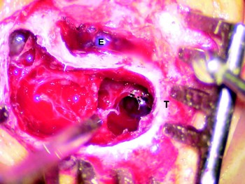
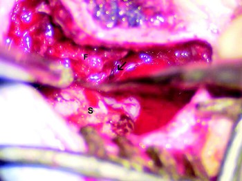
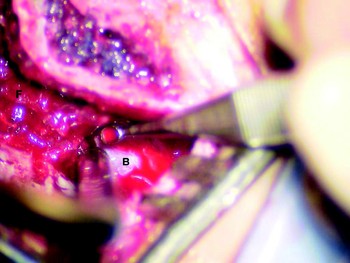
The patient was taken to the operating theatre and a right mastoid exploration was performed. Intra-operative findings were: osteolysis of the mastoid cortical bone and the temporal squama that could be easily curetted; wide, hyperpneumatised mastoid air spaces filled with a yellowish granulation tissue exuding clear, straw-coloured fluid, occasionally mixed with blood; and deossification of the otic capsule, which appeared egg shell blue in colour. Much of the osteolytic bone of the mastoid cortex was removed as well as the adjacent squamous temporal bone. The mastoid portion of the facial nerve was covered with osteolytic bone which, when carefully curetted, denuded the nerve from its bony covering but left the nerve intact. The granulation tissue was also removed and sent for histopathological analysis. Taking advantage of a wide retrofacial–infralabyrinthine cell tract, the petrous apex was reached and cleared of granulation tissue (Figures 4 to 6).
Intra-operative photograph of the right ear showing an open mastoid cavity filled with homogeneous granulation tissue. E = external auditory canal; T = mastoid tip

Intra-operative photograph of the right ear showing an open mastoid cavity with the facial nerve skeletonised (F) and an exposed sigmoid sinus (S). The arrow points to the retrofacial air cell tract.

Intra-operative photograph of the right ear showing an open mastoid cavity with the petrous apex approached through the retrofacial air cell tract. B = jugular bulb

Post-operatively, the patient regained consciousness with no untoward side effects, but continued to be febrile for three more days. Over the ensuing six weeks, he gradually improved, and finally returned to his normal state.
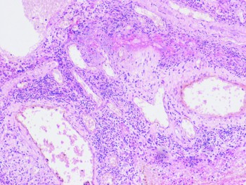
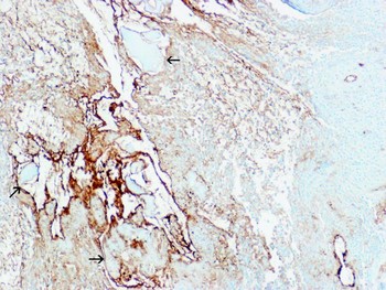
Histopathological analysis of the lesions returned a diagnosis of lymphangioma with firm confirmation of the negative differential of histocytosis, multiple myeloma and metastasis. Examined specimens showed multiple vascular spaces lined with endothelium, interspersed with chronic inflammatory cellular infiltrate. Necrotic bony spicules were also detected amidst an inflammatory cellular infiltrate denoting chronic osteomyelitis, with vascular spaces insinuated between the bony spicules and causing their lysis (Figures 7 to 9).
Photomicrograph of operative specimen showing multiple vascular spaces lined with a single layer of epithelium, interspersed with chronic inflammatory cellular infiltrate (H&E; ×100).

Photomicrograph of operative specimen showing necrotic bony spicules with chronic inflammatory cellular infiltrate, denoting chronic osteomyelitis. Note the vascular spaces insinuated between the bony spicules, causing their lysis (H&E; ×100).

Photomicrograph of factor VIII immunological staining of the endothelial lining of the vascular spaces, showing dilated, intercommunicating spaces filled with a pale, homogeneous, lymphatic fluid (arrows).

The final diagnosis was multiple lymphangiomatosis of the skull.
Discussion
Lymphangiomas are rare, congenital tumours. As many as 70 per cent of reported cases have occurred in the head and neck. First described in 1947 by Bickel and Broders,Reference Bickel and Broders1 it is estimated that fewer than 30 cases of bone lymphangiomas have been reported in the literature; in most such reported cases, lesions are disseminated throughout the skeleton.Reference Kaya, Colak, Kutlay and Gungor2
The histological, radiological and clinical characteristics of lymphangiomatosis overlap with those of haemangiomatosis and Gorham massive osteolysis, so that these entities are often grouped together as angiomatous bone lesions.Reference Brown, Pathria, Ruggieri, Jacobson, Craig and Resnick3 The cause of these disorders is unknown, but most investigators suggest that the multifocal, angiomatous bone lesions represent a congenital malformation of the vascular or lymphatic systems or both.Reference Latour, Simeons, Gryspeerdt, Samson, Mertens and Vandamme4
Histologically, lymphangioma of bone appears as a lymph-filled, multilocular cavity with an endothelial lining. Osseous lymphangiomatosis typically develops during childhood.Reference Nixon5 This may have been the pathology underlying our patient's childhood diagnosis of osteomyelitis affecting the left upper arm.
Lymphangiomas are known to result from failure of connection between the lymphatic capillaries and the lymph vessel trunks. Progressive accumulation of lymph fluid results in dilatation of the lymphatic capillaries in a cystic pattern. Although not performed in this case, lymphangiography in a case of diffuse lymphangioma of a lower extremity demonstrated multiple areas of osseous involvement.Reference Nixon5 In our case, the lymphangiomatous origin of osteolysis, observed during surgery, was supported by the histopathological findings. Moreover, histopathological evidence of osteomyelitis, with chronic inflammatory cells, was also detected, which may explain the clinical picture.
Long-standing, slow progression of disease is a consistent clinical feature of lymphangiomas. Most cases of lymphangiomatosis have bony and visceral involvement, and usually present with chylothorax, chylous ascites or acute symptoms related to the affected organ (commonly the lung, spleen or liver). Such cases are associated with a very poor prognosis and a high mortality rate.Reference Nishimura, Segami, Kaneyama and Sato6 However, visceral involvement was negative in our case. In contrast to visceral lesions, bony lesions (as demonstrated in our case) reportedly tend to remain stable and show no progression; they can even regress.Reference Nishimura, Segami, Kaneyama and Sato6
Treatment options for lymphangiomatosis include surgical therapy (i.e. curettage and excision), radiation therapy and chemotherapy (e.g. with interferon).Reference Nishimura, Segami, Kaneyama and Sato6 However, bony lesions often receive no treatment, as these lesions tend to remain stable and asymptomatic.
The final diagnosis of bony lymphangiomatosis is made histologically from a large biopsy specimen. Microscopic differentiation between lymphangioma (lymph-filled) and haemangioma (blood-filled) is difficult because of the similarity of the endothelial-lined, cystic lesions. In the present case, the diagnosis of lymphangiomatosis was made on clinical and histopathological grounds.
Conclusion
Lymphangiomatosis should be kept in mind in the differential diagnosis of lytic lesions of the skull.