




No CrossRef data available.
Article contents
Avian malaria in Brazilian passerine birds: parasitism detected by nested PCR using DNA from stained blood smears
Published online by Cambridge University Press: 19 October 2004
Abstract
The microscopical examination of Giemsa-stained thin blood smears and a nested PCR were performed to detect avian Plasmodium in 275 passerine birds from small and large fragments of Atlantic Forest, Minas Gerais, Brazil. The 275 blood smears were used both for the microscopical examination and nested PCR providing the DNA template used for the reactions. The sensitivity of the nested PCR assay was higher than that observed for blood smears through microscopical examination. High prevalence (39·6%) of Plasmodium infections was detected by nested PCR while the microscopical examination detected only 16·5% positive birds. Poor agreement was observed between the results of the two different tests. The PCR data obtained were correlated to the forest fragment size of the Atlantic Forest and also correlated to the biological characteristics of the birds (nest type construction, diet, participation in mixed-species flocks, age and sex). Birds captured in the large forest areas were more infected than birds captured in the small areas (51·9% and 28·5%, respectively). Diet and participation in mixed-species flocks were correlated to the Plasmodium parasitism. The insectivorous birds and those that participated in mixed-species flocks were more frequently infected (47% and 41·5%, respectively) than the other groups.
Keywords
Information
- Type
- Research Article
- Information
- Copyright
- 2004 Cambridge University Press
INTRODUCTION
Avian plasmodia have been extensively used for ecological modelling of host-parasite systems and their study is important for ecology, conservation and management of several species of birds in wild areas. Major surveys of Plasmodium prevalence in birds have been performed in many parts of the world, including North America, South America, Europe, Asia and Africa (van Riper III, Atkinson & Seed, 1994). In Brazil, the most recent studies of avian Plasmodium prevalence were conducted by Bennett & Souza (1980) and Woodworth-Lynas, Caines & Bennett (1989) in São Paulo state. These studies showed a very low level of parasitism (less than 10%) based on microscopical examination. Although the microscopical examination of blood smears remains the method of choice for the plasmodia identification, in recent years, considerable attention has been given to the molecular methods, including the PCR technique. PCR is considered a specific and sensitive method because it detects as few as 1 malaria parasite/μl of blood (Snounou et al. 1993a,b; Tirasophon et al. 1994; Oliveira et al. 1995; Seesod et al. 1997). In this study, the sensitivities obtained by light microscopy and by the polymerase chain reaction (PCR), were compared for Plasmodium detection in blood samples of passerine birds from Atlantic Forest, Minas Gerais state, Southeastern Brazil. Frequencies obtained by the nested PCR were also compared to the biological characteristics of the avian hosts, such as nest type construction, participation in bird mixed-species flocks, sex, age, and diet. Furthermore, the data were correlated to the forest fragment size, either large or small.
MATERIALS AND METHODS
Study areas, bird capture and blood sample collection
This study was conducted in 2 distinct areas of the Atlantic rainforest, Minas Gerais state, Southeastern Brazil, (1) ‘Reserva Particular do Patrimônio Natural (RPPN) Mata do Sossego’, Simonésia municipality (20°07′S, 4°00′W) and (2) ‘RPPN Mata do Jambreiro’ (19°58′S, 43°55′W), Nova Lima municipality. Passerine birds (n=275) were captured in fragments which had less than 30 ha of extension and in large areas with more than 1000 ha. Birds were caught between July and December 2000, by using 15–17 mist nets (36 mm mesh and 12 m long by 2·5 m high) which were placed close to the ground and opened between 7 a.m. and 4 p.m. during 3/4 days a week. During the dry season (August and September), 202 birds were captured and 73 birds during the rainy season (November).
Captured birds were identified with the help of field guides (Hilty & Brown, 1986; Dunning, 1987; Ridgely & Tudor, 1989). Each bird was weighed, metal- and colour-banded, measured (tail, wing, tarsus and beak), sexed when possible, and aged through the cranial ossification. The bird's biological characteristics such as dietary behaviour, participation in mixed-species flocks and nest type construction were also determined.
Blood samples were collected by a small puncture in the tarsal vein (left tarsus) using a disposable lancet. Thin blood smears (TBS) were prepared, fixed in methanol and stained in 3% Giemsa's solution. The slides were exhaustively examined by a well-trained microscopist. The parasite density was quantified after examination of 200 microscopic fields (approximately 150 erythrocytes/field) at 1000× magnification under oil-immersion.
DNA template preparation
Blood samples conserved as TBS were used for extraction of Plasmodium DNA as previously described (Kimura et al. 1997). Briefly, the Giemsa-stained TBS containing 10 μl of blood were de-stained, scraped using a clean scalpel blade and collected in a 1·5 ml microfuge tube containing 4 M guanidine. All the samples were extracted with phenol-chloroform and the DNA was precipitated with isopropanol. The DNA pellet was resuspended in 30 μl of double-distilled sterile water and kept at −20 °C until use.
Nested PCR assay
DNA samples were processed by nested PCR to amplify genus-specific sequences of the small subunit ribosomal ribonucleic acid (18S SSU rRNA) genes of Plasmodium sp., as previously described (Singh et al. 1999), with modifications. In the first step, a 1670 bp fragment of the Plasmodium sp. 18S gene was amplified using 15 pmoles of each genus-specific primer named rPLU1 (5′-TCA AAG ATT AAG CCA TGC AAG TGA-3′) and rPLU5 (5′-CCT GTT GTT GCC TTA AAC TCC-3′). Then 20 μl of a PCR mixture (200 μM of each dNTPs, 4 mM MgCl2, 1X Taq DNA polymerase buffer (10 mM Tris-Hcl, pH 8·5, 50 mM KCl) and 2·5 U of Taq DNA polymerase) were added to 5 μl of isolated DNA. The DNA amplification was carried out in a Thermocycle PTC-100, MJ Research, under the following conditions: 94 °C for 4 min, 39 cycles at 94 °C for 30 s, 53 °C for 1 min and 72 °C for 1 min, followed by a final extension at 72 °C for 4 min.
Two μl of the first amplified PCR product were used in the second amplification. The PCR procedure was performed using rPLU3 (5′-TTT TTA TAA GGA TAA CTA CGG AAA AGC TGT-3′) in combination with rPLU4 (5′-TAC CCG TCA TAG CCA TGT TAG GCC AAT ACC-3′). The conditions of DNA amplification used were the same as those used for the first amplification steps, except for the primers concentration (10 pmoles) and the annealing temperature (62 °C).
For the detection of malaria, in each experiment, positive and negative controls were included. Positive controls consisted of genomic DNA from Plasmodium falciparum (obtained from in vitro cultivation), Plasmodium gallinaceum and Plasmodium juxtanucleare (obtained from experimentally infected chickens), and Plasmodium berghei (obtained from experimentally infected mice). Negative controls were DNA samples obtained from 2 to 3-day-old chicks shown to be clear of infection by microscopical and repeated PCR screening. These controls were used for the 2 steps of nested PCR. DNA from Haemoproteus sp., obtained from naturally infected animals was also included as a negative control. The amplified products were visualized in 6% polyacrylamide gels stained with silver nitrate.
In order to prevent cross-contamination, different sets of micropipettes and distinct work areas for DNA extraction, mix preparation and PCR amplification were used. Moreover, 1 uninfected blood sample was included in every 10 samples processed. Twenty percent of positive-PCR samples were re-tested to confirm the presence of plasmodial DNA.
Statistical analysis
Individual data points were stored in a data base using Epi-Info package version 6.03 (Center for Disease Control and Prevention, Atlanta, GA). Differences in proportions were evaluated by the Chi-square test with Yates correction. The kappa index was used to measure agreement between the PCR results and microscopy. P values <0·05 were considered significant.
RESULTS
A total of 275 samples of birds from Atlantic forest, Southeastern Brazil, were screened: 131 (47·6%) birds were captured in large fragments while 144 (52·4%) were captured in small fragments. All birds were Passeriformes, representing 9 avian families: Conopophagidae, Dendrocolaptidae, Emberizidae, Furnariidae, Muscicapidae, Pipridae, Thamnophilidae, Tyrannidae and Vireonidae (Table 1).

Plasmodium sp. was detected in 44 (16%) of the 275 birds by the microscopical examination. However, the nested PCR using DNA extracted from stained blood smears showed a statistically higher prevalence of avian malaria (39·6%) (Table 1). This is also true for different families and species of birds (Table 1), except Muscicapidae and Pipridae which had more birds infected by microscopical examination than by PCR. It is important to note that all avian families analysed in this study were infected by plasmodia. However, 7 bird genera (Arremon, Attila, Campylorhamphus, Lochmias, Myiophobus, Schiffornis and Syndactila) comprising 5 families (Dendrocolaptidae, Emberezidae, Furnariidae, Pipridae and Tyrannidae) were entirely clear of malaria infection.
The nested PCR used seems to be genus-specific since no amplification was observed for Haemoproteus infection (Fig. 1, lane 8). Indeed, the Haemoproteus DNA was capable of being amplified using a PCR protocol previously described by Wozniak, Telford & McLaughlin (1996) which targets the 18S rRNA gene of both the host and the parasite. A positive result comprised a 590 bp DNA fragment (avian host) and one of 610 bp (Plasmodium/Haemoproteus) (data not shown). Then, the use of another set of primers to amplify the DNA of 2 genera of avian malaria confirms that the nested PCR used here did not detect Haemoproteus DNA.

Fig. 1. Electrophoresis after Plasmodium-genus nested PCR amplification. Lanes (1) and (9) 1 Kb Ladder molecular size marker (BRL); Lanes (2) and (3) field negative blood samples; Lanes (4) and (7) field positive samples; Lane (5) Plasmodium falciparum obtained from in vitro culture; Lane (6) Plasmodium juxtanucleare from experimentally infected chicken; Lane (8) Haemoproteus obtained from naturally infected bird.
The plasmodia 18S SSU rRNA gene was amplified from different vertebrate hosts as seen in Fig. 1. Interestingly, size polymorphisms were observed among distinct isolates obtained from these different hosts.
The nested PCR results were analysed in relation to the level of circulating parasites detected by microscopical examination. For this analysis only 44 birds with parasites detected by microscopical examination were included. The PCR-positive frequency tended to be higher in birds showing high levels of circulating parasites (Fig. 2). Nineteen of the birds, showing a mean parasitaemia of 2·3±2·1 parasites/200 microscopy fields (range: 1–10 parasites), were PCR-positive, whereas the 25 individuals that were PCR-negative had a mean parasitaemia of 1·8±0·8 parasites/200 fields (range: 1–4 parasites), even though this difference was not of statistical significance.

Fig. 2. Effect of parasite density on PCR positivity. The parasitaemias were expressed as number of parasites per 200 microscopic fields containing approximately 150 erythrocytes/field at 1000× magnification under oil-immersion.
Among the 231 microscopy-negative samples, the PCR detected as many as 90 samples containing Plasmodium DNA. On the other hand, for unknown reasons, Plasmodium DNA was not detected in the other 25 birds which had visually detectable parasites in their blood smears. Thus, the agreement between PCR and microscopy was poor (Kappa index= 0·017).
The malaria prevalence data obtained by microscopical examination and PCR analysis were correlated to the fragmentation level of the Atlantic Forest. More birds captured in large fragments were infected by Plasmodium sp. (58·8%) than birds from small fragments (39·5%) (P=0·002). In addition, we analysed the influence of seasonality on plasmodia infection prevalence. The highest prevalence values were observed at the month with the highest precipitation (November). Thus, among the 134 positive birds as detected by microscopy or PCR, 51 samples were collected during the rainy season (November) representing a rate of 70% of the 73 birds captured during this period. However, 41% of the 202 birds sampled during the dry season (August and September) were positive, a significantly different rate (P<0·0001).
Plasmodium prevalence varied significantly among bird dietary groups, being higher in insectivorous birds (Table 2) than in birds with other diets. Birds that participated in mixed-species flocks also had higher plasmodia prevalence than those that did not participate in this group. Birds nest type, host sex and age did not show any significant relationship with Plasmodium sp. infection.

DISCUSSION
Avian plasmodia are routinely identified by microscopical examination of Giemsa-stained blood smears. The frequency of Plasmodium infection detected by microscopy was low (16%), reflecting several difficulties in Haematozoa quantification in blood smears, such as the non-homogeneous distribution of non-infected and infected erythrocytes and the number of erythrocytes per field of view that must be counted for an adequate estimate of parasitism. However, the microscopical analysis of blood smears allowed the identification of morphological characteristics of different stages of the parasite, which is the parameter still used to classify avian plasmodia species. In this study, the Plasmodium specific identification was not done due to the very low parasitaemias observed. Since only 1 parasite was frequently observed in the slides (43% of the 44 microscopically positive samples), it would be difficult to morphologically characterize the Plasmodium species.
To overcome some of limitations of microscopy for detection and identification of avian malaria, PCR-based assays have been performed. Whereas the PCR assays have not been infallible, they have proven to be effective in detecting malarial infections in avian populations (Richard et al. 2002). The PCR using DNA template obtained from stained slides showed high sensitivity. In fact, the use of DNA obtained from thick or thin smears has produced good results by PCR in different studies involving Plasmodium (Ekala et al. 2000; Ivanova et al. 2001). This fact suggests that this method of DNA preservation could be considered adequate for field studies.
The nested PCR used was previously described by Singh et al. (1999) and its use has been proposed to screen samples for the presence of malaria before determining the Plasmodium species. Primers rPLU3 and rPLU4 have been used in single nested PCR assays to amplify DNA specifically from the four human malaria parasites (P. falciparum, P. vivax, P. malariae and P. ovale), from a simian malaria parasite (P. gonderi) and from rodent malaria (P. yoelli, P. berghei, P. chabaudi and P. v. petteri) (Hulier et al. 1996). Thus, these two nested genus-specific primers could be used successfully in nested PCR assays (in combination with nested 1 primers rPLU1 and rPLU5) to amplify DNA from avian malaria parasites as we can now observe.
The nested PCR assay was found to be more sensitive since additional malaria-positive samples were detected by PCR compared through microscopy. However, some slides in which Plasmodium sp. was observed by microscopical examination could not be amplified by PCR. The poor agreement between the microscopy and PCR results could be caused by a misidentification of the parasites on the slides. It is often difficult to discriminate avian haemoparasites using morphological criteria mainly between Plasmodium and Haemoproteus genera. In addition, the occurrence of false negatives by PCR was probably due to the reduced number of parasites in the samples, as some could have been lost during the process of scraping the slides. Furthermore, classical methods for fixing cells may be crucial factors in determining the rate of DNA degradation. It is well known that methanol fixation may affect the dissociation of DNA within the cell. Thus, fixation and Giemsa-staining could act negatively on the DNA stability.
The occurrence of false-negative results by PCR could be also caused by a failure of the DNA extraction itself. Thus, all samples classified as false-negatives were tested at least twice to ensure that the negative results were not caused by PCR failure.
Since the biological characteristics of the hosts can influence the frequency of haemoparasites, the prevalence data obtained by the nested PCR assay were compared to the biological characteristics of the avian hosts, such as the nest type, participation in mixed-species flocks, sex, age, and dietary behaviour. Birds that construct opened nests, for example, could be frequently in contact with the vectors if compared to the birds that construct closed nests. Birds that participate in mixed-species flocks might have less probability of infection, due to the low contact with vector-susceptible host inside a group of birds.
Here, we did not observe association between plasmodia infection and nest type, sex or age. However, birds that participate in mixed-species flocks were more frequently infected with Plasmodium than birds that do not participate in this group. These data suggest that plasmodia vectors transit frequently among different host species and that more than one host species are susceptible to plasmodia species.
Plasmodium infections occurred more frequently in insectivore birds, rather than granivorous, frugivorous or omnivorous ones. This fact suggests that those birds could be more exposed to infected vectors since the destruction of insects during feeding could release the sporozoites that would penetrate into the birds' mucosa. However, it is important to consider that the frequency of avian haemoparasites may be associated with the foraging strategies, that facilitate or not the bird-vector contact. Malaria infection may represent low immunity in some individuals, a decrease in health state and body condition, and a decrease in the bird's movement ability, which may increase the contact rate with vectors, causing a further increase in the infection probability.
Seasonal variation in malaria distribution can be generated by variation in host exposure to parasite infective stages which is often due to the fact that the development of plasmodia in their vectors is temperature dependent or sensitive to seasonal variation in humidity. To address this issue, we examined the temporal changes in the pattern of avian malaria distribution. The high level of plasmodia parasitism that we observed could be due to vector-related factors such as (1) relative high densities of avian malaria vectors; (2) increased breeding sites during the rainy season; (3) increased vector longevity due to high relative humidity. Indeed, the highest prevalence value was observed at the month with the highest precipitation rate (November). Additional studies are needed to confirm the association between seasonality and avian malaria transmission in the Brazilian rainforest.
The avian malaria frequency was compared to the Atlantic Forest fragmentation level. In this case, birds that were captured in large forests were more frequently infected with Plasmodium than birds captured in small fragment areas. Probably the occurrence of vectors in fragmented or disturbed habitats could be low, as previously suggested by Tella et al. (1999). Considering that different host species may have completely distinct malaria prevalence rates due to differences in tolerance and immunity, we also compared the plasmodia prevalence in one host species Conopophaga lineata. Among the 33 specimens captured, 16 (48%) were infected by Plasmodium species as detected by microscopical examination or PCR. However, no significant difference was observed between the prevalence rates obtained among birds captured in large or small fragments. It is important to know in greater detail the role of wild species, such as birds, in dispersion of avian malaria since epidemics occur easily when diseases move within new host populations which have not been exposed in their natural habitat.
In conclusion, the higher prevalence of Plasmodium species determined by the nested PCR compared with microscopy demonstrated that this assay is available to study avian malaria among different hosts. Furthermore, the DNA preservation as stained thin blood smears could be an excellent alternative to molecular studies when other blood sources are unavailable. In addition, it must be noted that the nested PCR assay used here cannot amplify DNA of Haemoproteus sp. and could be used to distinguish species between the two genera, an important contribution to the avian malaria studies.
This work was partiality supported by Fundação de Amparo à Pesquisa do Estado de Minas Gerais FAPEMIG (grant no. CBB177/01) and fellowship to Sérgio Ribeiro. Brazilian National Research Council CNPq granted scholarship to Fabiane Sebaio and Felipe Branquinho and research grant and fellowship to Miguel Marini. We thank the administrators who allowed our study in their conservation units, Instituto Brasileiro de Meio Ambiente IBAMA that gave the banding/collecting authorization and Leonardo Lopes and Alexandre Fernandes who helped considerably with field work.
References
REFERENCES
BENNETT, G. F. &
SOUZA, O. (
1980).
Blood parasites of some birds from São Paulo, Brazil.
Memórias do Instituto Oswaldo Cruz 75,
117–134.CrossRefGoogle Scholar
DUNNING, J. S. (
1987).
South American Birds. A Photographic Aid to Identification.
Harrowood Books,
Newton Square, UK.
EKALA, M. T.,
LEKOULOU, F.,
DJIKOU, S.,
DUBREULL, G.,
ISSIFOU, S. &
NOTOUMI, F. (
2000).
Evaluation of a simple and rapid method of Plasmodium falciparum DNA extraction using TBS from Gabonese patients.
Bulletin de la Societé de Pathologie Exotique et de ses Filiales 93,
8–11.Google Scholar
HILTY, S. L. &
BROWN, W. L. (
1986).
A Guide to the Birds of Colômbia.
Princeton University Press,
Princeton, USA.
HULIER, E.,
PETOUR, P.,
SNOUNOU, G.,
NIVEZ, M. P.,
MILTGEN, F.,
MAZIER, D. &
RENIA, L. (
1996).
A method for the quantitative assessment of malaria parasite development in organs of the mammalian host.
Molecular and Biochemical Parasitology 77,
127–135.CrossRefGoogle Scholar
IVANOVA, N. V.,
MOROZOV, E. H.,
KURKINA, I. V.,
MAKSAKOVSKAIA, E. V.,
RABINOVICH, A. S. &
POLTARAUS, A. B. (
2001).
IT1S, 5.8S and A-type ITS2 rDNA sequences from Plasmodium vivax and development of a method for restropective PCR diagnosis of malaria by stained TBS.
Molecular Biology 35,
515–525.Google Scholar
KIMURA, M.,
KANOEK, O.,
LIU, Q.,
ZHOU, M.,
KAWAMOTO, F.,
WATAYA, Y.,
OTANI, S.,
YAMAGUCHI, Y. &
TANABE, K. (
1997).
Identification of the four species of human malaria parasites by nested PCR that targets variant sequences in the small subunit rRNA gene.
Parasitology International 46,
91–95.CrossRefGoogle Scholar
OLIVEIRA, D. A.,
HOLLOWAY, B. P.,
DURIGON, E. L.,
COLLINS, W. E. &
LAL, A. A. (
1995).
Field evaluation of a polymerase chain reaction-based nonisotopic liquid hybridization assay for malaria diagnosis.
The Journal of Infectious Diseases 173,
1284–1287.Google Scholar
RICHARD, F. A.,
SEHGAL, R. N. M.,
JONES, H. I. &
SMITH, T. B. (
2002).
A comparative analysis of PCR-based detection methods for avian malaria.
Journal of Parasitology 88,
819–822.CrossRefGoogle Scholar
RIDGELY, R. S. &
TUDOR, G. (
1989).
The Birds of South America, 1 and 2.
University of Texas Press,
Austin,
USA.
SEESOD, N.,
NOPPARAT, P.,
HEDRUM, A.,
HOLDER, A.,
THAITHONG, S.,
UHLEN, M. &
LUNDEBURG, J. (
1997).
An integrated system using immunomagnetic separation, polymerase chain reaction, and colorimetric detection for diagnosis of Plasmodium falciparum.
American Journal of Tropical Medicine and Hygiene 56,
322–328.CrossRefGoogle Scholar
SINGH, B.,
BOBOGARE, A.,
COX-SINGH, J.,
SNOUNOU, G.,
ABDULLAH, M. S. &
RAHMAN, H. A. (
1999).
A genus and species-specific nested polymerase chain reaction malaria detection assay for epidemiologic studies.
American Journal of Tropical Medicine and Hygiene 60,
687–692.CrossRefGoogle Scholar
SNOUNOU, G.,
VIRIYAKOSOL, S.,
JARRA, W.,
THAITHONG, S. &
BROWN, K. N. (
1993 a).
Identification of the four human malaria parasite species in field samples by the polymerase chain reaction and detection of a high prevalence of mixed infections.
Molecular and Biochemical Parasitology 58,
283–292.Google Scholar
SNOUNOU, G.,
VIRIYAKOSOL, S.,
ZHU, X. P.,
JARRA, W.,
PINHEIRO, L.,
DO ROSARIO, V. E.,
THAITHONG, S. &
BROWN, K. N. (
1993 b).
High sensitivity of detection of human malaria parasite by the use of nested polymerase chain reaction.
Molecular and Biochemical Parasitology 61,
315–320.Google Scholar
TELLA, J. L.,
GUILLERMO, B.,
FOREROS, M. G.,
GAJO, A. L. &
DONA'ZAR, A. (
1999).
Habitat, world geographic range, and embryonic development of hosts explain the prevalence of avian hematozoa at small spatial and phylogenetic scales.
Proceedings of the National Academy of Sciences, USA 96,
1785–1789.CrossRefGoogle Scholar
TIRASOPHON, W.,
RAJKULCHAI, P.,
PONGLIKITMONGKOL, M.,
WILAIRAT, P.,
BOONSAENG, V. &
PANYIM, S. (
1994).
A highly sensitive, rapid, and simple polymerase chain reaction-based method to detect human malaria (Plasmodium falciparum and Plasmodium vivax) in blood samples.
The American Journal of Tropical Medicine and Hygiene 51,
308–313.CrossRefGoogle Scholar
VAN RIPER III, C.,
ATKINSON, C. T. &
SEED, T. M. (
1994).
Plasmodia of birds.
Parasitic Protozoa 7,
73–140.Google Scholar
WOODWORTH-LYNAS, C. B.,
CAINES, J. R. &
BENNETT, G. F. (
1989).
Prevalence of avian haematozoa in São Paulo, Brazil.
Memórias do Instituto Oswaldo Cruz 84,
515–526.CrossRefGoogle Scholar
WOZNIAK, E. J.,
TELFORD Jr, S. R. &
McLAUGHLIN, G. L. (
1996).
Molecular differentiation of two New World saurian Plasmodium species in different host species using the Polymerase Chain Reaction.
Journal of Parasitology 82,
372–375.CrossRefGoogle Scholar
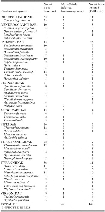
Table 1. Birds sampled for avian malaria from Atlantic Forest, Minas Gerais, Southeastern Brazil
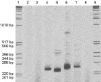
Fig. 1. Electrophoresis after Plasmodium-genus nested PCR amplification. Lanes (1) and (9) 1 Kb Ladder molecular size marker (BRL); Lanes (2) and (3) field negative blood samples; Lanes (4) and (7) field positive samples; Lane (5) Plasmodium falciparum obtained from in vitro culture; Lane (6) Plasmodium juxtanucleare from experimentally infected chicken; Lane (8) Haemoproteus obtained from naturally infected bird.
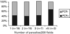
Fig. 2. Effect of parasite density on PCR positivity. The parasitaemias were expressed as number of parasites per 200 microscopic fields containing approximately 150 erythrocytes/field at 1000× magnification under oil-immersion.
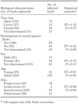
Table 2. Associations between biological characteristics of the 275 birds caught in the Atlantic Forest fragments of Minas Gerais, Brazil, and Plasmodium infection detected by microscopical examination or PCR
 You have
Access
- 45
- Cited by