Introduction
The anatomy of the temporal bone is extremely complicated. Intensive training is required before the surgeon can master tympanoplasty. We previously created a three-dimensional (3D) model using a selective laser sintering method and reported its validity in surgical training and medical education.Reference Suzuki, Ogawa, Kawano, Hagiwara, Yamaguchi and Ono1–Reference Suzuki, Hagiwara, Kawaguchi and Ono3 In the previous model, however, the stapes could not be reproduced because of its low radiopacity on computed tomography (CT) scanning. In this study, we attempted to replicate the stapes by enhancing its CT intensity.
The inner-ear model was made by simply reversing the CT value for bone replication.Reference Suzuki, Ogawa, Hagiwara, Yamaguchi and Ono2 This method eliminates data on the bone structure, and hence the model lacks the ossicles and the bony wall that covers the inner ear. In this study, we attempted to create both the inner ear and its surrounding bony structure by selecting both fluid and bone surface intensities.
Material and methods
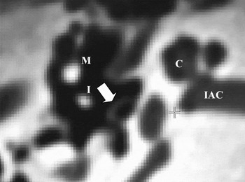
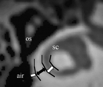
The methods used to construct the temporal bone model have been reported in detail previously.Reference Suzuki, Ogawa, Kawano, Hagiwara, Yamaguchi and Ono1 The derived 3D data were converted into the STL (standard triangular language) file. In order to create the stapes, the CT intensity of the stapes was enhanced by changing the threshold value (Figure 1). When the intensity value of the bone part was simply reversed, the inner-ear part, as well as all the air space, was created.Reference Suzuki, Ogawa, Hagiwara, Yamaguchi and Ono2 In the present study, the intensity value was adjusted so that both the bony part and the air space were eliminated, but the area with an intensity between that of bone and the air, such as the fluid or the bone surface, could be retained (Figure 2). The derived 3D data were converted into a STL file system.
Axial computed tomography showing the ossicles. The malleus (M) and the incus (I) are radiopaque, but the stapes (arrow) is far less so. The intensity value of the stapes was enhanced along each slice. C = cochlea; IAC = internal auditory canal

Determination of the computed tomography value on STL data. The most radiopaque bone area (black arrow) and the air were eliminated. The intensity of the fluid and the bone surface (white arrows) was selected. OS = ossicle; SC = semicircular canal

The powder material, polyamide nylon plus glass beads, was laser sintered according to the STL protocol. The created model was dissected using conventional otosurgical instruments.
Results
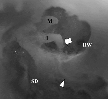
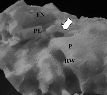
The appearance of the replicated stapes is shown in Figure 3. The posterior canal wall was removed. The malleus, incus and posterior crus of the stapes could be seen. Other structures, such as the mastoid air cells, surgical dome, facial nerve, promontory, auditory tube, round window niche, semicircular canal and cochlea, were recreated as in the previous model.Reference Suzuki, Ogawa, Kawano, Hagiwara, Yamaguchi and Ono1Figure 4 shows the replicated stapes seen from the external canal side after removing other ossicles. The neighboring structures, such as the facial nerve, pyramidal eminence and promontory, are demonstrated. Another stapes model is shown in Figure 5 after removing the posterior canal and incus. In this case, the stapes head and two arches can easily be identified.
Replicated model of right ear stapes. The posterior canal wall is removed. The posterior crus and incudostapedial joint are seen (arrow). M = malleus; I = incus; RW = round window niche; SD = surgical dome; arrow head = facial nerve

Replicated stapes seen from the external canal. The superstructure of the stapes is shown (arrow). FN = facial nerve; PE = pyramidal eminence; P = promontory; RW = round window niche

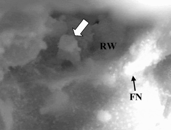
Replicated stapes of another case, right ear. The posterior canal wall and the incus are removed. The crura and the head of the stapes are seen (arrow). RW = round window niche; FN = facial nerve

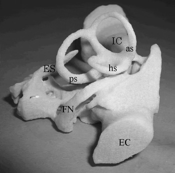
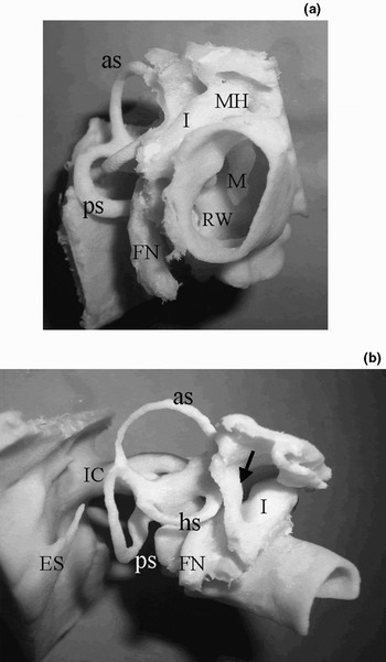
The whole appearance of the inner-ear model, replicated as per the previously published method,Reference Suzuki, Ogawa, Hagiwara, Yamaguchi and Ono2 is shown in Figure 6. In this model, the inner-ear parts as well as the air space were replicated, as only the radiopaque bony area was eliminated. The inner ear and the compact external ear canal were shown, but no ossicles were reproduced. In the present model, the inner ear as well as the surrounding bony wall and the ossicles were created, since both the radiopaque bone area and the lucent air space were eliminated (Figure 7). The auditory canal wall, promontory, window niches, malleus, incus, stapes, facial nerve, surgical dome, and epitympanic tegmen were reproduced. The inner ear, including semicircular canals, cochlea and endolymphatic sac, and the internal auditory meatus were also reproduced, as in the previous model. The present model allows easier demonstration of the labyrinth's anatomical relation to the ossicles, surgical dome, promontory and window niches.
Inner-ear model replicated using the previous method. Note the compact external canal (EC). No ossicles were created. IC = internal auditory canal; ES = endolymphatic sac; FN = facial nerve; as = anterior semicircular canal; hs = horizontal semicircular canal; ps = posterior semicircular canal

Inner-ear models replicated by the present method. (a) The contour of the external canal is recreated. The malleus, incus and round window niche (RW) can be seen through the canal. (b) In another model, the anatomical relationship between the semicircular canals and the incus, facial nerve and surgical dome (arrow) can be easily seen. The lower half of the external canal has been cut. IC = internal auditory canal; ES = endolymphatic sac; FN = facial nerve; MH = malleus head; I = incus; as = anterior semicircular canal; hs = horizontal semicircular canal; ps = posterior semicircular canal; M = malleus

Discussion
It is not easy to acquire the skills required to perform tympanoplasty, because of the anatomical complexity of the ear. In recent years, computerised 3D displays of the temporal bone and ‘virtual reality’ endoscopic images have been developed.Reference Morra, Tirelli, Rimondini, Cioffi, Russolo, Giacomarra and Pozzi-Mucelli4, Reference Begall and Vorwerk5 In cases with anatomical anomalies, safe surgery is extremely difficult, even after scrutinising the CT. Navigational surgery and robotic surgery allow good orientation of abnormal structures and ensure safety.Reference Selesnick and Kacker6, Reference Federspil, Geisthoff and Henrich7 However, these interventions are costly and require much space and equipment. Rapid prototyping is cost-effective and allows replication of anatomical details. In particular, the models created using selective laser sintering allow easy dissection and are particularly suited for pre-operative simulation.Reference Suzuki, Ogawa, Kawano, Hagiwara, Yamaguchi and Ono1
In the previous model, the stapes could not be replicated because of its low density on STL data, as shown in Figure 1, although the malleus and incus could be reproduced. In this study, we attempted to locally enhance the stapes' radiopacity by changing the threshold value on STL data. This allowed replication of the stapes, including both crura and head. The anatomical location of the stapes is extremely important, since it serves as a good landmark to locate other structures such as the horizontal segment of the facial nerve and the round window niche. The present model has more advantages as an educational resource, compared with the past model.
The inner-ear model serves as a good resource when teaching 3D orientation within the inner ear.Reference Suzuki, Ogawa, Hagiwara, Yamaguchi and Ono2 The structural features of the model, such as the internal auditory meatus, facial nerve and endolymphatic sac, allow easy understanding of the inner ear within its wider context. The model can be used as an anatomical guide while dissecting the temporal bone model. The present model is useful for teaching the relationship between the inner ear and the surrounding bone structure. There are clinically important landmarks around the labyrinth, such as the incus short process, surgical dome, promontory and window niches. The anatomical relations between the labyrinth and these structure can readily be demonstrated using this 3D model.
• The anatomy of the temporal bone is extremely complicated. Intensive training is required before the surgeon can master tympanoplasty
• This study investigates the validity of adjusting the computed tomography (CT) threshold to replicate a temporal bone model suitable for dissection training and education
• A simulated three-dimensional (3D) model of a human temporal bone was constructed using a selective laser sintering method. The powder layers were laser-fused based on detailed CT data and accumulated to create a 3D structure
• This technique has potential to develop a viable temporal bone substitute for surgical training
In the future, we will examine the extent to which medical trainees can develop their anatomical knowledge and surgical skills by using these models. We will also study replication of an ossicular anomaly, by adjusting the local threshold value of deformed ossicles. Three-dimensional evaluation of congenitally malformed ears will also give us new insights into the developmental process of the ear.
Conclusion
Using the rapid-prototyping technique, the CT threshold was modified in order to replicate the stapes. The temporal bone model, with stapes intact, has great advantages as an educational resource. By adjusting the intensity value of the CT, the bony labyrinth and the surrounding bone structure could be recreated. The model contributes to easy understanding of the relationship between the labyrinth and the surrounding structures.