It is widely recognized that healthcare workers’ hands are the main vectors of germ dissemination in the healthcare setting. The key role of caregivers’ hands in the transmission of germs has been clearly establishedReference Pittet, Allegranzi and Sax 1 and has led to the development of comprehensive and successful hand hygiene promotion strategies. 2 – Reference Luangasanatip, Hongsuwan and Limmathurotsakul 4 In contrast, the potential role of other vectors in germ dissemination, such as stethoscopes, remains poorly understood.
Numerous studies have shown that stethoscopes may be contaminated by various microorganisms, including methicillin-resistant Staphylococcus aureus (MRSA), Escherichia coli, and Pseudomonas aeruginosa.Reference Nunez, Moreno, Green and Villar 5 Recently, our group has demonstrated the presence of wide variations in the level of stethoscope contamination with MRSA following a single physical examination.Reference Longtin, Schneider and Tschopp 6 For example, contamination of the diaphragm with MRSA following examination of a MRSA-colonized patient can vary from 0 to more than 1,000 colony-forming units (CFU) per 25 cm2. In addition, a strong correlation was detected between contamination of caregivers’ hands and stethoscopes. Whether a similar correlation exists for other microorganisms, such as Enterobacteriaceae and nonfermenting gram-negative bacilli, is unclear. In addition, the reasons behind the strong correlation between the level of contamination of the physicians’ hands and stethoscopes remain to be elucidated. Numerous environmental, patient-related, and physician-related factors could conceivably be implicated, but these hypotheses have not been investigated yet.
Hence, we performed a study (1) to identify predictors of heavy stethoscope contamination and (2) to compare the recovery rates of different microorganisms from the physicians’ hands and stethoscopes following a single physical examination.
METHODS
Study Design and Patient Recruitment
We conducted a structured prospective study from August 20, 2009, through January 28, 2010, at the University of Geneva Hospitals, Geneva, Switzerland, which is a 2,200-bed primary and tertiary teaching hospital admitting 47,000 patients annually and with a long-standing experience in hand hygiene promotion.Reference Pittet, Hugonnet and Harbarth 7 A total of 56 patients were included. To obtain a study population heterogeneous with regard to multidrug-resistant pathogen colonization, we used 2 different recruitment strategies. First, 48 patients were recruited from the internal medicine and general surgery wards using a convenience-based strategy. Second, 8 additional patients were identified by querying the institutional infection control database of patients infected or colonized with the following pathogens: extended-spectrum β-lactamase–producing Enterobacteriaceae (ESBL-E), enterococci, Acinetobacter baumannii, and MRSA.
All study subjects gave written informed consent. The study was approved by the research ethics committee at University of Geneva Hospitals.
Standardized Physical Examination
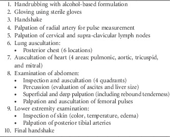
Following patient enrollment, a single physical examination was performed by 1 of 2 examiners (C.T. or A.S.). The physical examination was performed according to a standardized protocol to ensure reproducibility (Table 1). Physicians were allowed to adapt to unforeseen events (such as unfastening the patient’s gown or moving the bedside table) as long as the action was commonly encountered in routine clinical practice. The examiners changed their white coats every 6 hours and donned an isolation gown whenever the patient was under contact precautions. Examiners used sterile gloves to ensure that their hands were free of bacteria before starting the physical examination. A sterile stethoscope was handed to the examiner before the start of the examination. Physicians were allowed to wrap the stethoscope around the neck when not in use. To evaluate whether the brand of stethoscope was associated with the level of contamination, 1 of 4 different models of stethoscopes was used: Littmann Master Cardiology (3M), Littmann Classic II (3M), Maestro Adult (Colson), and Duplex (Riester). The stethoscope selection was randomized and performed after patient enrollment. Sterilization was performed using hydrogen peroxide gas plasma technology to preserve the integrity of the material (Sterrad 100NX Sterilizer; Advanced Sterilization Products).
Standardized Physical Examination

Parameters Assessment
Numerous patient- and environment-related parameters that may be associated with stethoscope contamination were collected. Variables included demographic and anthropomorphic variables (sex, age, weight, and height), comorbidities, antimicrobial use, and presence of drains. In addition, parameters that may impact the level of patient skin bacterial load, such as the type and moment of last bathing, were documented. The patient’s skin humidity was also assessed by the examiner and reported on an ordinal scale (dry/slightly humid/very humid). Finally, room temperature and ambient air humidity were measured.
Specimen Collection and Processing
Upon completion of the physical examination, 4 regions of the physician’s dominant gloved hand (fingertips, dorsum, and thenar and hypotenar eminences) and 2 regions of the stethoscope (diaphragm and tube) were sampled to assess the level of bacterial contamination. Sampling was conducted by gently pressing the region under study on 25-cm2 nonselective contact plates with trypticase soy agar (replicate organism detection and counting plates; bioMérieux) for 2 seconds.Reference Longtin, Schneider and Tschopp 6 Sampling of the stethoscope tube (performed at approximately 10 cm from the stethoscope head) was conducted by rolling it across the plate using a technique adapted from intravenous catheter culture.Reference Raad, Hanna and Maki 8 Contamination of the patient’s skin was determined by sampling 1 inguinal fold with a contact plate.
Following an incubation at 35°C for 18–24 hours, the total aerobic colony count on each plate was determined on digital photographs using the Photoshop CS4 counting tool (Adobe Systems) as previously described.Reference Longtin, Schneider and Tschopp 6 For the purposes of our study, we fixed the upper CFU limit of the aerobic colony count at 3,000 per 25 cm2. Beyond this number, colonies formed a confluent surface. Owing to high interobserver reliability (intraclass correlation coefficient, 0.99) in preliminary studies, each photograph was analyzed by a single observer.
To detect the presence/absence of different pathogens, the following method was employed: after taking the photograph to establish total aerobic colony count, a sterile inoculation loop was rubbed over the surface of each culture plate and the collected bacteria were suspended in 2 mL NaCl 0.5%. An aliquot of the suspension was then inoculated onto the following plates using a sterile swab: (1) S. aureus ID agar plate (SAID; bioMérieux), (2) MacConkey agar (to detect nonfermenting gram-negative bacilli and Acinetobacter baumannii), (3) plates to detect ESBL-E (BLSE ID; bioMérieux), and (4) colistin and nalidixic acid blood agar with a vancomycin disk to detect vancomycin-resistant gram-positive cocci. The plates were incubated at 35°C for 48 hours and then examined for the presence of the following organisms: S. aureus (either methicillin-susceptible S. aureus or MRSA); Enterococcus spp.; Enterobacteriaceae (including ESBL-E); and A. baumannii and other nonfermenting gram-negative bacilli. Microorganisms were identified by colony morphology, Gram stain, and matrix-assisted laser desorption/ionization time-of-flight mass spectrometer (Biotyper 2.0; Bruker). Methicillin-susceptible S. aureus and MRSA were identified by detection of femA-SA and MecA genes by duplex quantitative polymerase chain reaction assay.Reference Francois, Pittet and Bento 9
Statistical Analysis
Discrete variables are reported as number and proportions in each category; continuous variables are represented as mean (SD) or median (interquartile range [IQR]). Owing to skewed distributions, the degrees of contamination of different parts of the hands and stethoscopes were described as medians with 25th and 75th percentiles (ie, IQR) and depicted in a box plot. Contamination of different regions of hands and stethoscopes was compared using Wilcoxon rank sum tests for paired continuous variables. To identify predictors of stethoscope contamination, bacterial counts (in CFU) on diaphragms and tubes were dichotomized into heavy vs nonheavy growth. Heavy contamination was defined as a CFU count above the 75th percentile (ie, >512 CFU for the diaphragm and >322 CFU for the tube).
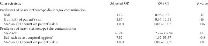
Predictors of heavy bacterial contamination of stethoscope diaphragm and stethoscope tube were assessed by logistic regression, χ2 test, and Fisher exact test, as appropriate. All variables found to be associated with heavy contamination (P<.05) by univariate analysis were considered for inclusion in a multivariate model to adjust for potential confounders. We built 2 different forced-entry models, 1 for the diaphragm and 1 for the tube. The variables “body mass index” (calculated as weight in kilograms divided by height in meters squared), “skin humidity,” and “patient skin CFU count” were included for the diaphragm contamination model, whereas the variables “male sex,” “patient skin CFU count,” and “bed bath” were included for the tube contamination model. All data related to a single respondent were excluded when any of the variables included in the model had missing values. The magnitude of the association between outcomes and explanatory variables was measured by odds ratios and corresponding 95% confidence intervals. The variable “skin humidity” was treated as an ordinal variable in the univariate analysis to facilitate reporting but as a continuous variable in the multivariate analysis. We used the Spearman rank correlation coefficient (ρ) to measure the correlation between the frequency of recovery of different microorganisms from the hands vs. the stethoscopes. All tests were 2-tailed and P<.05 was defined as statistically significant. Statistical analyses were performed with PASW Statistics, version 18.0 (SPSS).
RESULTS
Patient Characteristics
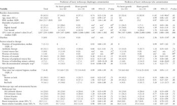
Patient characteristics are presented in Table 2. Eighteen patients (32.1%) were receiving antibiotic therapy at the time of physical examination and 7 (12.5%) were undergoing skin decolonization with an antiseptic soap. The median (IQR) total bacterial CFU count on their skin was 1,037 (255–3,000) per 25 cm2; a fifth of patients (21.4%) had very humid skin. Approximately half of patients had at least 1 wound and approximately half had at least 1 peripheral venous line.
Predictors of Heavy Stethoscope Contamination Following a Physical Examination

NOTE. Data are no. (%) of patients, unless otherwise indicated. BMI, body mass index (calculated as weight in kilograms divided by height in meters squared); CFU, colony-forming unit; IQR, interquartile range; OR, odds ratio.
a Not applicable because of absence of outcome in 1 group.
b By Fisher exact test.
c By χ2 test.
d For decolonization of methicillin-resistant Staphylococcus aureus carriers.
Levels of Contamination of Physician’s Hands and Stethoscopes
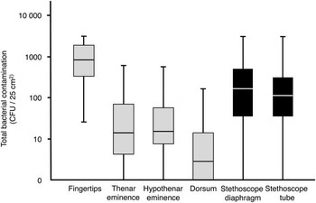
A total of 392 cultures from 4 hand sites, 2 stethoscope sites, and 1 skin site were taken to evaluate bacterial contamination. Figure 1 presents the levels of bacterial contamination of stethoscopes and physicians’ hands. Following a single examination, the most heavily contaminated region was the fingertips (median [IQR] contamination, 834 [331–1,838] CFU/25 cm2), followed by the stethoscope diaphragm (172 [36–535] CFU/25 cm2) and tube (116 [34–321] CFU/25 cm2). The contamination levels of the thenar and hypothenar eminences were comparable (14 [4–71] CFU/25 cm2 and 16 [8–58] CFU/25 cm2, respectively). The least heavily contaminated region was the dorsum of the hand (3 [1–15] CFU/25 cm2). When comparing the various regions, the levels of diaphragm and tube contamination were significantly lower than fingertip contamination, but significantly higher than contamination of the thenar eminence, hypothenar eminence, and dorsum of the hand (P≤.001 for each comparison).
Total aerobic colony count recovered from physicians’ gloved hands (gray boxes) and stethoscopes (black boxes) following a single physical examination. Results are presented on a logarithmic scale. The top and bottom of the box plots represent the interquartile ranges and the horizontal lines represent the median values. The bars extend to the maximum and minimum values.

Frequency of Recovery of Microorganisms From Hand and Stethoscope
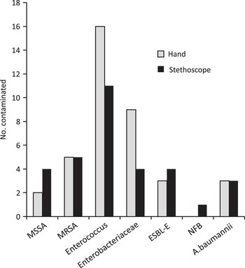
The frequency of recovery of microorganisms varied between genus/species (Figure 2). For both physicians’ hands and stethoscopes, the most frequently recovered microorganism was Enterococcus spp. (16/56 [29%] and 11/56 [20%], respectively), followed by Enterobacteriaceae (9/56 [16%] and 4/56 [7%], respectively). Methicillin-susceptible S. aureus, MRSA, ESBL-E, and A. baumannii were slightly less frequently found and were all recovered from 4%–9% of the samples from either the hands or stethoscopes. Finally, other nonfermenting gram-negative bacilli were not recovered from any physician’s hands but were recovered once from a stethoscope.
Bar chart showing the frequency of recovery of various microorganisms from stethoscopes and physicians’ hands following 56 standardized physical examinations. ESBL-E, extended-spectrum β-lactamase–producing Enterobacteriaceae; MSSA, methicillin-sensitive Staphylococcus aureus; MRSA, methicillin-resistant S. aureus; NFB, nonfermenting gram-negative bacilli.

Correlation Between Hand and Stethoscope Contamination
For all groups of microorganisms, there was a strong and significant association between the percentage of recovery from stethoscopes and the percentage of recovery from the physician’s hands (ρ=0.79; P=.04). Organisms that were more frequently recovered from the hand—such as Enterococcus spp. and MRSA—were also more frequently recovered from stethoscopes, whereas organisms that were infrequently recovered from hands—such as A. baumannii and other nonfermenting gram-negative bacilli—were also rarely recovered from stethoscopes.
Factors Predicting Heavy Stethoscope Contamination
By univariate analysis, the following variables were associated with heavy contamination of the stethoscope diaphragm (P<.05): greater body mass index, higher bacterial count on the patient’s skin, and higher humidity of the patient’s skin. A dry skin was associated with a decreased likelihood of heavy diaphragm contamination. Male sex, higher bacterial count of the patient’s skin, and reception of a bed bath rather than a shower or sink bath were significantly associated with heavy contamination of the stethoscope tube.
In multivariate analysis (Table 3), the level of the patient’s skin contamination was independently associated with both contamination of the diaphragm (adjusted odds ratio [aOR], 1.001; P=.007) and the tube (aOR, 1.001; P=.003). In addition, male sex (aOR, 28.24; P=.01) and reception of a bed bath (aOR, 7.52; P=.048) were associated with heavy tube contamination.
Predictors of Heavy Stethoscope Contamination by Multivariate Analysis

NOTE. BMI, body mass index (calculated as weight in kilograms divided by height in meters squared); CFU, colony-forming unit; OR, odds ratio.
a As opposed to corporal hygiene with a shower, with a bathtub, or at the sink.
DISCUSSION
Even though healthcare workers’ hands represent the main vector of cross-transmission in hospitals, the role of medical equipment is becoming increasingly recognized.Reference Matsuo, Oie and Furukawa 10 – Reference Whitehead, Thompson and Lewis 13 To our knowledge, this study is the first to investigate factors that influence contamination of stethoscopes following a physical examination, and the first to demonstrate that the main predictor of contamination is the level of bacterial contamination of the patient’s skin. Patients with more heavily colonized skin will contaminate the stethoscope more readily. This finding shows that “contagiousness” is not equivalent among patients and suggests that source control (ie, reducing the bioburden on the patient’s skin) may be useful to interrupt cross-transmission.Reference Vernon, Hayden, Trick, Hayes, Blom and Weinstein 14 Also, reception of a bed bath instead of a shower or a sink bath was associated with heavy stethoscope tube contamination. We hypothesize that this may be due to the lower efficacy of bed baths to remove bacteria from the patient’s skin.Reference Grice, Kong and Conlan 15 This could also be due to the rapid recolonization of the skin by bacteria present on the bed sheets following bathing. Furthermore, male sex was associated with heavy tube contamination. This unexpected finding may be related to the documented influence of gender on skin microbiome.Reference Callewaert, Kerckhof, Granitsiotis, Van Gele, Van de Wiele and Boon 16 Surely, more studies will be required to shed light on these findings.
We have previously shown the presence of a strong correlation between hand and stethoscope contamination for total aerobic count and MRSA. The present study shows that a similar correlation exists for other microorganisms. Taken together, all these findings reinforce the notion that stethoscopes have the potential to be a significant vector of transmission in hospitals.
Our study has some limitations. It was conducted in a single hospital with the participation of a limited number of physicians and patients. We used a convenience-based strategy to recruit patients. The high number of variables explored in this study may increase the risk of type 1 error. It was methodologically impossible to both detect the presence of multiple different pathogens and quantify them. This study also assessed gloved hand contamination, rather than bare hand contamination.Reference Lingaas and Fagernes 17 , Reference Vajravelu, Guerrero, Jury and Donskey 18 We assessed contamination of 4 regions of physicians’ dominant hands, 2 sections of stethoscopes, and only a single region of patient skin. Contamination of the entire surfaces of hands and stethoscopes was not assessed because these are technically difficult to evaluate.
In conclusion, this study shows that stethoscope contamination following a single physical examination is not negligible and is related to the level of contamination of the patient’s skin. Whether contamination of stethoscopes could be interrupted through source control should be investigated.
ACKNOWLEDGMENTS
Financial support. University of Geneva Hospitals (grant CGR 75308) and the Swiss National Science Foundation (grant 3200B0-122324/1 for hand hygiene research activities).
Potential conflicts of interest. All authors report no conflicts of interest relevant to this article.