Introduction
Over 800 000 people die by suicide each year, devastating families, communities and societies. At the time of suicide, 60% have a mood disorder, such as bipolar disorder (BD) or major depressive disorder (MDD) (Sher et al., Reference Sher, Oquendo and Mann2001), and the lifetime risk of suicide in these groups ranges from 5% to 11% (Angst et al., Reference Angst, Angst, Gerber-Werder and Gamma2005; Isometsa, Reference Isometsa2014). Suicidal ideation is the best predictor of imminent risk of suicide attempts and suicide (Du et al., Reference Du, Zeng, Liu, Tang, Meng, Li and Fu2017), but sometimes those at greatest risk deny ideation because they seek to avoid being thwarted in their attempt. Therefore a novel alternative approach is to determine a brain signature that underlies suicidal ideation, which may allow the detection of patients at high risk and inform the development of targeted prevention.
Suicidal thinking is usually ephemeral and therefore its neural substrate may be more closely related to functional alterations in brain circuitry than neuroanatomical ones (Balcioglu and Kose, Reference Balcioglu and Kose2018). Thus the majority of studies in this field have used functional magnetic resonance imaging (fMRI) coupled with cognitive/affective neuroscience paradigms to interrogate the relationships between cognitive, emotional, and behavioral components of suicide and neural function (Serafini et al., Reference Serafini, Pardini, Pompili, Girardi and Amore2016; Balcioglu and Kose, Reference Balcioglu and Kose2018). However, the integration of imaging findings and the derivation of robust conclusions has been difficult because of the variety of experimental tasks employed (Ordaz et al., Reference Ordaz, Goyer, Ho, Singh and Gotlib2018). To overcome this problem studies have started to focus on the intrinsic activity of the brain using resting state fMRI (rsfMRI).
Resting brain activity has been linked to higher order cognitive processes and several studies suggest that an impairment of cognitive control may underpin the high suicide rates in individuals with depression (Pu et al., Reference Pu, Setoyama and Noda2017). However, thus far, only a handful of studies have examined its relationship to suicide in the context of mood disorders (Fan et al., Reference Fan, Wu, Yao and Dong2013; Cao et al., Reference Cao, Chen, Chen, Ai, Gan, Wang, Lv, Zhang, Zhang, Wang, Kuang and Fang2016; Du et al., Reference Du, Zeng, Liu, Tang, Meng, Li and Fu2017; Kang et al., Reference Kang, Na, Choi, Kim, Son and Lee2017; Ordaz et al., Reference Ordaz, Goyer, Ho, Singh and Gotlib2018). In recent years a growing number of researchers have attempted to understand the dynamics of the brain at rest using a network approach (Calhoun et al., Reference Calhoun, Adali, Pearlson and Pekar2001), in which networks are thought to represent functional modules of the brain that may provide valuable diagnostic and prognostic information (Whitfield-Gabrieli and Ford, Reference Whitfield-Gabrieli and Ford2012). To date, as far as we know, only two studies have used this approach but both of these have focused on adolescents (Zhang et al., Reference Zhang, Chen, Kuang, Cao, Zhang, Ai, Wang, Zhang, Wang, Liu and Fang2016; Ordaz et al., Reference Ordaz, Goyer, Ho, Singh and Gotlib2018). The human brain is a complex dynamical system generating a multitude of oscillatory waves. Studies have demonstrated that multiple frequency bands within the 0.004–0.15 Hz range contribute to the signal of resting state networks (RSNs) (Balsters et al., Reference Balsters, Robertson and Calhoun2013), but no studies have investigated spectral properties of the BOLD signal to understand suicide in mood disorder patients. The functional connectivity between two brain regions (using seed-based analysis) has been used to examine differences between suicide attempters (SA) and non-attempters (NA) (Kang et al., Reference Kang, Na, Choi, Kim, Son and Lee2017) but no studies have investigated functional connectivity between RSNs to understand suicide in mood disorders even though impaired functional network connectivity (FNC) among RSNs has been reported in mood disorder patients (Das et al., Reference Das, Calhoun and Malhi2014).
A triple network model has been proposed to understand cognitive and affective disorders (Menon, Reference Menon2011). These networks are the default mode (DMN), central executive (CEN), and salience (SN) networks (Menon, Reference Menon2011). Using this network approach, one study has investigated this triad of networks and another study has interrogated the DMN to understand suicidal activity in depressed adolescents (Zhang et al., Reference Zhang, Chen, Kuang, Cao, Zhang, Ai, Wang, Zhang, Wang, Liu and Fang2016; Ordaz et al., Reference Ordaz, Goyer, Ho, Singh and Gotlib2018). Traditionally the basal ganglia are linked to motor functions and movement, but functional magnetic resonance imaging suggests that these nuclei are also involved in more complex cognitive and affective processes (Arsalidou et al., Reference Arsalidou, Duerden and Taylor2013). A large and diverse literature has implicated structural and functional abnormalities in striatal structures in mood disorders (Marchand and Yurgelun-Todd, Reference Marchand and Yurgelun-Todd2010), and more precisely, fronto-striatal dysfunction has been implicated in suicidal thoughts and behaviors (Marchand, Reference Marchand2012; Balcioglu and Kose, Reference Balcioglu and Kose2018).
Thus, to date, no studies have investigated functional connectivity specifically within (examining networks' spatial maps and time courses), and between (using FNC), these four key intrinsic networks (ECN, DMN, SN, and BGN) to explicate suicide in adult mood disorder patients.
Therefore in our study, we first examined how intrinsic activity within, and between all four of these networks (ECN, DMN, SN, and BGN), differed between healthy adults and depressed patients. It is important to note that the depressive phase of illness was chosen because suicidal acts occur predominantly when individuals are depressed (Sher et al., Reference Sher, Oquendo and Mann2001; Isometsa, Reference Isometsa2014). We then divided patients into two groups; those who have attempted suicide (at least once in their lifetime; SA), and those who have not (NA), and compared them to healthy control participants (HC). Having identified differences, we then examined whether these differences underlie patients' suicidal activity. Since gender influences the rate of suicide (Vijayakumar, Reference Vijayakumar2015), the effect of gender was also investigated.
Materials and methods
Participants
One hundred and forty-five participants aged 18–65 years (mood disorder patients: 79; healthy controls: 66) took part in the study. All participants underwent a structured clinical interview by a psychiatrist (GSM) to determine whether they met DSM-5 criteria for either bipolar disorder (BD) or major depressive disorder (MDD). Healthy controls were also assessed to ensure the absence of psychiatric symptoms and the absence of any history of a psychiatric disorder. Of the 79 patients 43 met criteria for a diagnosis of BD and 36 for MDD. The male to female ratio was 41:38 in patients and 32:34 in healthy controls. Twenty-five patients had attempted suicide at least once in their lifetime. Participants were recruited through the CADE Clinic (http://www.cadeclinic.com) at Royal North Shore Hospital and via local advertisements. Ethical permission for this study was granted by the University of Sydney and Royal North Shore Hospital. Written consent was obtained from all participants prior to testing in accordance with National Health and Medical Research Council guidelines.
Exclusion criteria included current hospitalization, substance abuse, a history of traumatic head injury, neurological illness (e.g. seizures or stroke), learning or developmental disorders, or poor English proficiency. Participants were excluded from analysis if their functional scans showed extreme motion (translation >3 mm, roughly one voxel and rotation >1°). All participants were instructed to refrain from alcohol or recreational drug use at least 24 h prior to the brain scan and from caffeine or nicotine within 1 h of the scan.
Questionnaires
A series of clinical questionnaires were administered including the Columbia-Suicide Severity Rating Scale (CSSRS) (Posner et al., Reference Posner, Brown, Stanley, Brent, Yershova, Oquendo, Currier, Melvin, Greenhill, Shen and Mann2011), which measures suicidal ideation and behavior over the past month and lifetime, the State and Trait Anxiety Inventory (STAI), and the Hamilton Depression (HAM-D) Scale.
MRI data acquisition
Imaging datasets (structural and functional) were acquired on a research-dedicated University of Sydney 3T Siemens Magnetom Trio Scanner, based at the Advanced Research and Clinical High-field Imaging (ARCHI) facility at Royal North Shore Hospital. A T2*-weighted gradient-echo echo-planar imaging (EPI) sequence [29 axial slices, slice thickness 4 mm with 1 mm gap, repeat time (TR) = 2000 ms, echo time (TE) = 35 ms, flip angle (FA) = 70°, 64 × 64 matrix] was used to acquire 150 whole-brain volumes of functional data. A high-resolution T1-weighted structural image was also acquired for precise localization of brain activity using a magnetization prepared rapid gradient echo (MPRAGE) sequence (TR = 1570 ms, TE = 3.22 ms, FA = 15°, matrix 256 × 256, 192 slices). Participants were instructed to keep their eyes open during the functional scan and stare passively at a foveally presented fixation cross, because this facilitates network delineation more so than eyes-closed conditions (Van Dijk et al., Reference Van Dijk, Hedden, Venkataraman, Evans, Lazar and Buckner2010) and mitigates against participants falling asleep. Head motion during scanning was restrained using foam pads inserted on each side, and all participants were awake and alert at the start and conclusion of scanning.
fMRI data analysis
Pre-processing of data
Pre-processing was conducted using SPM8 software (http://www.fil.ion.ucl.ac.uk/spm/software/spm8). Each participant's functional and structural images were first inspected visually for scanner artifacts and gross anatomical abnormalities, and then re-oriented so that the origin of the image lay within 3 cm of the anterior commissure. The initial five images were discarded to remove longitudinal equilibration effects. Rigid body motion correction was performed using INRIAlign – a motion correction algorithm (Freire and Mangin, Reference Freire and Mangin2001) which is unbiased by local signal changes. It was followed by a slice-timing correction to account for timing differences in slice acquisition where the middle slice was used as the reference frame. Then these images were spatially normalized to a common stereotactic space using the Montreal Neurological Institute (MNI) EPI template and spatially smoothed with a Gaussian kernel of 8 mm3 full width at half maximum. Following spatial normalization, the data (originally acquired at 3.75 × 3.75 × 5 mm3) were slightly subsampled to 3 × 3 × 3 mm3, resulting in 53 × 63 × 46 voxels.
Identification of resting state networks and visualization
Prior to performing group independent component analysis (ICA), each voxel time course was variance normalized as this approach has been shown to yield better decompositions of subcortical and cortical sources (Damaraju et al., Reference Damaraju, Allen, Belger, Ford, McEwen, Mathalon, Mueller, Pearlson, Potkin, Preda, Turner, Vaidya, van Erp and Calhoun2014). Pre-processed time series were analyzed using Group ICA (Calhoun et al., Reference Calhoun, Adali, Pearlson and Pekar2001; Erhardt et al., Reference Erhardt, Rachakonda, Bedrick, Allen, Adali and Calhoun2011) within the GIFT toolbox (http://mialab.mrn.org/software/gift, version 4.0b). The number of independent ‘sources’/components/networks was estimated to be 21 by using a minimum description length (MDL) criteria modified to allow for spatially correlated data (Li et al., Reference Li, Adali and Calhoun2007) and identified using the Infomax algorithm (Bell and Sejnowski, Reference Bell and Sejnowski1995). To ensure the stability of estimation, this algorithm was repeated 10 times in ICASSO (http://research.ics.tkk.fi/ica/icasso/) and the most central run was selected and analyzed further. Participant-specific spatial maps (SMs) and time courses (TCs) were then obtained using the spatio-temporal regression back reconstruction approach implemented in GIFT software.
Selection of components/networks of interest
A network was included in the analysis (1) if its peak activation clusters fell within gray matter and was now overlapping with known vascular, susceptibility, ventricular, and edge regions corresponding to head motion, (2) if it had more spectral power in the lower frequency range (0.01–0.10 Hz) compared to the high frequency range (0.15–0.25 Hz) (Allen et al., Reference Allen, Erhardt, Damaraju, Gruner, Segall, Silva, Havlicek, Rachakonda, Fries, Kalyanam, Michael, Caprihan, Turner, Eichele, Adelsheim, Bryan, Bustillo, Clark, Feldstein Ewing, Filbey, Ford, Hutchison, Jung, Kiehl, Kodituwakku, Komesu, Mayer, Pearlson, Phillips, Sadek, Stevens, Teuscher, Thoma and Calhoun2011), and (3) if it represents either one of the four networks such as ECN, DMN, SN, and BGN or any network showing primary activity in the prefrontal cortices (frontal network).
Comparison between patients and healthy controls
Three outcome variables describing within and between networks functional connectivity were examined.
Specifically, within network connectivity was assessed using:
(1) the intensities of the network spatial map, which represents the connectivity and degree of co-activation within a network and,
(2) the power spectra of the network time course, which represents the level of coherent activity within a network.
Between networks connectivity was determined using functional network connectivity (FNC) (Jafri et al., Reference Jafri, Pearlson, Stevens and Calhoun2008). Before conducting comparisons between groups, a participant's time course from each component/network was detrended, despiked and orthogonalized with respect to their motion parameters (Damaraju et al., Reference Damaraju, Allen, Belger, Ford, McEwen, Mathalon, Mueller, Pearlson, Potkin, Preda, Turner, Vaidya, van Erp and Calhoun2014). Analyses were performed using the MANCOVAN toolbox within GIFT. In the model, addition to diagnosis, mean framewise displacement was also entered as a nuisance covariate to account for any residual motion-related variance in ICA derived measures (Damaraju et al., Reference Damaraju, Allen, Belger, Ford, McEwen, Mathalon, Mueller, Pearlson, Potkin, Preda, Turner, Vaidya, van Erp and Calhoun2014).
First, a component's spatial map was thresholded based on the distribution of voxelwise t-statistics so that it represents voxels that have shown strong and consistent activation across participants (Allen et al., Reference Allen, Erhardt, Damaraju, Gruner, Segall, Silva, Havlicek, Rachakonda, Fries, Kalyanam, Michael, Caprihan, Turner, Eichele, Adelsheim, Bryan, Bustillo, Clark, Feldstein Ewing, Filbey, Ford, Hutchison, Jung, Kiehl, Kodituwakku, Komesu, Mayer, Pearlson, Phillips, Sadek, Stevens, Teuscher, Thoma and Calhoun2011) and includes regions that are most associated with the component's time course. Then these thresholded SMs were compared between groups. To determine differences between groups in a component's spectral power the time course of a component was transformed to 129 spectral bins using fast Fourier transform and the power in each bin was compared. For comparing differences between groups in functional connectivity between components, first FNC was calculated following the procedure described by Jafri et al. (Reference Jafri, Pearlson, Stevens and Calhoun2008). For this, the time course data associated with the selected components were filtered using a fifth-order Butterworth low-pass filter with a high-frequency cut-off of 0.15 Hz (Allen et al., Reference Allen, Erhardt, Damaraju, Gruner, Segall, Silva, Havlicek, Rachakonda, Fries, Kalyanam, Michael, Caprihan, Turner, Eichele, Adelsheim, Bryan, Bustillo, Clark, Feldstein Ewing, Filbey, Ford, Hutchison, Jung, Kiehl, Kodituwakku, Komesu, Mayer, Pearlson, Phillips, Sadek, Stevens, Teuscher, Thoma and Calhoun2011). Then for each participant, correlations between pair-wise combinations were calculated using Pearson's correlations and these values were then transformed to z-scores using Fisher's transformation to use as a measure of FNC. Then significant differences between groups in these measures were determined using the MANCOVAN utility within GIFT (Allen et al., Reference Allen, Erhardt, Damaraju, Gruner, Segall, Silva, Havlicek, Rachakonda, Fries, Kalyanam, Michael, Caprihan, Turner, Eichele, Adelsheim, Bryan, Bustillo, Clark, Feldstein Ewing, Filbey, Ford, Hutchison, Jung, Kiehl, Kodituwakku, Komesu, Mayer, Pearlson, Phillips, Sadek, Stevens, Teuscher, Thoma and Calhoun2011) and results were corrected for multiple comparisons for univariate analyses using the false discovery rate (p < 0.05) and cluster size of minimum 10 voxels.
Comparisons among suicide attempters (SA), non-attempters (NA) and healthy controls (HC)
In order to understand whether an attempt of suicide altered brain activity patterns the neural activity of HC was compared against that of SA and NA. We then compared SA and NA directly across the three key outcome measures. All results were corrected using a false discovery rate of p < 0.05 and a cluster size minimum of five voxels.
Correlation analyses
To assess for any association between brain networks and suicidality the observed differences among patients and healthy controls (identified in the sections ‘Comparison between patients and healthy controls’ and ‘Comparisons among suicide attempters (SA), non-attempters (NA) and healthy controls’) were correlated with the suicidal activity (suicidal ideation or behavior) patients experienced over the past month (recent) and lifetime (lifelong) (measured using the CSSRS). Since mood state could be an important confounding factor depression severity (measured using HAM-D) was controlled for in the correlation analyses. These analyses were performed in SPSS.
Also to understand whether gender plays a role in these associations the patient groups (SA and NA) were further subdivided according to gender, and these associations were investigated in each group.
For differences in spatial maps a mask was created from the cluster showing differences between groups, and then activity from the masked region was determined for each participant. For differences in spectral power the component's frequency was divided into six equal bins (0.04, 0.08, 0.12, 0.16, 0.20, and 0.24 Hz) and spectral power from the bin showing significant differences between groups was chosen.
Results
Participants
There were no statistically significant differences between patients and healthy controls in terms of age (HC: 37.32 ± 13.7; MDP; 37.5 ± 13.5), gender (HC: male 32, female 34; MDP: male 41, female 38), and education level. Among 79 patients 12 were not taking any pharmacological agent for mood disorders, 14 were taking one type of agent and the rest were on a combination of agents. In patients, mean suicidal activity (ideation or behavior) over lifetime was (4.05 ± 2.5) and over the past month was (1.1 ± 1.1) (Table 1).
Demographic and clinical variables of patients and healthy controls

Networks of interest
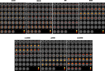
Out of 21 networks, seven networks were chosen as our networks of interest. These included two ECN networks – left ECN (lECN) and right ECN (rECN); three DMN networks – posterior DMN (pDMN), dorso-medial DMN (dmDMN), and ventro-medial DMN (vmDMN); and two networks reflecting the SN and BGN. These networks have previously been identified from resting data acquired from a large number of healthy participants (Allen et al., Reference Allen, Erhardt, Damaraju, Gruner, Segall, Silva, Havlicek, Rachakonda, Fries, Kalyanam, Michael, Caprihan, Turner, Eichele, Adelsheim, Bryan, Bustillo, Clark, Feldstein Ewing, Filbey, Ford, Hutchison, Jung, Kiehl, Kodituwakku, Komesu, Mayer, Pearlson, Phillips, Sadek, Stevens, Teuscher, Thoma and Calhoun2011). The cluster stability/quality (Iq) index for these chosen networks was very high (Iq > 0.9) (determined by running ICASSO 10 times). To visualize the spatial maps of a component, all participants' maps for that particular component were entered into a random-effect analysis model (one sample t test in SPM8) corrected for multiple comparisons using the family-wise error (FWE, p < 0.05) and an extent threshold of 50 voxels. Chosen networks are shown in Fig. 1 and primary regions within each network are provided in online Supplementary Table S1 of the Supplementary Materials.
Chosen networks of interest: left executive control network (LECN), right executive control network (RECN), salience network (SN), basal ganglia network (BGN), ventro-medial default mode network (vmDMN), posterior default mode network (pDMN), and dorso-medial default mode network (dmDM).

Differences between patients and healthy controls in within and between network functional connectivity
Groups differed only in within network functional connectivity, there was no difference in between network connectivity. Spatial Maps of the pDMN and BGN differed between depression and healthy control groups (see Fig. 2, panel A). HC had greater activity in the upper ventral striatum of the BGN (see Fig. 2, panel A, top) and lower activity in posterior cingulate cortex/precuneus region of the pDMN compared with depressed patients (see Fig. 2, panel A, bottom). Groups also differed (t = 3.72, p = 0.0003) in the spectral power of the dmDMN in the lower frequency range (0.04–0.08 Hz) (see Fig. 2, panel B) and overall HC had greater spectral power than the depressed group.
Panel A: Spatial map (SM) differences between healthy controls (HC) and mood disorders patients (MDP). Compared to MDP, HC displayed increased activity in the caudate region of the basal ganglia network (top left panel) and decreased activity in the posterior cingulate/precuneus region of the default mode network (bottom left panel). Panel B: Spectral power (SP) differences between healthy controls (HC) and mood disorder patients (MDP). Compared to MDP, HC displayed increased low frequency (0.04–0.08 Hz) spectral power in the dorso-medial default mode network (dmDMN).

Comparisons among suicide attempters (SA), non-attempters (NA) and healthy controls (HC)
The division of patients according to whether they had attempted suicide (SA) or not (NA) revealed that, compared to HC, SA had greater activity in the precuneus region of the pDMN and in the medial prefrontal region of the dmDMN but reduced activity in the inferior frontal region of the SN (see Fig. 3, panel A) whereas NA had reduced activity in the caudate region of the BGN and diminished spectral power in the low-frequency range of the dmDMN (see Fig. 3, panel B). Differences between SA and NA patients did not survive multiple comparison correction but at an uncorrected level SA had greater midline circuitry activity encompassing the precuneus and medial prefrontal brain regions compared to SA (not shown).
Differences in neural activity between suicide-attempters (SA) and non-attempters (NA) compared to healthy controls (HC). SA had greater precuneus activity (panel A), whereas NA had reduced activity of the caudate and diminished spectral power in the low-frequency range (panel B). SA also had greater activity in the medial frontal region of the dorso-medial default mode network (dmDMN; panel A) and reduced activity in the inferior frontal region of the salience network (SN; panel A).

Correlation analyses
Whole patient group
Only differences in the default mode networks (pDMN, dmDMN) displayed differential positive correlations with the scores of suicidal activity (ideation or behavior) depending on whether activity is recent (over the past month) or lifelong (measured over lifetime). Specifically, neural activity in the pDMN had positive correlations with scores of recent suicidal activity (r = 0.307, p = 0.006) whereas low frequency spectral power in the dmDMN displayed positive correlations with scores of lifelong suicidal activity (r = 0.282, p = 0.012).
Suicide attempters (SA)
In SA, only neural activity in the pDMN displayed positive correlations with scores of recent suicidal activity (r = 0.521, p = 0.009). This association was only significant in males at a trend level (N = 7; r = 0.646, p = 0.060).
Suicide non-attempters (NA)
In NA, only low-frequency spectral power in the dmDMN displayed significant positive correlations with scores of lifelong suicidal activity (r = 0.391, p = 0.004). This association was significant in females (N = 20; r = 0.479, p = 0.024) and at a trend level in males (N = 28; r = 0.347, p = 0.060). In females, lifelong suicidal activity also displayed positive associations with activity of the caudate region of the BGN (r = 0.429, p = 0.047) (Fig. 4).
Differences in DMN activity that displayed association with suicidal activity suicidal in patients. In suicide attempters (SA): greater activity in the posterior default mode network (pDMN) was associated with increased suicidal activity in the past month (recent suicidal activity). In non-attempters (NA)s: greater spectral power in the dorso-medial default mode network (dmDMN) was associated with increased lifetime suicidal activity.

Discussion
The primary aim of the present study was to elucidate neural mechanisms underlying suicidal activity (ideation and/or behavior) in mood disorder patients. It also investigated to what extent these mechanisms were determined by a previous suicide attempt and whether gender plays a role. To this end, the study investigated the intrinsic functional connectivity both within, and between, networks known to be involved in higher-order cognition and compared healthy controls and mood disorder patients with and without suicide attempts.
Three central findings emerged. First, relative to HC, mood disorder patients were characterized by greater functional activity in the posterior components of the DMN (pDMN) and lower functional activity in the BGN along with low-frequency spectral power in the dorsal medial frontal regions of the DMN (dmDMN). Second, increased activity in the pDMN is primarily evident in SA whereas reduced BGN activity and less spectral power of dmDMN was more pronounced in NA. Third, pDMN activity had a positive association with recent (past month) suicidal activity in SA, whereas dmDMN spectral power had a positive association with lifelong (lifetime) suicidal activity in NA.
Our finding of DMN abnormalities in mood disorder patients and their association with suicidal activity is in keeping with the findings of another important study where abnormalities in the DMN differentiated suicidal ideators and healthy controls (Chase et al., Reference Chase, Segreti, Keller, Cherkassky, Just, Pan and Brent2017). The DMN (pDMN, dmDMN) involves brain regions (posterior and frontal medial cortices) that form part of the Cortical Midline Structures (CMS), collective brain regions that have been repeatedly implicated in the neuropathology of suicide-related thoughts and behaviors (Marchand, Reference Marchand2012). Our study has found that patients who have attempted suicide have greater activity in these structures compared to both HC (Fig. 3) and non-attempters (at an uncorrected level). The involvement of the CMS in suicide is predicated on their key role in self-referential thinking (Northoff et al., Reference Northoff, Heinzel, de Greck, Bermpohl, Dobrowolny and Panksepp2006), and within the CMS it is specifically the dorso-medial prefrontal cortex that is thought to be involved in the evaluation and appraisal of self-referential stimuli. This contrasts then with the posterior medial cortex which is more so involved in temporally integrating new self-referential information with one's emotional and autobiographical self (Northoff and Bermpohl, Reference Northoff and Bermpohl2004). This anterior–posterior dissociation within the CMS has also been shown to vary according to the focus that self-referent thought processing adopts at any given time. Specifically, the anterior medial cortex shows greater activity when cued to engage in hopeful and aspirational thinking, whereas the posterior medial cortex shows greater activity when cued to think about one's duties and obligations (Johnson et al., Reference Johnson, Nolen-Hoeksema, Mitchell and Levin2009). Hence the finding of an association between activity in these brain regions of the CMS with suicide-related thoughts and behaviors in patients, suggests that suicidal ideation in patients with mood disorders perhaps stems from abnormal self-referential processing, which may be a consequence of altered integrity within the DMN. The DMN has been repeatedly shown to be active when engaged in self-reflection (rumination) – and in turn it is known that ruminative self-reflection is associated with suicidal ideation and behavior (Rogers and Joiner, Reference Rogers and Joiner2017). In other words, difficulty in regulating negative thinking, which can result in dysregulation of emotion, may in turn contribute to suicidality – in keeping with a recently posited model (Malhi et al., Reference Malhi, Outhred, Das, Morris, Hamilton and Mannie2018).
Interestingly, abnormalities in the DMN encompassing anterior and posterior medial cortices have also been associated with suicidal ideation and behavior in adolescents with mood disorders (Zhang et al., Reference Zhang, Chen, Kuang, Cao, Zhang, Ai, Wang, Zhang, Wang, Liu and Fang2016; Ordaz et al., Reference Ordaz, Goyer, Ho, Singh and Gotlib2018). Specifically, Ordaz and colleagues (Ordaz et al., Reference Ordaz, Goyer, Ho, Singh and Gotlib2018) found that lower coherence in the anterior DMN predicts greater lifetime severity of suicidal ideation. Furthermore, according to Zhang and colleagues (Zhang et al., Reference Zhang, Chen, Kuang, Cao, Zhang, Ai, Wang, Zhang, Wang, Liu and Fang2016) abnormal connectivity in the posterior medial cortex may be the substrate for suicidal behavior in depressed adolescent patients.
Thus, abnormal activity in the DMN and its association with suicidal activity both in adolescence and adulthood suggests that the DMN plays a critical role in suicidal activity in depressed patients with mood disorders, and that this perhaps first emerges and is ongoing as the brain matures from youth through to adulthood.
In addition to the important role of cortical midline structures, basal ganglia dysfunction has long been implicated in mood disorders (Marchand and Yurgelun-Todd, Reference Marchand and Yurgelun-Todd2010), and our finding of reduced activity in the BGN in depressed patients further substantiates this. Clinically, impaired ventral striatum functioning has been shown to contribute to depressive episodes (Pan et al., Reference Pan, Sato, Salum, Rohde, Gadelha, Zugman, Mari, Jackowski, Picon, Miguel, Pine, Leibenluft, Bressan and Stringaris2017) that feature symptoms such as hopelessness, negative affect and recurrent thoughts about death and suicide (Renteria et al., Reference Renteria, Schmaal, Hibar, Couvy-Duchesne, Strike, Mills, de Zubicaray, McMahon, Medland, Gillespie, Hatton, Lagopoulos, Veltman, van der Wee, van Erp, Wittfeld, Grabe, Block, Hegenscheid, Volzke, Veer, Walter, Schnell, Schramm, Normann, Schoepf, Konrad, Zurowski, Godlewska, Cowen, Penninx, Jahanshad, Thompson, Wright, Martin, Christensen and Hickie2017). However, our study did not find any direct association between activity in the BGN and suicidal ideation and behavior in the whole patient group. But functional connections between the DMN and BGN are necessary for exercising cognitive flexibility, and a compromise of this capacity through the development of cognitive rigidity is thought to be a problem for individuals with suicidal thinking (Marzuk et al., Reference Marzuk, Hartwell, Leon and Portera2005). Specifically, cognitive rigidity is thought to diminish their capacity to alter and adjust their thinking, or line of action, according to environmental demands. This is critical because it limits the ability of individuals with suicidal thinking, to generate alternative solutions once they are in a predicament. Such adaptation is necessary in order to be able to modify the negative schema and extinguish hopeless and helpless self-referent ideation, which is a strong feature and precursor of suicide (Surrence et al., Reference Surrence, Miranda, Marroquin and Chan2009; Stange et al., Reference Stange, Hamilton, Burke, Kleiman, O'Garro-Moore, Seligman, Abramson and Alloy2015). Though we did not observe any differences in functional connectivity between these networks in patients and healthy controls, the division of patients according to suicide attempts displayed differential correlation between BGN and DMN activity. In those who had not attempted suicide, their BGN activity correlated positively with both posterior (precuneus of pDMN) (r = 0.365, p = 0.007) and anterior (medial prefrontal of dmDMN) (r = 0.503, p = 0.000) medial cortivity, but in those who had attempted suicide at any time in their lifetime there was a positive correlation between the BGN and anterior medial cortex (r = 0.553, p = 0.005). This suggests that either the process of undertaking a suicide attempt impairs cognitive flexibility, or that it occurred because of a lack of cognitive flexibility in the first place. Clearly, causality in this regard is difficult to determine without conducting a longitudinal study.
Our finding of differential associations of recent and lifelong suicidal activity with the different regions of the DMN in groups that have and have not attempted suicide is interesting. These results suggest that an attempt at suicide alters the focus of subsequent self-referent thought processing (Marzuk et al., Reference Marzuk, Hartwell, Leon and Portera2005; Surrence et al., Reference Surrence, Miranda, Marroquin and Chan2009; Stange et al., Reference Stange, Hamilton, Burke, Kleiman, O'Garro-Moore, Seligman, Abramson and Alloy2015). Specifically, in those who have not attempted suicide, their suicidal activity is modulated by a region which shows greater activity when cued to engage in hopeful and aspirational thinking, but in those who have attempted suicide it is governed by a region which shows greater activity when cued to think about one's duties and obligations (Johnson et al., Reference Johnson, Nolen-Hoeksema, Mitchell and Levin2009). This intriguing finding requires further inquiry.
Collectively our findings are in line with the suggestion that CMS and striatal function play a role in suicide-related thoughts and behaviors. This is important because it suggests that interventions targeting self-referential thinking may target core deficits and therefore be particularly beneficial (Marchand, Reference Marchand2012). Training in this regard leading to an enhancement of cognitive flexibility may be of help to patients with suicidal ideation and behavior.
Several limitations of the present study must be considered. First, there may be other networks that may play an equally important role in suicidal ideation/behavior that have not been considered in this study. Second, there is a potential effect of psychoactive medications in rsfMRI and this could not be controlled for because the majority of patients were using a number of combinations of medications. Third, currently the origin of rsfMRI spectral power at different frequencies is not fully understood and therefore its significance is speculative, although similar findings of reduced spectral power in the low-frequency range in the dorsal anterior cingulate cortex (part of dmDMN) in suicidal ideators have been reported previously (Chase et al., Reference Chase, Segreti, Keller, Cherkassky, Just, Pan and Brent2017). This study provides some useful insights into the networks involved in suicidal thinking and behaviors and how suicide attempts affect these networks but only in the context of depression in patients with mood disorders. It assessed patients only when they were depressed, and so in future studies, it would be useful to adopt a longitudinal approach and examine how functioning of these networks changes when patients are no longer depressed or suicidal. Finally, it is worthwhile noting that our findings of the influence of gender are based on modest sample sizes. And therefore, although our findings indicate that the effect of lifelong suicidal activity and suicide attempt together may change brain activity patterns differentially in males vs females this finding is at best preliminary and needs replication using larger numbers of both males and females.
Author ORCIDs
Gin S. Malhi, 0000-0002-4524-9091; Pritha Das, 0000-0003-4371-7960
Supplementary material
The supplementary material for this article can be found at https://doi.org/10.1017/S0033291719001132.
Financial support
This research has been made possible with funding from an NHMRC Program Grant (APP1073041) and The American Foundation for Suicide Prevention (PRG-0-090-14).
Conflict of interest
Gin S. Malhi has received grant or research support from National Health and Medical Research Council, NSW Health, Ramsay Health, American Foundation for Suicide Prevention, AstraZeneca, Eli Lilly & Co, Organon, Pfizer, Servier, and Wyeth; has been a speaker for AstraZeneca, Eli Lilly & Co, Janssen Cilag, Lundbeck, Pfizer, Ranbaxy, Servier, and Wyeth; and has been a consultant for AstraZeneca, Eli Lilly & Co, Janssen Cilag, Lundbeck, and Servier. Pritha Das, Tim Outhred, Richard Bryant, John Mann and Vince Calhoun have no disclosures.