I. INTRODUCTION
Insect cuticle is a fibrous composite material, which is designed to maintain and support the insect shape; it is also a means of locomotion and provides defense against the environment all with as little weight as possible. Cuticles can be distinguished as hard, soft, or rubberlike according to the amount of solid materials from which they are comprised. Indeed, the orientation and volume ratio of its main constituents (highly crystalline nanofibers of chitin, lipids, polyphenols, water content, type of protein) and degree of cross-linking give the composite different mechanical properties. Soft cuticle contains an equal weight fraction of chitin and protein and 40–75% water, whereas hard cuticle contains 15–30% weight fraction of chitin and 12% water. Thus, the stiffness of the different types of cuticle will vary from 1 kPa for the most hydrated structures to 20 GPa for the most tanned one.Reference Vincent and Wegst1
Rubberlike cuticle exhibits high elasticity, transparency, and is colorless. Previous works have shown that the great deformability and long-range elasticity of these structures are due to the presence of a large amount of an elastomeric protein known as resilin.Reference Weis-Fogh2 Resilin is secreted by epidermal cells into the subcuticular space where it is cross-linked forming a three-dimensional network. The protein binds with chitin within the structure forming an extracellular composite matrix.Reference Vincent3 Such a structure assists muscles specifically by storing and releasing energy with great efficiency; it acts as a shock absorber, a power amplifier, and a force control.Reference Vincent3–Reference Shewry, Tatham and Bailey5 For instance, the locust uses resilin to open its spiracle during flight.Reference Miller6 Also, resilin is of great importance in the jumping mechanism of insects such as fleas, beetles, or froghoppers. Specifically, jumping involves the ability of the ligament present in the jumping leg to release energy rapidly, which has been stored during a prolonged muscular action.Reference Burrows7 In cockroaches, resilin has been detected in joints connecting segments of the legs. In that particular location, resilin would function as an elastic antagonist to the actions of leg muscles.Reference Neff, Frazier, Quimby, Wang and Zill8 The rubberlike cuticle therefore is clearly involved in the running mechanism.
Although resilin was discovered in 1960 and has been investigated since then, many aspects of the mechanical characteristics of this elastomeric protein have yet to be investigated.Reference Weis-Fogh2 The study of rubberlike cuticle will provide knowledge about this constituent and about the function of the elastomeric protein within the structure. This is especially the case for comparisons of the properties of resilin in phenotypes that differ in function that are expected to be influenced by this protein.
Wing polymorphism is a common feature of many insect species in which morphs exist that are specialized for very different functions. In many insects, including Gryllus firmus, the focus of the present study, morphs differ in aspects of dispersal. One morph has fully developed wings and is capable of flight, whereas the alternate morph has shortened wings and cannot fly. In addition, these morphs also differ in walking and jumping ability, in some species. Thus, investigation of the structure–function relationship of the rubberlike joint of the leg of two-wing morphs of the sand field crickets Gryllus firmus is expected to give further insight regarding engineered, optimized bioinspired rubbery materials. In the present study, the dynamic mechanical properties of rubberlike cuticle displayed are identified and characterized in two morphs [long-wing (lw) and short-wing (sw)] of sand field crickets Gryllus firmus using dynamic nanoindentation techniques.
Nanoindentation has already shown its great applicability for characterizing biological structures such as arthropods. Enders et al.Reference Enders, Barbakadze, Gorb and Arzt9 illustrated that nanoindentation was sensitive enough for determining mechanical properties (i.e., hardness and elastic modulus) of compliant and highly structured biological composite materials such as the pitcher plant, the head part of the head–thorax articulation system of beetles, and parts of the wing-arresting system of dung beetles. A study of the beetle Pachnoda marginata highlighted the influence of a multilayered structure on nanoindentation data. Indeed, hardness and elastic modulus values of the layered material are dependent on indentation depth, on the hydration of the sample, and on the chemical treatment.Reference Barbakadze, Enders, Gorg and Artz10 More recently, Sun et al.Reference Sun, Tong and Ma11 showed nanoindentation as a powerful tool to detect variations in the reduced modulus and hardness of singular segments of three different kinds of dung beetle. Insect cuticle exhibits a wide range of mechanical properties with respect to the skeleton structure. Here we show that dynamic nanoindentation can capture the differences in the viscoelastic properties of rubberlike cuticle with respect to structure as well as gender and functional adaptation (i.e., differences between morphs) of Gryllus firmus.
II. MATERIALS AND METHODS
A. Insect material rearing conditions, and background on wing polymorphism
Gryllus firmus is wing-polymorphic, existing as a lw morph that is capable of flight, or as an obligately flightless sw morph. Wing polymorphism occurs commonly in many insect groups and is an important adaptation for locomotory ability at the expense of reproductivity: the dispersing form has considerably reduced egg production compared to the flightless morph. Importantly for the present study, morphs differ in many characteristics other than flight ability, namely their ability to disperse by walking as well as by flight, and in differences in jumping ability.
Adult male and female lw and sw morphs of the sand field cricket, G. firmus were used in this study. All morphs and genders were raised under the same environmental conditions and were handled under exactly the same conditions. Crickets were raised at the same density (40–60 adults per 38 L aquarium) under a light/dark (LD) photoperiod of 14:10 h at 28–30 °C. They were fed a dry diet mixture of casein, yeast, wheat germ, ascorbic acid, and mold initors. The crickets were raised until 10–12 days after the final moult. After that time they were collected and stored in a −20 °C freezer until dissection. Four experimental groups of 10–12 days old sw and lw adults of both genders were tested. Each group consisted of at least 15 insects.
B. Identification of the rubberlike cuticle
Resilin is a colorless isotropic rubberlike protein present within the cuticle. It is birefringent when strained and swells isotropically and reversibly when immersed in aqueous media. While this protein is hard and brittle when dry, resilin exhibits low stiffness, high strain, efficient energy storage, and long fatigue life time when hydrated. The significant properties of resilin are due to the covalent cross-linkage between tyrosine residues, generating di-and trityrosines. Because of the presence of tyrosine, resilin stains a strong blue color with methylene blue or toluidine blue and can therefore be identified by a simple staining test. Also, under ultraviolet (UV) light, it autofluoresces a bright blue color.Reference Andersen and Weis-Fogh12
To detect fluorescence, untreated specimens were viewed under an upright fluorescence microscope (Olympus AX70, 4× objective) with a digital camera. The UV excitation light wave length was 395 nm, and the detected emission wave lengths were between 420–440 nm.
For identification of the rubberlike cuticle by staining, the procedure of Andersen and Weis-Fogh was used. Living crickets were killed by immersion in hot buffered water (pH 6–7) for 2 min. The walking leg was detached from the cricket body and then immersed in a mixture of glycerol and water (1:1 mixture) to which was added 5 drops of methylene blue and 2.5 mg of thymol (to prevent microbial growth).Reference Andersen and Weis-Fogh12 The tissue was stained for 24 h and then passed into distilled water for 2 h before being observed under a stereo fluorescence microscope (Nikon SMZ800), (Nikon Instruments Inc., Melville, NY) with a digital camera.
C. Microstructure of the rubberlike cuticle
1. Scanning electron microscopy
The samples were taken out of the freezer and broken in order to obtain a fresh and planar fracture of the membrane in the direction of the thickness. The cross-section of the cuticle was mounted on a metallic plate and examined without further preparation in a Quanta 200 FEG scanning electron microscope (FEI, Hillsboro, OR).
2. Transmission electron microscopy
The sample was dissected, then fixed at 4 °C for 12 h in 2.5% glutaraldehyde, postfixed at 2 °C for 1 h in 1% osmium tetroxide in phosphate buffer, and washed in distilled water as described by Barbakadze et al.Reference Barbakadze, Enders, Gorg and Artz10 Then the prepared sample was stained at 4 °C for 1 h in 0.1% aqueous uranyl-acetate solution, washed again, dehydrated in several baths of increasing ethanol concentration, and embedded in a low-viscosity resin. Finally, ultrathin sections were collected on copper grid slots and observed at a resolution of 1 k through a Hitachi H7500 transmission electron microscope (TEM, Hitachi, Tokyo, Japan).
D. Characterization of the rubberlike cuticle by nanoindentation
All the crickets used in this study were freshly frozen and stored at −20 °C. After thawing, the structures of interest were dissected, freed from hairs using a razor blade, glued on a metallic plate, and then tested using nanoindentation.
The nanoindentation technique consists of the well-controlled application of a hard tip, typically diamond, into the sample. The load (or displacement) is controlled, while the displacement (or load) is monitored.Reference Odegard, Gates and Herring13,Reference Syed Asif, Wahl and Colton14 The nanoindentation tests were performed at room temperature using a Hysitron Bio-Ubi® nano-dynamic mechanical analysis (nano-DMA) system (Hysitron, Eden Prairie, MN) with a 120° angle Berkovich indenter tip (a three-sided pyramidal tip). The tip was engaged with the sample in the direction normal to the surface of the cuticle. The cuticle was loaded at 10, 100, and 190 Hz to a quasistatic load ranging from 100 to 1000 µN. Superposed on this load was a dynamic load of 1 µN covering a range of contact depth from 420 to 2686 nm. Hence, the displacement response is measured at the same frequency as the applied oscillating force, at a point on the viscoelastic sample, giving a local measurement of properties such as the storage modulus, the loss modulus, and the percent of resilience. The storage modulus, E′ (also called the dynamic stiffness) is a measure of the energy stored by the sample during a cycle of loading. Any resulting phase lag between the force applied and the displacement is related to a loss of energy known as the loss modulus or damping, E″. Overall, in viscoelastic solids, the storage and the loss moduli represent the stored energy in the elastic portion and the energy dissipation.
The elastic modulus was calculated using the Oliver and Pharr model.Reference Oliver and Pharr15 The storage modulus, E′, and loss modulus, E″, are given by
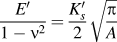 $${{E'} \over {1 - {{\rm{\nu }}^2}}} = {{{{K'_s}}} \over 2}\sqrt {{{\rm{\pi }} \over A}} \quad$$
and
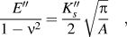 $${{E''} \over {1 - {{\rm{\nu }}^2}}} = {{{{K''_s}}} \over 2}\sqrt {{{\rm{\pi }} \over A}} \quad ,$$
which relate the measured storage stiffness (K′) and loss stiffness (K″) to the storage modulus (E′) and loss modulus (E″) of the sample. In Eqs. (1)–(2), A defines the area of contact between the indenter tip and the sample and ν is the Poisson’s ratio. From E′ and E″, it is possible to determine the percent resilience as
 $$R = 100{e^{\left( { - {{\rm{\pi }} \over 2}\tan {\rm{\delta }}} \right)}}\quad ,$$
where tan δ = E″/E′. The resilience defines the ability of a rubber to recover the stored energy after loading. Finally, the hardness is defined by
 $$H = {{{P_{\max }}} \over A}\quad ,$$
where P max is the maximum force achieved during loading.
E. Statistical analysis
First, a two-tailed paired student's t-test was applied on the results to quantify the significance of changes with respect to frequency, morph, and gender of the cricket. Changes with respect to frequency were quantified by pairing the frequencies for each gender and for each morph. Also, to detect the changes with respect to contact depth, the contact depth results were paired for each gender and for the morphs. The mean value for each data set is given within a confidence level of 95 percent.
III. RESULTS
A. Identification of rubberlike cuticle on crickets
The blue fluorescence characteristic of resilin has been identified in different locations of the walking leg of the crickets. The wider patch of resilin was located at the trochanteral–femoral (TrF) joint as shown in Fig. 1. In addition, the presence of resilin at this location was positively identified using methylene blue staining. After dying, the joint appeared stained in deep blue although it is translucent in nature.
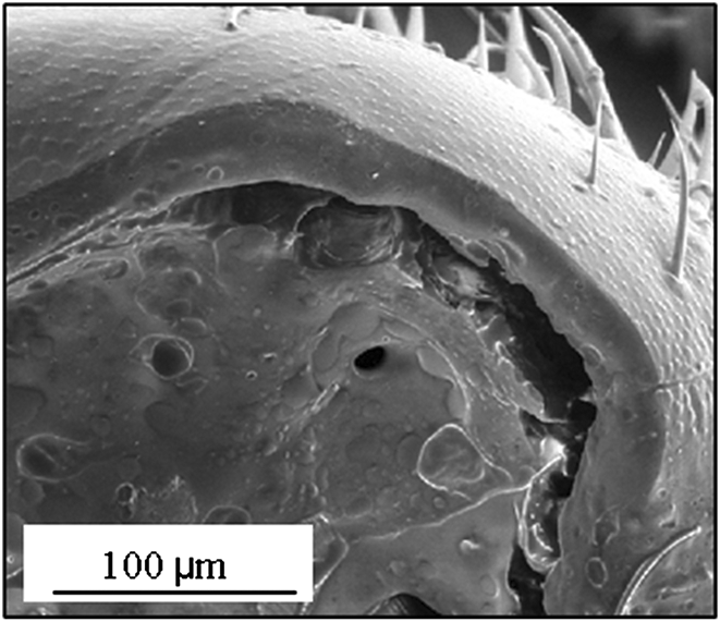
(a) Schematic of the cricket walking leg; (b) composite image from upright fluorescent microscope and light microscope of cricket walking leg (excitaton light at 395 nm and measured at 420–440 nm for the emitted fluorescence light); (c) SEM image of the cross-section of the trochanteral–femoral joint of Gryllus firmus.

B. Microstructure of the rubberlike cuticle
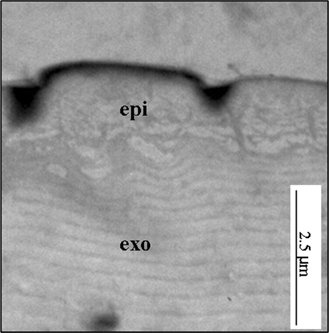
The scanning electron microscopy image of the cross-section of TrF Joint (Fig. 2) revealed a rather smooth and hairless surface. The thickness of the membrane is about (∼32 μm). The TEM micrograph of the TrF joint revealed a layered structure of the fiber composite (Fig. 3). A thin and dense epicuticle (epi ∼ 1 μm), followed by a thick exocuticle (exo > ∼15 μm) can be distinguished. The fibers are aligned horizontally and oriented parallel to the surface along the cross-section.
SEM image of the cross-section of the trochanteral–femoral joint of Gryllus firmus.
TEM micrograph of the cross-section of the trochanteral–femoral joint of Gryllus firmus. Abbreviations: epi, epicuticle; exo, exocuticle.
To ensure that the effect of the substrate was insignificant, the indentation depth was maintained between 400 and 2800 nm (no greater than 10% of the sample thickness). Because the focus of the measurements here is on the exocuticle, indentations depths that were sufficiently deep are of primary interest. We expected to see an initial depth-dependent behavior as the epicuticle is compressed. For larger depths, we expected the behavior of the exocuticle to dominate the overall response.
C. Viscoelastic properties of the rubberlike cuticle
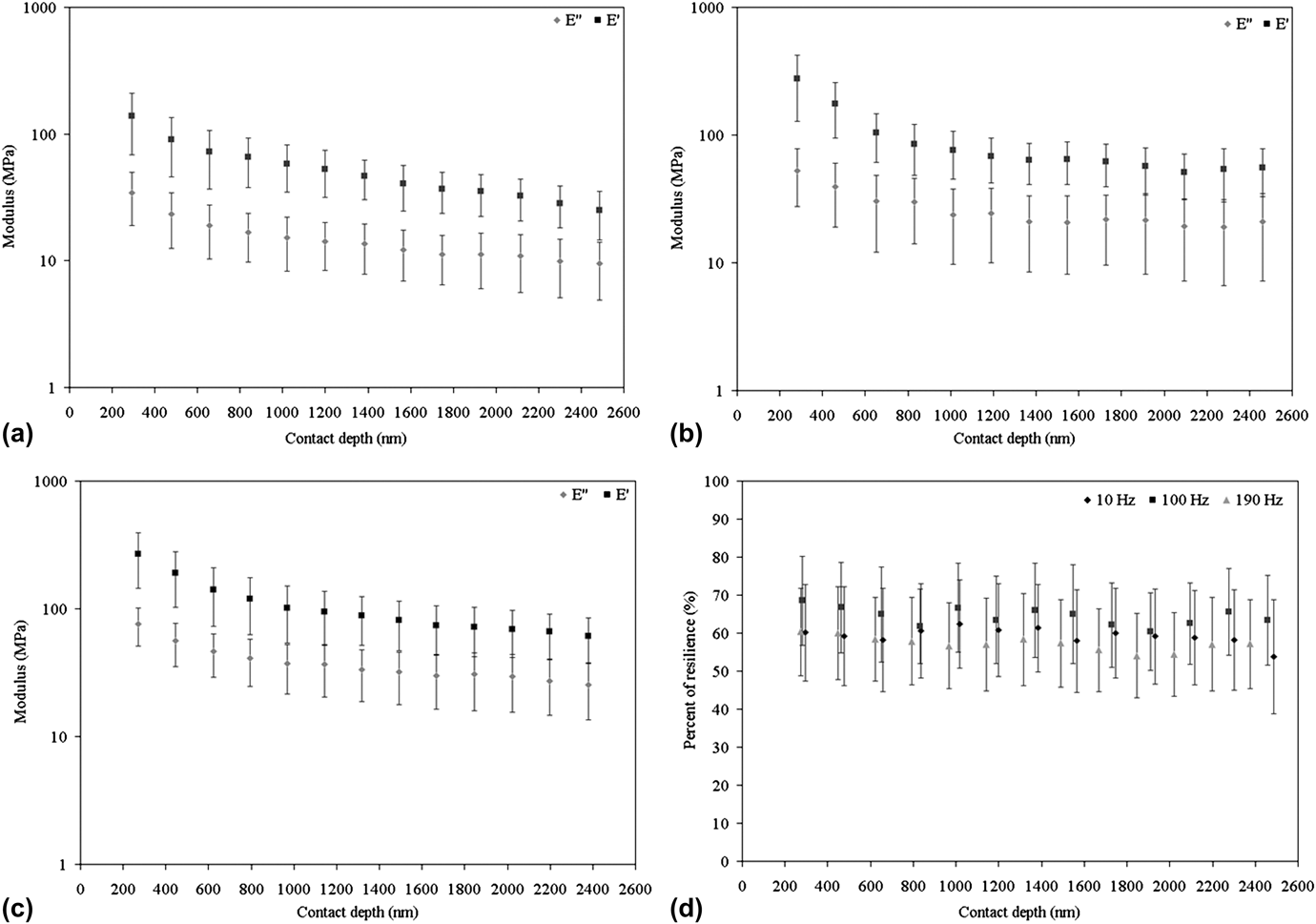
The storage and loss moduli, as well as the percent of resilience and hardness were determined as a function of depth at three frequencies (10, 100, and 190 Hz) for females and males of the lw and sw morphs. Similar trends of the moduli, percent of resilience, and hardness were observed for the different morphs and genders with respect to contact depth at the three different frequencies. For clarity, only the moduli and percent of resilience for the lw female (lwf) at 10, 100, and 190 Hz are shown in Fig. 4.
Changes in the storage modulus, E′; loss modulus, E″ at (a) 10 Hz, (b) 100 Hz, and (c) 190 Hz; and (d) percent of resilience, R of the trochanteral–femoral joint of the lw morph female at 10, 100, and 190 Hz.
1. Moduli profile
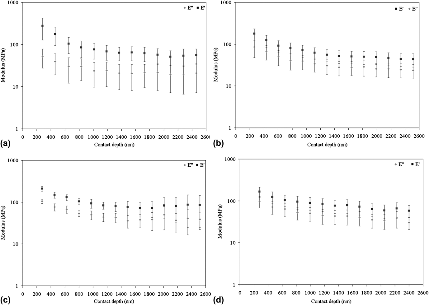
Each morph and gender revealed significant (P < 0.05) moduli changes with depth up to 1000 nm at 10, 100, and 190 Hz due to the layered microstructure of the joint. After this depth, the mechanical response of the exocuticle begins to dominate the behavior and no further depth dependence is observed. For clarity, only the data at 100 Hz are shown in Fig. 5.
Changes in the storage modulus, E′ and the loss modulus, E″, of the cuticle of: lw (a) female, (b) male; and sw (c) female, (d) male at 100 Hz.
Pairing the results at different frequencies with respect to contact depth for the different morphs and genders revealed no frequency dependence of the storage modulus (thus, only data at 100 Hz are discussed here). Also, no difference between morphs and genders was observed for the storage modulus. At 1500 nm, where the mechanical properties of the exocuticle are expected to be predominant, the mean (±95% confidence interval) storage modulus for the lw male (lwm) was 52 ± 18 MPa, for the lwf was 64 ± 23 MPa, for the sw male (swm) was 78.5 ± 28 MPa, and for the sw female (swf) was 76 ± 28 MPa.
Contrary to the storage modulus, the pairing of loss modulus results with respect to frequency (at 1500-nm depth) for the different morphs and genders revealed a frequency dependence. Indeed, at 10 Hz the corresponding loss moduli are: lwm = 7.19 ± 6.95 MPa, lwf = 12.19 ± 5.24 MPa, swm = 16.29 ± 7.20 MPa, swf = 17.54 ± 6.98 MPa. At 100 Hz, the loss moduli are: lwm = 28 ± 10 MPa, lwf = 20 ± 12 MPa, swm = 44 ± 17 MPa, swf = 36 ± 12 MPa. Finally, at that same depth at 190 Hz, the loss moduli are: lwm = 49.18 ± 14.38 MPa, lwf = 31.88 ± 14.20 MPa, swm = 62.36 ± 24.20 MPa, swf = 39.58 ± 8.64 MPa. The loss modulus increases significantly with frequency for each morph and gender. In addition, significant differences were observed between the loss modulus of females from the lw and sw morphs. The loss modulus of sw females is significantly higher (p < 0.05) than the loss modulus of lw females. The cuticle organization of sw females seems to dissipate more energy than lw females. This difference can reflect the variation in the volumetric concentrations of the protein, the chitin and water, or changes of the structural interactions between those constituents or their organization.
2. Percent of resilience
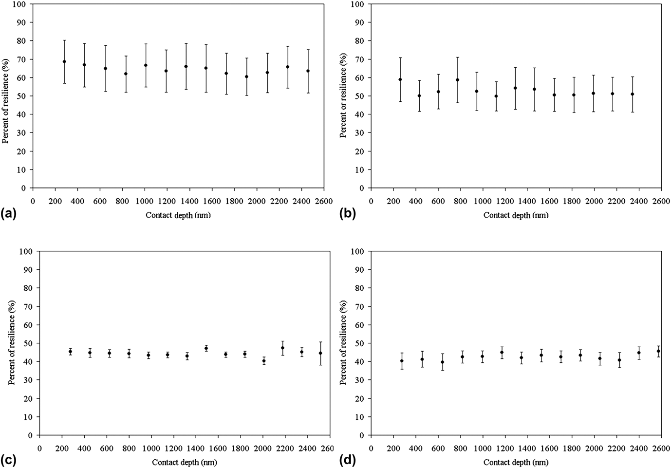
Interestingly no significant changes with depth were observed for the percent of resilience for each morph and gender at 10, 100, and 190 Hz as shown in Fig. 6 (only 100 Hz results are shown for clarity). Thus, the behavior observed for the storage and loss moduli, from which the percent of resilience is calculated [Eq. (3)], have the same dependence such that it cancels. Also, as expected, pairing the frequencies with respect to contact depth for the different morphs and genders revealed significant frequency dependence (p < 0.05) of percent of resilience primarily because of the behavior of the loss moduli. At 10 and 190 Hz, no difference between morphs and genders was observed, whereas at 100 Hz, significant differences were observed between females from the lw and sw morphs. The cuticle of lw females exhibits a higher percent of resilience than that of sw for males and females at 100 Hz. Lw females appear to have a better ability to restore energy than sw females. No differences were observed between genders of same morphs or between males of the two morphs. At 1500 nm, the percent of resilience is: lwm = 53%, lwf = 65%, swm = 43%, swf = 47%.
Changes in percent of resilience, R, of the cuticle of: lw (a) female, (b) male; and sw (c) female, (d) male at 100 Hz.
D. Hardness profile of the rubberlike cuticle
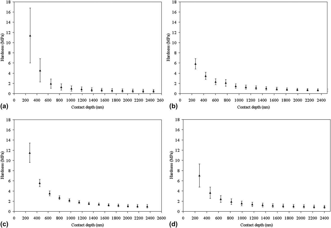
The hardness that was determined as a function of depth at three frequencies (10, 100, and 190 Hz), for females and males in the lw and sw morphs, is depth and frequency dependent in a similar manner as the loss modulus and the percent of resilience (hardness results shown in Fig. 7 are only for 100 Hz, for clarity). Each morph and gender revealed significant (p < 0.05) changes with depth up to 1000 nm at 10, 100, and 190 Hz. At 10 and 190 Hz, no difference between morphs and genders was observed. At 100 Hz, the percent of resilience shows significant differences between lw versus sw females. At 1500 nm, lwm = 0.9 ± 0.3 MPa, lwf = 0.71 ± 0.38 MPa, swm = 1.2 ± 0.36 MPa, swf = 1.4 ± 0.2 MPa. It is suggested here that the strength of chemical and physical bonds in the cuticle is the same between morphs but different between genders of the lw morphs.
Changes in hardness of the cuticle of: lw (a) female, (b) male; and sw (c) female, (d) male at 100 Hz.
IV. DISCUSSION
A. Comparison with previous studies
In the present study, the effect of the layered material on mechanical properties is once again highlighted. Because the elastic modulus and hardness decrease with indentation depth, it is clear that the properties of the structure at shallow depths (<1000 nm) are influenced by the entire thickness of the composite material, but as we go deeper (>1000 nm), the influence of the exocuticle increases and becomes more and more predominant. This result is in agreement with the Barbakadze et al.Reference Barbakadze, Enders, Gorg and Artz10 study in which deeper cuticle layers appeared to be softer and more compliant than the outer layer. Similar to their study, we can deduce that joints are soft materials which are coated by a stiffer film.
Other studies on cuticle mechanical properties were performed using quasistatic nanoindentation and only a few experiments were performed using the dynamic mode but at a macroscale. The elastic modulus of the dry membrane of beetle (E = 7.5 ± 1.8 GPa) and dry wing membrane of dragonfly (E = 1.5 ± 0.5 GPa) are very high when compared with those of the joint of the cricket at 100 Hz at 1500 nm depth (lwm-E′ = 52 ± 18 MPa, lwf-E′ = 64 ± 23 MPa, swm-E′ = 78.5 ± 28 MPa, swm-E′ = 76 ± 28 MPa).Reference Barbakadze, Enders, Gorg and Artz10,Reference Kreuz, Kesel, Kempf, Göken, Vehoff and Nachtigall16 Furthermore, the elastic modulus of the biological structure made of pure resilin was reported to be about 2 MPa for the tendon from the hindwing of a dragonfly and for the ligament of locust.Reference Weis-Fogh17,Reference Jensen and Weis-Fogh18 Similarly, King in his thesis shows that the storage modulus of fully hydrated resilin was 1.6 MPa but it increases to 30 MPa in the glassy state.Reference King19 Therefore, at this depth, the results showed that the presence of resilin in the structure of the leg joint reduced the cuticle stiffness, whereas chitin strengthened the resilin matrix. Also, the prealar arm of the locust (R = 85%) and the tendon of the dragon fly (R = 98%), both made of pure resilin, are more resilient than those of the trochanteral–femoral joint of the cricket (lwm-R = 53%, lwf-R = 65%, swm-R = 43%, swf-R = 47%).Reference Andersen and Weis-Fogh12,Reference King19 Therefore it is not surprising to observe the same trend when comparing the hardness of the beetle (H = 0.49 ± 0.14 GPa) and the locust membrane (H = 0.2 GPa) with those of the cricket joint (lwm-H = 0.9 ± 0.3 MPa, lwf-H = 0.71 ± 0.38 MPa, swm-H = 1.2 ± 0.36 MPa, swf-H = 1.4 ± 0.2 MPa).Reference Barbakadze, Enders, Gorg and Artz10,Reference Kreuz, Kesel, Kempf, Göken, Vehoff and Nachtigall16
B. Effect of dimorphism
As mentioned previously, morphs of wing-polymorphic insects differ in many characteristics that are expected to be influenced by the content and organization of the joint. Of particular importance for the present study are functions involving the legs. Although differences in walking have not been studied in G. firmus, Socha and Zemek revealed differences in the pattern of locomotor activity and dispersal by walking between morphs and gender in the flightless linden bug, Pyrrhocoris apterus. The macropterous morph of the Pyrrhocoris apterus exhibits higher locomotor activity than brachypterous. During the day, macropters have been shown to walk three times further than brachypters. This result indicates that contrary to sedentary brachypterous, the macropterous morphs might exhibit dispersal behavior. Furthermore, analysis of the path of P. apterus revealed that the lw morph has a less tortuous path, a longer spatial displacement, and a higher walking velocity than the sw morph.Reference Socha and Zemek20,Reference Socha and Zemek21 Walking legs of the lw morph will undergo more deforming force than those of the sw morph. Also it has been shown that legs of arthropods are highly resilient. Therefore, it is reasonable to assume that long wings' legs will exhibit more resilience and higher energy efficiency than short wings' in order to withstand more strain.
With coordination of the movement of different legs, the motion of the joints of the leg has been shown to be responsible for controlling insect motion such as running, climbing, and walking.Reference Dudek and Full22–Reference Cruse and Bartling24 Due to the location of trochanteral–femoral (TrF) joint proximally on the leg, small motion of the TrF joint generates larger rotation at the tibia and tarsus.Reference Watson, Ritzmann, Zill and Pollack23 Also, the orientation and location of the joint affects the amount of work and energy involved in motion. For dispersal purposes, it is reasonable to think that females of the lw morph have developed a more efficient energy-saving mechanism than females of the sw morph because of the presence of more or less resilient rubberlike joint in their walking leg.
Finally, the flight-capable lw morph of a closely related cricket species, Gryllus lineaticeps, has recently been shown to have greater jumping ability than the sw form.Reference Mitra25 The greater jumping ability is thought to be advantageous with regard to escaping predators. Thus morph differences in performance unrelated to dispersal may also be influenced by variation in joint characteristics. Indeed, a close relationship exists between the structural organization of biological materials and their functionality and thus their mechanical properties. For instance, the study of exoskeleton of the American lobster, H. americanus, by Raabe et al.Reference Raabe, Sachs and Romano26 revealed a mechanical gradient, through the hierarchical organization of the mineralized fibrous chitin-based exoskeleton, which is attributed to the changes in the plywood pattern and the stacking density of the chitin-protein layers across the structure. Meyers et al.,Reference Meyers, Chen, Lin and Seki27 brought into light the effect of hierarchical organization (at a meso-, micro-, and nanoscale) of biological structures such as bone, abalone shell, and crab exoskeleton to accumulate functions such as support, attachments of muscles, environmental protection, etc. Also, investigation of the mechanical properties of fibrolamellar bone has shown that there is a high degree of mechanical anisotropy at the microscale. This behavior was related to the existence of weak interfaces between fibrolamellar units at the mesoscale.Reference Seto, Gupta, Zaslansky, Wagner and Fratzl28 Finally, a previous study aimed at the characterization of limpet teeth have highlighted the importance of the structural organization of the tooth to assure load-bearing function. The short goethite nanofibers, which are embedded in a polymeric matrix made of chitin, appeared to have an ideal length, leading to an optimization of the transfer of stress from the matrix to the fiber within the teeth.Reference Lu and Barber29 Thus, it should be clear that quantitative conclusions about joint performance for a given species must include information about the joint geometry and material organization before conclusions about material performance can be made—movement by animals is a complex dynamic process.
V. CONCLUSION
Nanoindentation has been used to detect significant changes in the mechanical properties of rubberlike cuticle from different morphs and genders of Gryllus firmus. The mechanical properties of the trochanteral–femoral joint of the crickets reveal a dependence of the joint with respect to its layered microstructure. The contribution of the protein resilin and the chitin fibers in the mechanical properties of the joint remains to be quantified with respect to the amount of each constituent present in the material. Furthermore, lw females exhibit higher elasticity and resilience than sw females. This result reflects a trade-off between dispersal ability by walking and reproduction for males and females of the two morphs of G. firmus. It is believed that highlighting the relationship between the structure and mechanical property of the trochanteral–femoral joint of different morphs and genders provides knowledgeable data so that new composite materials can be developed with improved mechanical properties.
ACKNOWLEDGMENTS
This work was partially supported by the Department of Education ATLANTIS program, the FACE-PUF foundation, and the National Science Foundation (Grant No. CMMI-1050685).