Introduction
Bone Morphogenetic Proteins (BMPs) are a highly conserved family of proteins present across a diverse range of organisms, from primitive species to more complex animals, including both vertebrates and invertebrates. They play a crucial role in regulating the formation of bones and cartilage in vertebrates, while also influencing numerous biological processes and developmental pathways, such as body patterning, organ formation, and cellular communication in both groups (Katagiri and Watabe Reference Katagiri and Watabe2016; Kondo Reference Kondo2007; Lochab and Extavour Reference Lochab and Extavour2017). BMP signaling pathways are essential for proper development and the maintenance of normal physiological functions. Dysregulation of BMP activity can lead to developmental abnormalities and diseases (Bandyopadhyay et al. Reference Bandyopadhyay, Yadav and Prashar2013; Wang et al. Reference Wang, Green, Wang, Deng, Qiao, Peabody, Zhang, Ye, Yan, Denduluri, Idowu, Li, Shen, Hu, Haydon, Kang, Mok, Lee, Luu and Shi2014)
As members of the Transforming Growth Factor-beta (TGF-β) superfamily, BMPs share conserved structural domains and features. They are synthesized as large precursors, which typically consist of an N-terminal region that contains both a signal peptide and a non-conserved pro-peptide domain, followed by a conserved TGF-β domain at the C-terminus. A consensus proteolytic cleavage site, Arg-X-X-Arg, located between the pro-peptide and the mature domain, facilitates the release of mature protein (Anderson and Wharton Reference Anderson and Wharton2017; Ducy and Karsenty Reference Ducy and Karsenty2000; Goldman et al. Reference Goldman, Hackenmiller, Nakayama, Sopory, Wong, Kulessa and Christian2006). Mature BMPs, which are approximately 100–140 amino acids in length, contain seven conserved cysteine residues that are essential for forming intra- and inter-disulfide bonds (Goebel et al. Reference Goebel, Corpina, Hinck, Czepnik, Castonguay, Grenha, Boisvert, Miklossy, Fullerton, Matzuk, Idone, Economides, Kumar, Hinck and Thompson2019; Hinck Reference Hinck2012; Hinck et al. Reference Hinck, Mueller and Springer2016). These bonds contribute to the formation of a cysteine knot structure, a crucial element for the dimerization process. The biological activity of BMPs relies on this dimerization, where two processed C-terminal ‘mature’ domains of the precursor proteins form active homodimers or heterodimers, which are essential for their functional roles.
More than 20 BMP ligands, existing as either homodimers or heterodimers, have been identified in vertebrates (Kishigami and Mishina Reference Kishigami and Mishina2005; Madamanchi et al. Reference Madamanchi, Mullins and Umulis2021). These ligands are classified into five subgroups based on their sequence similarity, structural features, and functional roles: (i) BMP2/4, (ii) BMP5/6/7/8, (iii) BMP9/10, (iv) BMP12/13/14, and (v) BMP1/3/11/15 (Akiyama et al. Reference Akiyama, Raftery and Wharton2024; Carreira et al. Reference Carreira, Lojudice, Halcsik, Navarro, Sogayar and Granjeiro2014; Chen et al. Reference Chen, Dong, Kong, Zhang, Li and Xu2017; Katagiri and Watabe Reference Katagiri and Watabe2016; Mueller and Nickel Reference Mueller and Nickel2012; Zhang et al. Reference Zhang, Yu, Han, Ma, Zhang, Li, Tang, Nie and Zhou2024). In invertebrates, BMPs exhibit considerable diversity and can be classified into distinct subgroups. For example, in Drosophila, three BMP homologs have been identified: Decapentaplegic (Dpp), Glass Bottom Boat (Gbb), and Screw (Scw). Dpp is categorized as a member of the BMP2/4 subgroup, while Gbb and Scw belong to the BMP5/6/7/8a/8b subgroup (Akiyama et al. Reference Akiyama, Raftery and Wharton2024; Van der Zee et al. Reference Van der Zee, da Fonseca and Roth2008; Wotton et al. Reference Wotton, Alcaine Colet, Jaeger and Jimenez-Guri2013). Similarly, in Caenorhabditis elegans, two BMP homologs have been documented: Dbl-1, which corresponds to the homologs of mammalian BMP2/4 and D. melanogaster’s Dpp, and the ligand Tig-2, which is homologous to Drosophila’s Gbb and Scw, placing it in the BMP5/6/7/8a/8b subgroup (Savage-Dunn and Padgett Reference Savage-Dunn and Padgett2017).
The study of BMPs in parasitic helminths, particularly within trematodes, is limited but essential for understanding their developmental biology and potential roles in host interactions. To date, BMPs have been characterized only in Schistosoma mansoni (Freitas et al. Reference Freitas, Jung and Pearce2009) and S. japonicum (Liu et al. Reference Liu, Q-p, Ye, Xiong, C-l, H-f and M-s2013). Exploring BMPs in helminths could contribute to the development of novel therapeutic or control strategies for trematodiasis, as targeting BMP signaling pathways may effectively disrupt the growth and survival of these parasites.
Fasciola gigantica is an important liver fluke affecting livestock and, consequently, the agricultural economy. Infection with this parasite can lead to significant health issues in animals, such as decreased productivity and liver damage, and it poses a zoonotic risk to humans (Mas-Coma et al. Reference Mas-Coma, Valero and Bargues2023; Siles-Lucas et al. Reference Siles-Lucas, Becerro-Recio, Serrat and González-Miguel2021). While key components of the TGF-β/BMP signaling pathway have been identified in the genomes of Fasciola species (Japa et al. Reference Japa, Hodgkinson, Emes and Flynn2015; Japa et al. Reference Japa, Phuangsri, Klinbumrung and Prakhammin2025; Japa et al. Reference Japa, Phuangsri, Klinbumrung, Prakhammin and Flynn2024; Japa et al. Reference Japa, Prakhammin and Flynn2022; Wu et al. Reference Wu, Kong, Zhang and Di2023), a specific BMP-like ligand in the liver fluke F. gigantica has yet to be clearly identified and characterized. This lack of information creates a significant gap in our understanding of BMP signaling in this parasitic species.
To address this, the present study aimed to identify and characterize a BMP-like ligand in F. gigantica. A genome-wide screening approach was employed to detect potential BMP-like sequences within the F. gigantica genome. The identified candidate underwent comprehensive analysis, including conserved domain prediction, homology-based annotation, gene structure organization, multiple sequence alignment, and phylogenetic analysis to determine its evolutionary relationship with known BMP family members. In addition, structural modeling and predictions of physicochemical properties were performed to further characterize the protein. The expression profile of the identified BMP -like gene was also assessed across different developmental stages of F. gigantica using quantitative real-time PCR (qRT-PCR).
Materials and methods
Ethics approval
All animal-related procedures conducted in this research were approved by the Institutional Animal Care and Use Committee at the University of Phayao, Thailand (Reference No. 63 01 04 006).
Parasites
Adult and unembryonated eggs
Adult F. gigantica specimens were collected from the bile ducts and gall bladders of naturally infected buffaloes at an abattoir in Phayao, Thailand. The flukes were maintained in RPMI-1640 medium supplemented with 10 μg/ml gentamycin, then thoroughly washed with sterile phosphate-buffered saline (PBS) and preserved in Trizol reagent (Life Technologies) for RNA extraction. Eggs released into the transport medium were collected, rinsed with sterile distilled water, and designated as unembryonated. A portion of these eggs was reserved for embryonation, while the remainder was preserved in Trizol for subsequent RNA extraction.
Embryonated eggs and miracidia
Embryonated eggs and miracidia of F. gigantica were generated under controlled laboratory conditions following the protocols described by Moxon et al. (Reference Moxon, LaCourse, Wright, Perally, Prescott, Gillard, Barrett, Hamilton and Brophy2010) and Japa et al. (Reference Japa, Prakhammin and Flynn2022). Unembryonated eggs were incubated in darkness at 30°C for two weeks to promote embryonic development. Fully developed embryonated eggs were carefully selected and preserved in Trizol reagent for experimental use. To obtain miracidia, the embryonated eggs were exposed to natural light at room temperature to induce hatching. Freshly hatched miracidia were monitored, immediately collected, and either preserved in Trizol reagent for RNA extraction or directly used to infect Radix (Lymnaea) rubiginosa snails.
Cercariae, rediae, and metacercariae
The larval stages of F. gigantica were obtained by experimentally infecting R. (L.) rubiginosa snails with freshly hatched miracidia, following the protocol of Japa et al. (Reference Japa, Prakhammin and Flynn2022). Infected snails were regularly monitored, and successful infection was confirmed by the emergence of cercariae. Cercarial shedding was induced by exposing the snails to natural light, and the emerging cercariae were collected using a micropipette. To produce metacercariae, cercariae were allowed to encyst on clean cellophane sheets under control conditions. Rediae were recovered by carefully dissecting infected snails under a stereomicroscope and isolating them from the snail tissues. All larval stages including rediae, cercariae, and metacercariae were preserved in Trizol reagent for subsequent RNA extraction.
Newly excysted juveniles (NEJs)
Newly excysted juveniles (NEJs) of F. gigantica used for life stage expression analysis were prepared through in vitro excystation of metacercariae, following the protocols of McVeigh et al. (Reference McVeigh, McCammick, McCusker, Morphew, Mousley, Abidi, Saifullah, Muthusamy, Gopalakrishnan, Spithill, Dalton, Brophy, Marks and Maule2014) and Japa et al. (Reference Japa, Prakhammin and Flynn2022). The process began with the removal of the outer cyst, after which the metacercariae were treated with 0.5% bleach for 4 min. They were then thoroughly rinsed five times with distilled water. The cleaned metacercariae were incubated at 37°C for 1 hour in an excystation solution containing sodium tauroglycocholate, NaCl, NaHCO3, L-cysteine, and HCl. Excystation was monitored at 30-min intervals. Once NEJs emerged, they were transferred to RPMI-1640 medium containing 10 μg/ml gentamicin, washed with 1× D-PBS, and subsequently preserved in Trizol reagent for RNA extraction.
RNA extraction
For RNA extraction, parasite samples were collected in three biological replicates for each life stage, with the following quantities per replicate: 1,000 unembryonated eggs, 1,000 embryonated eggs, 1,000 miracidia, 100 rediae, 100 cercariae, 100 metacercariae, 100 newly excysted juveniles (NEJs), and one adult fluke. Following collection, samples from each life stage underwent multiple washes with PBS before being preserved in TRIzol reagent.
The preserved parasites were then homogenized and subsequently mixed with chloroform, followed by centrifugation. The clear aqueous phase was carefully collected and further purified using the RNeasy Micro Kit (Qiagen, Germany), according to the manufacturer’s instructions. The purified RNA was eluted and stored at -80°C until further use.
Genome-wide identification of BMP genes
BMP protein-like sequences from Schistosoma mansoni [ACG60602.1] and S. japonicum [ADM48811.1] were retrieved from the National Center for Biotechnology Information (NCBI) database. These sequences were used as query sequences to identify BMP genes in the F. gigantica genome using a tBLASTn search against the cDNA database on WormBase Parasite (https://parasite.wormbase.org/).
Rapid amplification of cDNA ends (RACE)
5′ and 3′ RACE cDNA libraries were synthesized from 1 μg of total RNA extracted from adult F. gigantica using the SMARTer™ RACE cDNA Amplification Kit (Takara, Japan). The 5′ and 3′ ends of the F. gigantica BMP -like cDNA were then amplified by nested PCR using SeqAmp DNA Polymerase (Takara, Japan). The primers used in this study included the Universal Primer Mix (UPM) and Universal Primer Short (UPS), both supplied with the kit, as well as gene-specific primers (GSPs) designed based on conserved regions identified in the F. gigantica genome via a tBLASTn search. The primer sequences are listed in Table 1, and all primers were synthesized by U2Bio (Korea).
Primers used for rapid amplification of cDNA ends (RACE) and developmental stage-specific PCR analysis of FgDpp in Fasciola gigantica

PCR amplification for both the first and second rounds was performed under the following conditions: an initial denaturation at 95°C for 5 min, followed by 35 cycles of denaturation at 95°C for 15 s, annealing at 65°C (first PCR) or 63°C (second PCR) for 30 s, and extension at 72°C for 3 min. A final extension was carried out at 72°C for 15 min. The resulting PCR products were separated on a 1.5% agarose gel pre-stained with Nancy-520 (Sigma-Aldrich, UK) and visualized using a gel documentation system to confirm the presence and expected size of the amplified fragments.
Cloning and sequencing
Positive 5′ and 3′ RACE products were purified and ligated into the pRACE vector (Takara, Japan), followed by transformation into Stellar competent cells (Takara, Japan) according to the manufacturer’s instructions. Recombinant clones were screened, and plasmid DNA was extracted for sequencing. Nucleotide sequencing was performed by the FastNGS service (Tsingke, China).
The raw sequencing data obtained from cloned 5′ and 3′ RACE products of the BMP gene were initially assembled by the sequencing service provider using their in-house pipeline. The resulting sequences were then inspected and analyzed using BLASTn and BLASTx against both the NCBI non-redundant (nr) database and the F. gigantica genome to confirm their identity as BMP transcripts. Vector sequences and overlapping regions were trimmed prior to final sequence assembly using BioEdit software (Hall Reference Hall1999; Hall et al. Reference Hall, Biosciences and Carlsbad2011). The accuracy of the assembled sequence was confirmed through a BLAST search against the F. gigantica genome. The F. gigantica BMP -like cDNA sequence identified in this study has been deposited in the GenBank database under accession number PV552021.
Sequence analysis of the F. gigantica BMP
Sequence analysis of the F. gigantica BMP was first conducted to identify open reading frames (ORFs) using the NCBI ORF Finder tool (https://www.ncbi.nlm.nih.gov/orffinder). The identified ORF was then translated into the corresponding amino acid sequence using the ExPASy translation tool (https://web.expasy.org/translate/). Propeptide cleavage sites in eukaryotic protein sequences were predicted using the ProP 1.0 tool (https://services.healthtech.dtu.dk/services/ProP-1.0/) (Duckert et al. Reference Duckert, Brunak and Blom2004). The signal peptide was identified using SignalP 6.0, accessible at https://services.healthtech.dtu.dk/service.php?SignalP-6.0 (Teufel et al. Reference Teufel, Almagro Armenteros, Johansen, Gíslason, Pihl, Tsirigos, Winther, Brunak, von Heijne and Nielsen2022). To assess conserved domains, the F. gigantica BMP sequence was analyzed with the CD-Search tool on the NCBI website (https://www.ncbi.nlm.nih.gov/Structure/cdd/wrpsb.cgi). The general physicochemical properties of the protein were determined using the ProtParam tool on the ExPASy website (https://web.expasy.org/protparam/).
Gene structure prediction
The full-length cDNA sequence of F. gigantica BMP was analyzed using BLASTn searches against the F. gigantica genome via the WormBase ParaSite database. The identified matching regions were used to map the locations of exons and introns. The gene organization of F. gigantica BMP was then visualized and represented graphically using GeneMapper 2.5.
Sequence homology and conservation analysis
To evaluate sequence similarity, PSI-BLAST (Position-Specific Iterated BLAST) was used to compare both the precursor and mature forms of the F. gigantica BMP protein with homologs from related species. The analysis included sequences from platyhelminths available in GenBank as well as those from mammalian hosts (human and bovine). Sequence identity percentages were calculated to evaluate the degree of homology across species.
To further assess conserved sequence patterns, a multiple sequence alignment of the mature BMP regions was conducted using the MUSCLE algorithm (Edgar Reference Edgar2004) implemented in Seaview (Gouy et al. Reference Gouy, Tannier, Comte and Parsons2021). This analysis included sequences from trematodes, cestodes, free-living flatworms, and mammalian hosts. The alignment was visually inspected and graphically presented using BioEdit, with emphasis on identifying conserved and functionally similar residues across species.
Evolutionary analysis
Following the sequence conservation assessment, a phylogenetic analysis was conducted to examine the evolutionary relationships of the F. gigantica BMP protein. BMP sequences from a diverse set of organisms – including mammals, nematodes, and platyhelminths – were included in the analysis. The conserved region of the F. gigantica BMP was aligned with those from other species using the same MUSCLE-based approach described earlier. After removing gaps and incomplete positions, a final alignment of 179 amino acid residues was used to construct a phylogenetic tree using the Maximum Likelihood (ML) method implemented in MEGA11 (Tamura et al. Reference Tamura, Stecher and Kumar2021), applying the Whelan and Goldman (WAG) substitution model. The robustness of the tree topology was evaluated using 1,000 bootstrap replicates.
F. gigantica BMP 3D modeling
The folding structures of the precursor and mature F. gigantica BMP proteins were predicted using the Phyre2.2 online tool with its intensive mode (http://www.sbg.bio.ic.ac.uk/~phyre2) (Powell et al. Reference Powell, Islam, David and Sternberg2025). The resulting PDB models were then processed and visualized using UCSF Chimera 1.16 (Pettersen et al. Reference Pettersen, Goddard, Huang, Couch, Greenblatt, Meng and Ferrin2004).
Life stage expression analysis
To determine the expression profile of F. gigantica BMP across eight life stages of F. gigantica (adult, unembryonated egg, embryonated egg, miracidium, redia, cercaria, metacercaria, and newly excysted juvenile), quantitative real-time PCR (qRT-PCR) was performed with three biological and three technical replicates per stage.
cDNA synthesis was performed using the GoScript™ Reverse Transcription System (Promega, UK) with oligo-dT primers, following the manufacturer’s instructions. Primers for life stage detection of F. gigantica BMP in qPCR were designed to span intronic regions, with F. gigantica glyceraldehyde 3- phosphate dehydrogenase gene (FgGAPDH) used as the endogenous control, as described by Japa et al. (Reference Japa, Prakhammin and Flynn2022). The primer sequences used for life stage-specific PCR and controls are provided in Table 1.
Each qPCR reaction consisted of 1 μl of cDNA from each life stage, 1 μl of each primer (0.5 pmol), 10 μl of 2X QuantiNova SYBR Green master mix (Qiagen, Germany), and RNase-free water to a final volume of 20 μl. Amplification was performed on a Bio-Rad CFX96 Real-Time PCR System using the following thermal cycling conditions: an initial denaturation at 94°C for 2 min, followed by 40 cycles of 94°C for 5 s, 57°C for 10 s, and 72°C for 30 s, with a final extension at 72°C for 5 min. A melting curve analysis was carried out after amplification to ensure the specificity of the products.
Statistical analysis
The relative expression levels of F. gigantica BMP compared to FgGAPDH at various life stages were calculated using the 2−ΔCT method (Silver et al. Reference Silver, Best, Jiang and Thein2006) and presented as the mean ± standard error of the mean (SEM) from three independent experiments. Data visualization was performed with a bar chart using GraphPad Prism 8.0.1 (GraphPad Software, San Diego, CA, USA). Statistical analysis was conducted using one-way ANOVA followed by Tukey’s post hoc test, with significance determined at p < 0.05.
Results
Identification of BMP gene family members in the F. gigantica Genome
Four candidate gene or transcript sequences were initially identified from the F. gigantica genome database based on domain architecture and sequence similarity: FGIG_07517 (Lectin_legB domain-containing protein), FGIG_12664 (TMEM132 domain-containing protein), FGIG_00622 (Beta-lactamase domain-containing protein), and FGIG_01550 (Inhibin beta B chain). Among these, FGIG_01550 was excluded from further analysis as it had previously been characterized as FgTLM, a member of the TGF-β subfamily (Japa et al. Reference Japa, Prakhammin and Flynn2022), and therefore not considered a BMP candidate.
The remaining sequences were subjected to further domain analysis to evaluate their potential classification within the BMP family. Based on the presence of a conserved TGF-β propeptide superfamily domain, FGIG_12664 was selected as the most likely BMP gene candidate in F. gigantica.
Subsequently, the candidate gene FGIG_12664 was selected for further analysis and designated FgDpp (F. gigantica Decapentaplegic) based on its sequence similarity to Dpp, a well-characterized member of the BMP subgroup in other species. Gene-specific primers (GSPs) were designed based on conserved regions of the FgDpp sequence and used for 5′ and 3′ rapid amplification of cDNA ends (RACE) to obtain the full-length cDNA.
Isolation, sequence assembly, and characterization of FgDpp cDNA
RACE amplification yielded approximately 1 kb and 2.8 kb fragments from the 5′ and 3′ ends of the FgDpp cDNA, respectively. Sequence assembly generated a full-length FgDpp cDNA of 3,765 bp. Initial verification using BLASTn against the F. gigantica cDNA database revealed that a 1,253 bp segment (positions 112–1,364) aligned with nucleotides 179,510–180,762 of FGIG_00622, while a 789 bp segment (positions 1,363–2,151) matched nucleotides 640–1,428 of FGIG_12664, based on data from the WormBase ParaSite database (Figure 1). Notably, the remaining regions of the FgDpp cDNA showed no alignment with annotated genomic sequences, suggesting possible gaps or unannotated regions in the current genome assembly.
Verification of the full-length FgDpp cDNA sequence by BLASTn analysis. BLASTn results showing regions of sequence identity between the newly assembled 3,765 bp FgDpp cDNA and annotated cDNA sequences in the Fasciola gigantica genome. (A) Alignment with the FGIG_00622 gene (1,253 bp segment, positions 112–1, 364; nucleotides 179,510–180,762); (B) Alignment with the FGIG_12664 gene (789 bp segment, positions 1,363–2,151; nucleotides 640–1,428).

The complete FgDpp cDNA consisted of a 27 bp 5′ untranslated region (UTR), a 2,811 bp coding sequence (CDS) encoding a 936-amino acid protein, and a 927 bp 3′ UTR followed by a poly (A) tail. The start codon (ATG) was located at positions 28–30, and the stop codon (TAG) appeared at positions 2,836–2,838.
Structural features and physicochemical properties of FgDpp
The translated FgDpp protein consisted of 936 amino acid residues, with serine (10.0%), leucine (9.7%), and arginine (8.0%) being the most abundant. Sequence analysis revealed two conserved domains: a TGF-β pro-peptide domain spanning residues 452–545 and a TGF-β domain located between residues 835–936. No signal peptide sequence was identified; however, four furin cleavage sites – RHHR (441–443), RTRR (662–664), RKRR (684–687), and RCVR (749–751) – were detected, defining a mature FgDpp protein of 185 amino acids. Additionally, seven conserved cysteine residues were located within the C-terminal domain at positions 835, 864, 868, 900–901, 933, and 935, indicating the potential for disulfide bond formation and structural stability (Figure 2).
The open reading frame (ORF) of the putative FgDpp gene and its translated protein consisting of 936 amino acids. The amino acid sequence is displayed beneath the corresponding nucleotide sequence, with numbering shown at both ends. The start codon (ATG) and stop codon (TAG) are indicated in bold and underlined text. The TGF-β pro-domain is highlighted in light brown, and the TGF-β domain is marked in green, with seven conserved cysteine residues shown in red. Four predicted furin cleavage sites are shaded in pink.

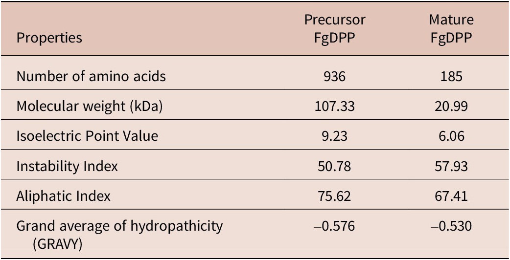
The predicted molecular weights of the FgDpp precursor and its mature protein were 107.33 kDa and 20.99 kDa, respectively, with corresponding pI of 9.23 and 6.06. The physiological analysis predicted that both precursor and mature forms of FgDpp were likely to be hydrophilic and unstable. The Grand Average of Hydropathicity (GRAVY) scores were -0.576 for the precursor and -0.530 for the mature protein, with instability indices of 50.78 and 57.93, respectively. The predicted physiological properties of FgDpp are summarized in Table 2.
Predicted physicochemical properties of the FgDpp protein from Fasciola gigantica, as analyzed using the ExPASy ProtParam tool

Gene structure of FgDpp
The FgDpp gene spanned approximately 48.89 kb and consisted of five exons and four introns. The exon sizes varied from 123 bp to 1,366 bp, while the introns ranged from 20 bp to 44,090 bp. Exon 1 included a 5’ UTR and a CDS, while Exon 3 contained both a CDS and a 3’ UTR. Coding sequences (CDS) were present in Exons 1 through 3. The organization of the FgDpp gene is illustrated in Figure 3.
Diagram illustrating the genomic structure of FgDpp. Coding sequences (CDS) are shown as striped boxes, and untranslated regions (UTRs) are represented by empty boxes. Introns are depicted as connecting lines, with longer introns abbreviated using “//”. The length of each segment is labeled in base pairs (bp).

Homology and conservation of FgDpp
Homology searching of the FgDpp precursor revealed the highest matches with trematode sequences in the GenBank database, particularly within the genera Fasciola and Schistosoma. The homology percentages ranged from 34.15% to 94.44%. The greatest similarity was observed with the F. hepatica sequence [THD23991.1], which exhibited a 94.44% similarity (884 out of 936 amino acids). In contrast, the FgDpp precursor showed lower similarity with Schistosoma species, including S. japonicum [ADM48811.1], S. mansoni [ACG60602.1], and S. haematobium [XP_051071960.1], with similarity percentages ranging from 33.01% to 35.40%.
Sequence analysis of the mature FgDpp protein revealed the highest similarity with F. hepatica Dpp [THD23991.1], showing 92.22% identity (178/185 amino acids), followed by Fasciolopsis buski Dpp [KAA0189347.1] at 77.30% (143/185), and DVR-1 from Clonorchis sinensis [KAG5453466.1] at 62.21% (107/172). Among Schistosoma species, a comparable level of homology was observed, with 87.39% identity (97/111) to BMPs from S. japonicum [ADM48811.1], S. mansoni [ACG60602.1], S. haematobium [XP_051071960.1], and DVR-1 from S. japonicum [TNN14071.1]. In contrast, similarity to BMPs from cestodes was lower, ranging from 53.78% to 54.62%, as observed in sequences from Echinococcus granulosus [KAH9280384.1, CDS19307.1] and E. multilocularis [CDS37359.1]. Among free-living species, the FgDpp protein showed higher similarity to human BMP2 [ACV32586.1, 1ES7_A, 1REW_A] with 51.89% identity (55/106 amino acids) compared to planarian BMPs from Dugesia japonica [BAA32087.1] with 48.48% identity (64/132) and Schmidtea mediterranea [ABV04322.1] with 52.38% identity (55/105).
The final 105 amino acids of the FgDpp sequence were found to be highly conserved among other flatworm species and even in humans. Multiple sequence alignment revealed that within this region, the sequence identity with BMPs from trematodes ranged from 87.62% to 89.52%, with 100% identity observed for F. hepatica Dpp (FhDpp) and F. buski Dpp (FbDpp). In comparison, free-living planarians showed slightly lower identity levels, ranging from 51.43% to 53.33%, while cestodes displayed 58.65% and 59.62% identity. The similarity further declined when compared with human BMP2 and BMP4, which showed 49.04% to 51.92% identity. The multiple sequence alignment identified 34 fully conserved amino acids (*) and 24 amino acids with similar properties (:). The multiple sequence alignment is shown in Figure 4.
Multiple sequence alignment of the C-terminal 105 amino acids of the FgDpp protein with BMP-like homologs from diverse organisms, including trematodes, cestodes, free-living flatworms, and mammalian hosts. Identical residues are highlighted by color shading. In the consensus sequence, asterisks (*) indicate fully conserved residues, colons (:) represent strong similarity, and dots (.) represent weak or low similarity. Positions without a symbol reflect no detectable similarity.

Evolutionary analysis
The phylogenetic relationship between FgDpp and BMP homologs from other organisms was inferred using a PhyML tree constructed in MEGA11, based on a conserved 179-amino-acid region (Figure 5). BMPs from parasitic platyhelminths formed a distinct clade, clearly separated from those of free-living flatworms (planarians), nematodes, and mammals. Within this clade, FgDpp clustered closely with BMPs from other members of the Fasciolidae family, including F. hepatica and F. buski, forming a well-supported trematode lineage. In contrast, BMPs from Schistosoma species grouped into a distinct subclade within the trematodes, while the DVR protein from Clonorchis sinensis also formed a separate branch. Additionally, BMPs from Echinococcus spp. constituted a distinct subgroup within the cestode lineage. BMPs from planarians, representing free-living flatworms, were positioned apart from those of parasitic platyhelminths.
Phylogenetic analysis of the FgDpp protein (PV552021) from Fasciola gigantica and BMP-like proteins from other available sequences, including trematodes, cestodes, free-living flatworms, nematodes, and related mammalian hosts. The phylogenetic tree was constructed based on 179 amino acid positions using the maximum likelihood method in MEGA11 with 1,000 bootstrap replicates. Bootstrap values are shown at the nodes, with only those above 50% displayed.

The mammalian BMP sequences from humans and bovines were grouped into five well-supported clusters: cluster I, BMP1; cluster II, BMP3 and BMP15; cluster III, BMP5, BMP6, and BMP7; cluster IV, BMP10; and cluster V, BMP2 and BMP4. Notably, BMP sequences from nematodes were also found with clusters III and V.
Predicted 3D structure of FgDpp
The 3D structure of the FgDpp protein was predicted using intensive analysis with Phyre2.2, based on a search of the Protein Data Bank (PDB). The resulting models demonstrated significant structural homology to human BMP-2 (d2h62a1). The precursor FgDPP model was constructed using 177 amino acid residues, accounting for approximately 19% of the full-length protein, while the mature protein model included 183 residues, representing 99% of the mature sequence. Both models shared 52% sequence identity with their template and had a confidence level of 100%. The predicted 3D structures are presented in Figure 6,
Predicted 3D models of FgDPP from Fasciola gigantica generated using Phyre2.2 based on structural homology to human BMP-2 (PDB ID: d2h62a1). (A) precursor protein, (B) mature protein, and (C) human BMP-2 used as the reference model. Key domains are color-coded: the TGF-β pro-peptide domain is shown in brown, and the TGF-β domain in green. Furin cleavage sites are highlighted in pink, while conserved cysteine residues are labeled in red, indicating potential disulfide bond formation sites.

FgDpp Expression profile analysis
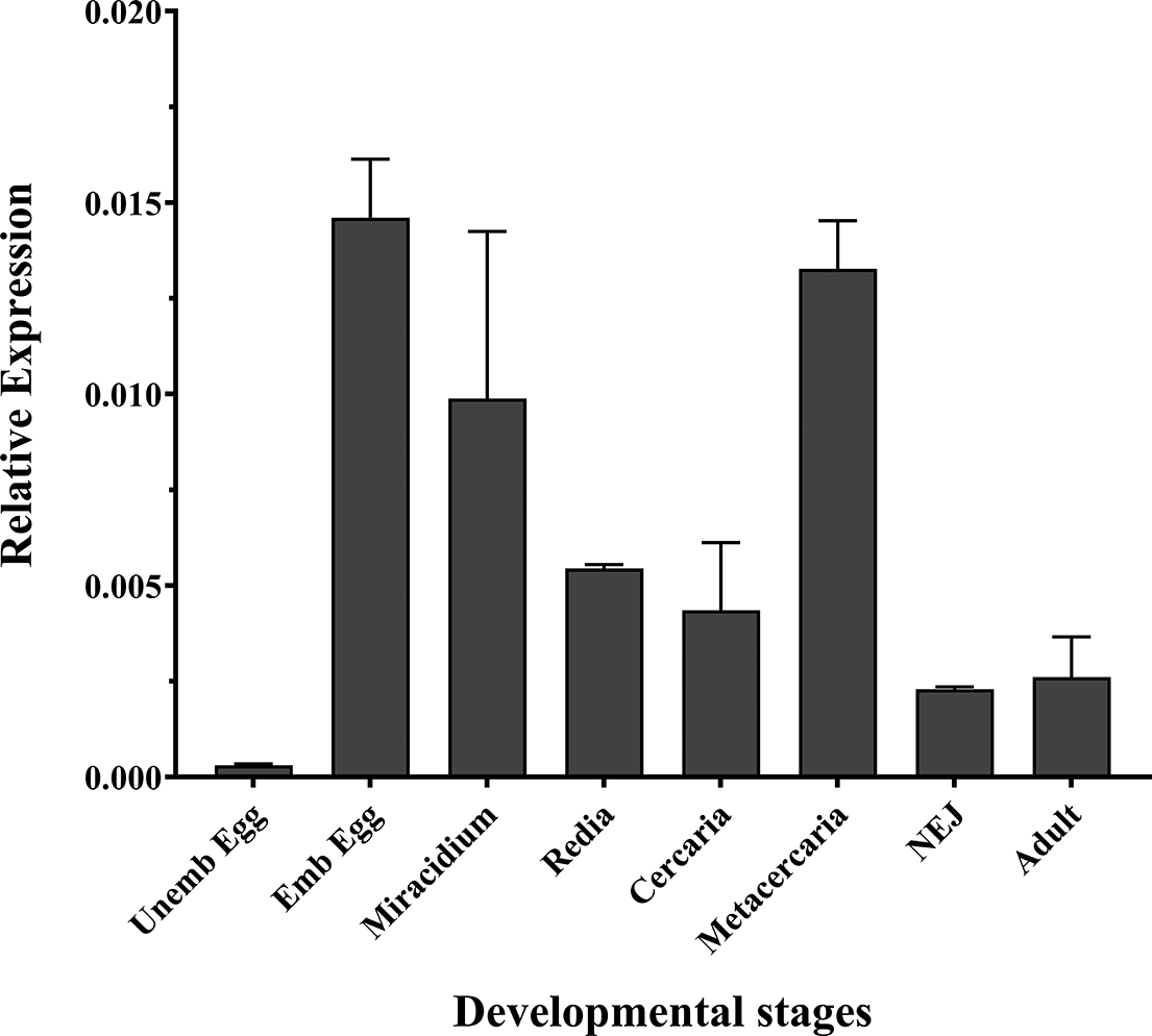
The expression profile of FgDpp was analyzed across the life cycle stages of F. gigantica using qRT-PCR. The comparative relative expression of FgDpp for each life cycle stage, normalized to FgGAPDH, is presented in Figure 7.
Expression analysis of FgDpp mRNA across different developmental stages of Fasciola gigantica. Relative expression levels were quantified by qPCR, normalized against FgGAPDH as the internal control. The bar chart represents the mean ± standard error of the mean (SEM) from three independent biological replicates.

According to the graph, FgDpp was predominantly expressed in the embryonated egg, metacercaria, and miracidium stages, with the lowest expression observed in the unembryonated egg. There was a dramatic increase in FgDpp expression during embryo development, with levels being 47.8 times higher in the embryonated egg and 32.4 times higher in the miracidium stage compared to the unembryonated egg. NEJ and adult exhibited similarly low levels of FgDpp expression, although these levels were still higher than those observed in the developing eggs. Notably, FgDpp expression in the metacercaria stage was significantly greater than the parasitic stages found in the first intermediate host (redia and cercaria) and the mammalian host (NEJ and adult).
Discussion
In this study, we screened the F. gigantica genome for BMP protein sequences using known BMP sequences from S. mansoni and S. japonicum as queries. tBLASTn analysis identified matching sequences corresponding to the gene IDs: FGIG_00622 and FGIG_12664 within the genome. Using 5’ and 3’ RACE, we obtained a full-length 3,765 bp cDNA encoding a 936-amino-acid BMP protein, which we named FgDpp. The cDNA and its translated sequence aligned with FGIG_00622, a Lectin_legB domain-containing protein, from positions 103 to 448, and with FGIG_12664, a TMEM132 domain-containing protein, from positions 446 to 708. These results suggest that FgDpp transcription involves both FGIG_00622 and FGIG_12664. However, the remaining portion of the sequence, spanning positions 709–936, did not align with any known sequences in the current F. gigantica genome, likely due to incomplete or missing genomic data, indicating that key regions of the mature FgDpp sequence remain unannotated.
The FgDpp protein shares several key features with BMP sequences from S. mansoni (Freitas et al. Reference Freitas, Jung and Pearce2009), S. japonicum (Liu et al. Reference Liu, Q-p, Ye, Xiong, C-l, H-f and M-s2013), and S. haematobium (XP_051071960.1). Unlike their mammalian counterparts, the precursor forms of FgDpp and schistosome BMPs are significantly longer, comprising nearly 1,000 amino acids due to an extended N-terminal region. Freitas et al. (Reference Freitas, Jung and Pearce2009) proposed that this extended region may play a crucial role in proper protein folding, which is necessary for directing BMP homologs to the extracellular matrix (Gregory et al. Reference Gregory, Ono, Charbonneau, Kuo, Keene, Bächinger and Sakai2005). It likely also contributes to protein stability (Constam and Robertson Reference Constam and Robertson1999) and promotes efficient dimerization and folding (Gray and Mason Reference Gray and Mason1990), processes essential for BMP function. Additionally, trematode BMPs, including FgDpp, lack a signal peptide – a structural feature typically involved in protein secretion – which further distinguishes them from BMPs in non-parasitic organisms. Furthermore, the FgDpp precursor contains multiple proteolytic processing sites, including four potential furin cleavage sites, which is comparable to the five sites observed in S. mansoni BMP (Freitas et al. Reference Freitas, Jung and Pearce2009). This similarity suggests that trematode BMPs may undergo complex proteolytic processing, potentially generating multiple BMP isoforms.
Notably, FgDpp contains a unique pro-TGF-β domain in its N-terminal region, as identified by the NCBI conserved domain search tool. Similarly, the FhDpp sequence deposited in GenBank [THD23991.1] also contains this domain, as confirmed by the same tool. In contrast, the domain was not detected in any recorded Schistosoma BMPs, despite their reported sequence lengths and annotations suggesting otherwise (Freitas et al. Reference Freitas, Jung and Pearce2009; Liu et al. Reference Liu, Q-p, Ye, Xiong, C-l, H-f and M-s2013). This absence may indicate functional divergence between BMPs in liver flukes and schistosomes. The conservation of the pro-TGF-β domain in Fasciola species suggests it may play a critical role in developmental or physiological processes specific to trematodes, distinguishing them from Schistosoma BMPs. Further investigation into the functional implications of this domain could reveal insights into the evolutionary adaptations of F. gigantica.
Based on sequence similarity, the FgDpp precursor exhibits relatively low conservation compared to BMPs from other species. In contrast, the mature region containing the bioactive domain essential for signaling functions is highly conserved. In particular, the final 105 amino acids demonstrate strong conservation across trematodes, such as Schistosoma, with up to 89.52% identity and complete identity with BMPs from the Fasciolidae family, including F. hepatica and F. buski. A similar pattern is evident among Schistosoma BMP sequences, where S. japonicum, S. mansoni, and S. haematobium exhibit complete conservation in this region. However, when FgDpp is compared to BMPs from non-parasitic organisms, such as human BMP2 (51.92%), human BMP4 (49.04%) and the free-living planarian BMPs (51.43–53.33%), the sequence identity is significantly lower. This strong conservation among closely related species underscores the evolutionary significance of BMPs in trematodes, highlighting their specialized roles in parasitic biology. Conversely, the observed differences with more distantly related species point to functional diversification driven by the distinct biological needs and evolutionary pressures faced by parasitic versus free-living organisms.
The expression of FgDpp varied across the life cycle stages of F. gigantica, with the highest levels observed in the embryonated egg, metacercaria, and miracidium stages, which are free-living and non-parasitic. A similar pattern was observed in S. japonicum, where the greatest expression of SjBMP was also recorded in the embryonated egg stage (Liu et al. Reference Liu, Q-p, Ye, Xiong, C-l, H-f and M-s2013). In contrast, the lowest expression of FgDpp was observed in the developing eggs, differing from the high levels of FhTLM and FgTLM found in the unembryonated eggs of F. hepatica and F. gigantica (Japa et al. Reference Japa, Hodgkinson, Emes and Flynn2015; Japa et al. Reference Japa, Prakhammin and Flynn2022). While FgDpp belongs to the BMP subfamily, distinct from the FgTLM and FhTLM proteins, our results suggest that FgDpp plays a more crucial role in late embryo developmental stages and free-living phases. Conversely, FhTLM and FgTLM are likely more important during the earlier stages of embryo development.
During the metacercaria stage of F. gigantica, FgDpp expression was significantly elevated compared to its levels during the intramolluscan stages. This observation aligns with our previous findings of increased BR-Smad and Co-Smad expression at the same stage (Japa et al. Reference Japa, Phuangsri, Klinbumrung and Prakhammin2025; Japa et al. Reference Japa, Phuangsri, Klinbumrung, Prakhammin and Flynn2024), reinforcing the involvement of BMP signaling. Similarly, FgTLM also showed high expression during this stage (Japa et al. Reference Japa, Prakhammin and Flynn2022), suggesting that both genes may operate in a coordinated manner. The concurrent upregulation of FgDpp and FgTLM signifies a shared role in maintaining biological stability and homeostasis during this resting, infective phase – a critical period that prepares the parasite for host invasion and subsequent life cycle progression.
Although Wu et al. (Reference Wu, Kong, Zhang and Di2023) reported an upregulation of FgBMPs during the parasitic stages of F. gigantica based on transcriptomic data, our analysis showed low levels of FgDpp expression in both NEJs and adult flukes. This discrepancy may be attributed to differences in dataset coverage, analytical methods, or the fact that FgDpp had not been specifically identified and characterized in previous studies. Overall, our findings suggest that FgDpp has a limited role in the later parasitic stages but may be critically involved in early developmental regulation, particularly during the metacercaria and embryonated egg stages, while exhibiting minimal activity in the unembryonated egg stage.
In conclusion, FgDpp, a member of the BMP subclass in F. gigantica, was successfully identified and characterized, marking the first annotation of this sequence in the species. Our analysis revealed several conserved features shared with other trematode BMP proteins, including a TGF-β domain and multiple proteolytic cleavage sites. Uniquely, FgDpp contains a pro-TGF-β domain, a feature likely specific to F. gigantica and possibly F. hepatica. Expression profiling showed that FgDpp expression was highest in the embryonated egg and metacercaria stages, with minimal expression in the unembryonated egg stage. This expression pattern indicates a likely role for FgDpp in early developmental processes and biological stability. However, further studies, including functional assays and detailed temporal expression analyses, are needed to clarify its specific role in the F. gigantica life cycle and to assess whether FgDpp is involved in critical developmental transitions or key biological pathways.
Data availability
All data generated or analyzed during this study are included in this published article.
Acknowledgements
The authors extend their gratitude to Ms. Chorpaka Puangsri for her valuable technical assistance.
Author contribution
OJ conceived and designed the study. OJ and CS collected and maintained the parasites. OJ performed the molecular works and bioinformatics analyses. OJ and CS collected, analyzed, and interpreted the data. OJ drafted the main manuscript. OJ and CS revised the manuscript. All authors read and approved the final version of the manuscript.
Financial support
This research project was supported by the Thailand Science Research and Innovation Fund and the University of Phayao (grant no. FF64-RIB007) and the School of Medical Sciences (grant no: MS241003), Phayao, Thailand.
Competing interests
The authors declare that they have no competing interests.