Implications
The objective of this study was to assess the utility of acute-phase protein (APP) measurements in saliva samples as an alternative to serum for longitudinal studies, in which repeated samples have to be used, and to monitor APP changes at the time of seroconversion of animals to different viruses under field conditions. The study could be considered as a contribution to knowledge about the application of non-invasive methods of APP quantification for monitoring animal health and welfare.
Introduction
Serum acute-phase proteins (APPs) increase or decrease within hours or days after the onset of inflammatory, infectious or traumatic reactions. These proteins have several potential applications in pig production, being of use for the identification of clinical disease, monitoring therapy and, overall, the assessment of animal health status (Millet et al., Reference Millet, Cox, Buyse, Goddeeris and Janssens2005). Complementary to the increasing use of APP in pig production, non-invasive sampling methods, such as the collection of saliva, are now being demanded (Guzik et al., Reference Guzik, Matthews, Kerr, Bidner and Southern2006). In a field study, salivary levels of C-reactive protein (CRP) and haptoglobin (Hp) were higher in clinically diseased animals with porcine reproductive and respiratory syndrome (PRRS) compared with healthy ones and showed high correlations with serum values (Gutiérrez et al., Reference Gutiérrez, Martínez-Subiela, Soler, Pallarés and Cerón2009a); thus, saliva samples could be used as an alternative to serum for APP measurement. CRP is considered a major fast APP in pigs and it is a clinical marker of acute systemic inflammation (Päiväniemi et al., Reference Päiväniemi, Maasilta, Vainikka, Alho, Karhunen and Salminen2009), whereas Hp is a moderate slow APP and could be used to assess the immune status of animals under different conditions (Millet et al., Reference Millet, Cox, Buyse, Goddeeris and Janssens2005; Le Floch et al., Reference Le Floch, Jondreville, Matte and Seve2006).
Longitudinal studies have been used to assess the evolution of selected APP in serum at specific times during the clinical course of experimentally induced viral diseases such as African Swine Fever and Aujeszky's disease (Carpintero et al., Reference Carpintero, Alonso, Piñeiro, Iturralde, Andrés, Le Potier, Madec, Alava, Piñeiro and Lampreave2007), PRRS (Díaz et al., Reference Díaz, Darwich, Pappaterra Pujols and Mateu2005) and also in bacterial infections with Streptococcus suis (Sorensen et al., Reference Sorensen, Tegtmeier, Andersen, Piñeiro, Toussaint, Campbell, Lampreave and Heegaard2006), Haemophilus parasuis (Martín de la Fuente et al., Reference Martín de la Fuente, Gutiérrez Martín, Pérez Martínez, García Iglesias, Tejerina and Rodríguez Ferri2009) or Actinobacillus pleuropneumoniae (Lauritzen et al., Reference Lauritzen, Lykkesfeldt, Skaanild, Angen, Nielsen and Friis2003). Under field conditions, APP have been monitored in pigs during the clinical course of post-weaning multisystemic wasting syndrome (PMWS; Grau-Roma et al., Reference Grau-Roma, Heegaard, Hjulsager, Sibila, Kristensen, Allepuz, Piñeiro, Larsen, Segalés and Fraile2009) and in pigs with PRRS virus (PRRSv) chronic infections (Asai et al., Reference Asai, Mori, Okada, Uruno, Yazawa and Shibata1999).
The main current practical limitation of longitudinal studies is that repeated serum sampling could cause an increase in stress and aggressive behaviors in animals. Moreover, multiple-repeated blood sampling at short time intervals could be difficult because of the clots produced in the sampling area. The use of saliva samples instead of serum might be beneficial for practical purposes because of the simple and minimally stressful sampling methodology required (Guzik et al., Reference Guzik, Matthews, Kerr, Bidner and Southern2006).
Despite the interest and advantages that the use of saliva for APP measurement can have in longitudinal studies, to the authors’ knowledge, no studies have been published on this topic. Therefore, the purpose of this study was to evaluate the usefulness of saliva for APP monitoring in longitudinal studies conducted under field conditions. To this end, we performed a longitudinal study monitoring salivary CRP and Hp levels in three different farms around the time of seroconversion of animals to porcine circovirus type 2 (PCV2) and/or PRRSv. We selected PCV2 and PRRSv as both infectious pathogens are major causes of economic losses for swine producers worldwide and affect late nursery and fattening pigs, causing growth retardation, respiratory distress and mortality as major clinical findings (Segalés et al., Reference Segalés, Piñeiro, Lampreave, Nofrarías, Mateu, Calsamiglia, Andrés, Morales, Piñeiro and Domingo2004).
Material and methods
Animals
Thirty male pigs, conventional Duroc × (Landrace × Large White), were included in the study. Animals were from three different farms in the Southeast of Spain, which previously had high health status of animals, but recently had several sanitary disorders:
1. Farm 1 had PCV2 infections at the mid-fattening period for at least a 1-year period.
2. Farm 2 had PRRSv infections in animals at the post-weaning stage including long-term growth retardation.
3. Farm 3 had outbreaks of PMWS and/or PRRS during the mid-fattening period.
Ten animals from each farm, without clinical signs of disease at the onset of sampling, were randomly selected and were ear-tagged at the beginning of the experiment. Animals were sampled once a week for 6 weeks (from 11 to 17 weeks of age in farms 1 and 3, and from 4 to 10 weeks of age in farm 2) to obtain serum and saliva samples. Selected ages of animals were different in each farm in order to include the period around the expected time of infection, as PCV2 infection typically affects pigs between 8 and 16 weeks of age (Harms et al., Reference Harms, Sorden, Halbur, Bolin, Lager, Morozov and Paul2001), whereas PRRS infection has higher virus replication in animals of 4 to 8 weeks of age (Cho et al., Reference Cho and Dee2006).
Animals were vaccinated twice against Mycoplasma hyopneumoniae (Stellamune Mycoplasma, inactivated M. hyopneumoniae NL 1042, Pfizer Animal Health, Madrid, Spain; 1st and 3rd weeks of life) and twice against Aujeszky disease virus (Porcilis Begonia, Intervet, Caracas, Venezuela; at 11 and 14 weeks of age).
Sample collection
Procedures involving animals were approved by the Murcia University Ethical Committee. All sampling was carried out by the same personnel in order to familiarize the animals to the procedure.
To obtain saliva samples, pigs were allowed to chew a sponge, which was clipped onto a flexible metal rod. After 5 min, when the sponge was fully soaked, sponges were placed in Salivette tubes (Sarstedt, Aktiengesellschaft & Co., Nümbrecht, Germany) and centrifuged for 10 min at 3000 × g. Saliva was collected from the bottom of the tube and stored at −20°C until analysis.
After saliva collection, blood samples were collected from the jugular vein into vacutainer tubes (Becton Dickinson Vacutainer Systems, Plymouth, United Kingdom). Then, blood samples were allowed to clot for 1 h at room temperature and the serum was separated by centrifugation (2000 × g for 15 min) and frozen at −20°C until testing.
Serology analysis
Serum samples from each animal were analyzed for the detection of antibodies to the most common porcine diseases in the area:
1. PRRS, which was analyzed using an enzyme-linked immunosorbent assay technique (HerdChek* Porcine Reproductive and Respiratory Syndrome Antibody Test Kit; IDEXX Laboratories Inc., Westbrook, ME, United States). In addition, PRRSv was detected by PCR using a high-purity nucleic acid kit (Roche Diagnostic GMBH, Roche Applied Sciences, Mannheim, Germany). Once DNA was extracted, PCR was performed following a protocol described previously (Donadeu et al., Reference Donadeu, Arias, Gómez-Tejedor, Aguero, Romero, Christianson and Sánchez-Vizcaíno1999).
2. PMWS, caused by PCV2, that was diagnosed using a capture immuno-enzymatic assay for specific immunoglobulin (Ig)G and IgM of the circovirus (INGEZIM CIRCOVIRUS IgG/IgM; Ingenasa, Madrid, Spain).
3. Enzootic pneumonia, caused by M. hyopneumoniae, that was diagnosed by the detection of specific antibodies against Mycoplasma (CIVTEST™ SUIS MYCOPLASMA HYOPNEUMONIAE; Hipra Laboratories, Gerona, Spain).
4. Aujeszky's disease caused by Pseudorabies virus, which was tested by a method based on the detection of specific antibodies against glycoprotein E of the virus that allows differentiation between vaccinated and infected pigs with a field strain of the virus (CIVTEST SUIS ADVgE; Hipra Laboratories, Gerona, Spain).
APP measurements
Saliva and serum samples collected each week were analyzed for Hp and CRP concentrations in a single analytical run 1 day post-collection using in-house time-resolved immunofluorometric assays (TR-IFMA) previously developed and validated in our laboratory. Both assays are non-competitive sandwich immunoassays using species-specific antibodies. The CV for the intra- and inter-assay precisions were lower than 12% in both assays and the limit of detection was 0.52 and 0.47 ng/ml for Hp and CRP, respectively (Gutiérrez et al., Reference Gutiérrez, Martínez-Subiela and Cerón2009b and Reference Gutiérrez, Martínez-Subiela, Eckersall and Cerón2009c).
Statistical treatment of results
For the comparison of serum and saliva concentrations of both APPs, a non-parametric Spearman correlation analysis was used. Median and upper and lower limit values were obtained using standard methods (Graph Pad Prism 5, Graph Pad Software Inc., La Jolla, CA, United States). As APP concentrations did not meet the normal distribution criteria, the Friedman non-parametric test and Dunn's multiple comparison test, as a post-hoc t-test, were used to evaluate CRP and Hp levels. Differences between APP concentrations before and after the seroconversion of pigs to specific viruses were compared on each farm. The statistical significance level was set at P < 0.05 and was determined using the statistical program mentioned above.
Results
Serological analysis
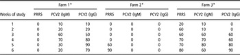
Summary results from serological analyses are shown in Table 1. All animals were seronegative to M. hyopneumoniae and P. virus during the entire period of study. Animals from farm 1 were also seronegative to the PRRSv, but on the other hand seroconversion to PCV2 took place during the 3rd week of the study in 5 out of 10 pigs and the maximum number of animals seroconverted by week 5 of the study (90%).
Percentage (%) of seroconverted pigs against the pathogens studied in each week of the study

PRRS = porcine reproductive and respiratory syndrome; PCV2 = porcine circovirus type 2; Ig = immunoglobulin.
*Pigs seronegative to Pseudorabies virus and Mycoplasma hyopneumoniae.
Animals from the second farm were negative to all viruses studied up to week 5 of the study, in which 6 out of 10 animals had a high level of specific antibodies against the PRRSv based on serology analysis. A total of nine animals had seroconverted to PRRSv by week 6. According to PRRS PCR, 80% of pigs suffered from PRRS viremia at the 4th week of the study, whereas the results decreased in the following week to 50%.
Pigs from farm 3 were positive to PCV2, based on the serological analysis, in 6 out of 10 animals at 3 weeks from the beginning of the study and in 9 out of 10 animals at week 6. In addition, six pigs from this third farm were also seropositive to PRRS at week 2, seven at weeks 4 to 5 and eight at week 6. The viremia to the PRRSv took place during the 1st week of the study in 90% of the animals and then decreased in the 2nd week to 40% according to PCR analysis.
No severe clinical signs of disease were reported in any pig throughout the entire period of study in the three farms. Animals from farm 1 developed lethargy at week 3 of the study. In addition, animals from farm 3 had a cough during the last 2 weeks of the study in which adverse climatology conditions were recorded.
APP measurements
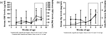
The Spearman R between the concentrations obtained in serum and saliva in the entire study was 0.68 for CRP, whereas for Hp determinations, the coefficient obtained was slightly higher at 0.78. These similarities could also be observed in the kinetic curves obtained for each body fluid across the study (Figures 1 to 3) with the exception of CRP levels from farm 3, in which the kinetics obtained for serum and salivary CRP measurements were slightly different. For example, salivary CRP levels peaked significantly at week 3 of the study, whereas serum concentrations changed over a short range during the entire period of study.
Median salivary (black squares) and serum (black circles) longitudinal levels of acute-phase proteins in pigs from farm 1. Animals aged 11 to 16 weeks of age from weeks 1 to 6 of the study. Vertical bars correspond to upper and lower limits. (a) C-reactive protein concentrations; (b) haptoglobin levels. Rectangle with rounded corners: seroconversion of animals to porcine circovirus type 2. Asterisks: saliva level of statistical significance *P < 0.05; **P < 0.01; ***P < 0.0001.

Median salivary (black squares) and serum (black circles) longitudinal levels of acute-phase proteins in pigs from farm 2. Animals aged 4 to 9 weeks of age from weeks 1 to 6 of the study. Vertical bars correspond to upper and lower limits. (a) C-reactive protein concentrations; (b) haptoglobin levels. Rectangle with rounded corners: seroconversion of animals to porcine reproductive and respiratory syndrome*: saliva level of statistical significance *P < 0.05; **P < 0.01.

Median salivary (black squares) and serum (black circles) longitudinal levels of acute-phase proteins in pigs from farm 3. Animals aged 11 to 16 weeks of age from weeks 1 to 6 of the study. Vertical bars correspond to upper and lower limits. (a) C-reactive protein concentrations; (b) haptoglobin levels. Rectangle: seroconversion of animals to porcine reproductive and respiratory syndrome. Rectangle with rounded corners: seroconversion of animals to porcine circovirus type 2. Asterisks: saliva level of statistical significance *P < 0.05; **P < 0.01.

Time-course analysis of APP concentrations in saliva from pigs belonging to farm 1, which were naturally in contact with the PCV2 virus, revealed statistically significant variations (Figure 1). Higher CRP concentrations (2 to 3 times higher) were obtained in weeks 1 and 3 than those found at the end of the experiment, when the percentage of seropositive animals began to decrease. In addition, significantly higher Hp concentrations (1.7 to 2.2 times higher) were obtained in weeks 2 and 5 than those obtained during the last week of the study.
The overall levels of salivary APP in pigs from farm 2 were lower than those observed in the other two farms. The highest levels of APP were obtained during the last week of the study, when 90% of animals were seropositive to the PRRSv (Figure 2). The concentrations of CRP were significantly higher at week 5, reaching maximum values in week 6 of approximately 38 ng/ml (1.9 times, relative to week 1, in which all animals were seronegative). Hp concentrations showed statistically significant differences over the course of the study, being increased at week 6 in comparison with those obtained at the beginning of the study (1.7 times higher).
The results of APP determinations in saliva of pigs from farm 3, which were naturally in contact with the PCV2 and PRRSv, showed the highest levels of CRP at week 3 of the study. These levels were significantly higher than those obtained at the end of the study, when animals started becoming seronegative again (2.5 times higher). Hp concentrations appeared high at week 4, with statistically significant differences from the 1st week, in which 80% of animals were already seronegative to both viruses, and from the end of the study (3.1 times).
Discussion
In this study, levels of Hp and CRP were measured in saliva samples collected weekly from 30 apparently healthy pigs from three farms, with different sanitary status, in order to obtain a longitudinal profile of APP. Under field conditions, APPs have been monitored in serum samples during the clinical course of PMWS (Grau-Roma et al., Reference Grau-Roma, Heegaard, Hjulsager, Sibila, Kristensen, Allepuz, Piñeiro, Larsen, Segalés and Fraile2009) and in pigs chronically infected with PRRSv (Asai et al., Reference Asai, Mori, Okada, Uruno, Yazawa and Shibata1999). To the authors’ knowledge, our study showed for the first time a longitudinal monitoring of CRP and Hp in saliva samples of pigs.
The farms used in this study were certified as having high sanitary status, but recently had outbreaks of different viral infections: one farm had PCV2 infections, another had PRRS outbreaks and the third was seropositive to both PRRS and PCV2. Serology was used to assess the evidence of contact with one or more pathogens. CRP was used as a fast APP and Hp as a slow one for a general overview of the levels of APP in different health environments of pigs. The sampling period in animals from farms 1 and 3 coincided with the routine vaccination against Aujeszky disease virus; thus, animals were sampled just before vaccination, and then once a week, starting 7 days post vaccination, in order to try to avoid the possible effect on APP levels in the 1st week following vaccination (Wimmers et al., Reference Wimmers, Schellander and Ponsuksili2004). Longitudinal salivary APP monitoring could be of great practical interest in detecting sub-clinically infected pigs that often play an important role in disease transmission and may represent a health hazard for humans (Eckersall, Reference Eckersall2000; Sorensen et al., Reference Sorensen, Tegtmeier, Andersen, Piñeiro, Toussaint, Campbell, Lampreave and Heegaard2006). Previous studies have shown high levels of APP in the saliva of diseased animals (Gutiérrez et al., Reference Gutiérrez, Martínez-Subiela, Soler, Pallarés and Cerón2009a), but there have been no reports on salivary APP concentrations in sub-clinically infected pigs until now.
During the time course of the study in farm 1, in which animals were in contact with PCV2, specific IgM antibodies were detected in 6 out of 10 animals at week 3, with a maximum of seven pigs in week 4. Specific IgG levels against PCV2 reached the maximum of 90% of animals in week 5; thus, seroconversion took place at around weeks 3 to 5 of the study. As it has been reported that seroconversion of animals takes place 3 to 4 weeks after contact with PCV2 (Opriessnig et al., Reference Opriessnig, Yu, Gallup, Evans, Fenaux, Pallarés, Thacker, Brockus, Ackermann, Thomas, Meng and Halbur2003), and taking into account our serological analysis, it could be postulated that our pigs from farm 1 were in contact with PCV2 in the 1st week of the study, when animals were 11 weeks of age.
It could be postulated that the two peaks, at weeks 1 and 3, of high salivary CRP levels obtained in pigs of farm 1 could represent the time of infection and the beginning of seroconversion, respectively, according to the chronology of PCV2 experimental infections (Opriessnig et al., Reference Opriessnig, Yu, Gallup, Evans, Fenaux, Pallarés, Thacker, Brockus, Ackermann, Thomas, Meng and Halbur2003). Similarly, two peaks of high protein levels were obtained for Hp determinations, appearing 1 and 2 weeks after CRP increments, respectively, and showing the later/slow behavior of Hp after being in contact with PCV2. A slow behavior of Hp in comparison with CRP after an inflammatory stimulus has been reported before in serum samples (Petersen et al., Reference Petersen, Nielsen and Heegaard2004), with Hp being characterized as a second-line APP. However, neither kinetic comparison of different APP in saliva samples nor weekly longitudinal experiments, which allow comparison with our results, have been reported until now, to the authors’ knowledge.
Overall, an increase in Hp and CRP was detected in pigs sub-clinically infected with PCV2, in line with the results obtained in serum samples of the same animals and with previously reported studies (Grau-Roma et al., Reference Grau-Roma, Heegaard, Hjulsager, Sibila, Kristensen, Allepuz, Piñeiro, Larsen, Segalés and Fraile2009). However, our results were in contrast to other studies that have suggested that the development of signs of PMWS, in PCV2-infected animals, is the unique cause of the possible increment of APP (Segalés et al., Reference Segalés, Piñeiro, Lampreave, Nofrarías, Mateu, Calsamiglia, Andrés, Morales, Piñeiro and Domingo2004). These differences between studies could be because of the time of sampling, as we evaluated the evolution of APP every week around the time of natural infection of animals with PCV2 (from 11 to 17 weeks of age), whereas in the other study only selected weeks (3, 7, 11 and 28 weeks of age) were monitored (Segalés et al., Reference Segalés, Piñeiro, Lampreave, Nofrarías, Mateu, Calsamiglia, Andrés, Morales, Piñeiro and Domingo2004). Furthermore, the different assays used to quantify each APP should be taken into account as a possible cause of difference, because highly sensitive methodologies could be required to measure slight variations of APP at very low levels, frequently next to the limit of detection, such as in sub-clinically infected animals (Eckersall, Reference Eckersall2000).
Regarding the concentrations of APP obtained in farm 2, in which animals were naturally infected with PRRS at an early state of production, salivary CRP concentrations started to increase significantly at week 5, corresponding to the beginning of seroconversion of pigs, whereas Hp levels increased 1 week after CRP. These results were similar to those obtained in experimental PRRS infections, where the highest levels of serum Hp were found at the time of seroconversion of pigs (4 times higher than those obtained at the beginning of the experiment; Díaz et al., Reference Díaz, Darwich, Pappaterra Pujols and Mateu2005). Moreover, in previous field investigations, serum Hp levels were also markedly increased after exposure to the PRRSv in pigs at 8 to 10 weeks of age from a farm with chronic PRRSv infection (Asai et al., Reference Asai, Mori, Okada, Uruno, Yazawa and Shibata1999). The percentage of infected pigs in the report of Asai (Reference Asai, Mori, Okada, Uruno, Yazawa and Shibata1999) was similar to that found in our study (85% v. 90%, respectively) at the time of Hp increment. The overall lower concentrations of APP obtained in farm 2, in comparison with the other two farms studied, could be because of the young age of animals, as it has been reported that APP in healthy animals increase with age in saliva samples (Gutiérrez et al., Reference Gutiérrez, Martínez-Subiela, Soler, Pallarés and Cerón2009a).
The acute-phase response monitored in animals from farm 3 has to be interpreted with caution, as animals were concomitantly in contact with PRRSv and PCV2; thus, viremia and seroconversion from each infection would have been overlapping. Serological analysis of PCV2 revealed an initial immune reaction during the 1st week of the study, as specific IgM were detectable. A high number of pigs started seroconversion at week 3 of the study, however the maximum number of animals seroconverted around the 5th and 6th weeks of the study. On the other hand, PRRS antibodies were detected from the 2nd week of the study in a high number of animals but seroconversion peaked at week 6; thus, the immune response to both viruses overlapped in pigs at 13 to 16 weeks of age. In this farm, the highest salivary levels of CRP were obtained at week 3, which corresponds to the initial overlapping of seroconversion of pigs to both viruses, according to the serological analysis performed. In addition, the highest levels of Hp were obtained 1 week after the CRP peak, similar to the kinetics of Hp in the other two farms, with CRP increments being faster than those recorded for Hp.
The overall levels of APP obtained in both serum and saliva samples in this study were highly correlated, with R similar to those reported previously (Gutiérrez et al., Reference Gutiérrez, Martínez-Subiela and Cerón2009b and Reference Gutiérrez, Martínez-Subiela, Eckersall and Cerón2009c). However, kinetic discrepancies in the CRP levels in serum and saliva were observed in pigs of the third farm; thus, further studies regarding the behavior of CRP in saliva samples from pigs with concomitant infections are required to explain these findings.
According to the results of this study, CRP and Hp seem to be increased around the time of seroconversion of animals to different viruses. It could be postulated that CRP peaks 1 week before Hp, because the highest increases were observed with this time difference. Nevertheless, several kinetic factors could be interfering with our results, because in farm 1 a second peak of Hp concentration was obtained 2 weeks after an increase of CRP. If daily, instead of weekly, sampling had been performed, more concrete results would have been obtained.
The concentrations of CRP obtained in sub-clinically infected animals in this study were lower than those obtained in previous studies in diseased animals of similar ages, location and breed; meanwhile, Hp levels were similar to those reported for diseased animals (Gutiérrez et al., Reference Gutiérrez, Martínez-Subiela, Soler, Pallarés and Cerón2009a). Additional studies with larger populations of well-characterized pigs should be performed in order to further study the behavior of salivary APP under sub-clinical conditions.
In conclusion, in a longitudinal study, CRP and Hp concentrations in saliva were highly correlated with those in serum, and showed similar patterns in different sub-clinical infections, with increases around the time of seroconversion of pigs. Hence, using TR-IFMA, saliva could be used for APP monitoring in longitudinal analysis involving a non-invasive and minimally stressful sampling methodology that could be performed by personnel with minimal training.
Acknowledgment
This work was supported by a grant from the Spanish Ministry of Education and Science (AGL 2006-05701).