Case report
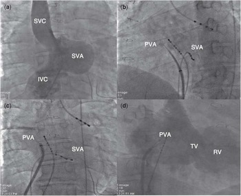
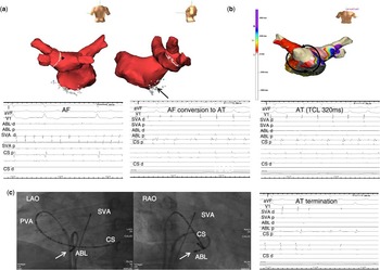
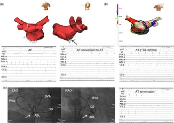
A 31-year-old man underwent a ventricular septation and Mustard operation with a bovine pericardium at 7 years of age because of a univentricular heart, which is a double inlet left ventricle combined with transposition of the great arteries. He presented with palpitations and shortened breathing at 20 years of age. A surface electrocardiogram revealed atrial tachycardia and atrial fibrillation. Despite the administration of antiarrhythmic medications, atrial tachyarrhythmias were not controlled well. After obtaining written informed consent, an electrophysiology study was conducted. A substrate map of the systemic venous atrium (functional right atrium) was created using a three-dimensional electroanatomical mapping system (NavX, St Jude Medical Inc., Minnetonka, Minnesota, United States of America). An open-irrigated 7-Fr 4-mm tip ablation catheter was used for electroanatomical mapping. With programmed atrial stimulation, sustained atrial tachycardia with 2:1 atrioventricular conduction at a tachycardia cycle length of 300 ms was induced. This tachycardia was, however, terminated after entrainment pacing at the interatrial septum of the systemic venous atrium followed by a long sinus pause. Then, atrial fibrillation was spontaneously induced by triggering a premature atrial beat. A double trans-septal puncture of the baffle was performed from the lower lateral portion of the baffle. After puncture, the needle location in the pulmonary venous atrium was confirmed by a hand injection of contrast (Fig 1). Intravenous heparin was used to maintain the activated clotting time of 300–350 seconds during the procedure. We empirically performed the left-sided pulmonary vein isolation first. During ablation of the posterior wall of the left superior pulmonary vein, atrial fibrillation was converted into atrial tachycardia (Fig 2a). An electroanatomic activation map revealed macro re-entrant atrial tachycardia with the wave-front circulating around the tricuspid valve annulus on the pulmonary venous atrium side (Fig 2b). Concealed entrainment criteria were achieved in the cavo-tricuspid isthmus of the pulmonary venous atrium side. A duodecapolar catheter was positioned into the coronary sinus via trans-septal puncture. Radiofrequency catheter ablation was performed in this area, and atrial tachycardia was finally terminated (Fig 2c). We confirmed a bidirectional block of the cavo-tricuspid isthmus ablation by pacing at the proximal coronary sinus and low systemic venous atrium. After the ablation procedure, no atrial tachyarrhythmias were induced by programmed atrial stimulation. We confirmed no reconnection of the left-sided pulmonary vein potentials. No procedure-related complications were noted. After successful ablation, the patient has remained arrhythmia-free for 24 months at follow-up.
Trans-septal puncture in a patient with Mustard baffle. ( a ) Angiography of the inferior limb of the baffle reveals the anatomic delineation of the systemic venous atrium. ( b ) A trans-septal needle is directed superiorly and anteriorly. ( c ) An 8-Fr sheath is advanced into the pulmonary venous atrium. ( d ) Right anterior oblique projection of angiography in the pulmonary venous atrium shows neo-left atrium, systemic ventricle, and cavo-tricuspid isthmus anatomy. IVC=inferior vena cava; PVA=pulmonary venous atrium; RV=right ventricle; SVA=systemic venous atrium; SVC=superior vena cava; TV=tricuspid valve.

Successful ablation site for atrial tachycardia and atrial fibrillation. ( a ) An electroanatomic map of the pulmonary venous atrium (PVA) and an intracardiac electrogram of atrial fibrillation (AF) and converted atrial tachycardaia (AT). White dots indicate the ablation site. The green dot (arrowhead) marks the conversion point from AF to AT, and the black dot (arrow) marks the AT termination site. ( b ) Electroanatomic activation map of intra-atrial reentrant tachycardia. AT cycle length was 320 ms. Anterior–posterior projection combined activation times from the PVA, especially the cavo-tricuspid isthmus (CTI), which proved to be a critical part of the tachycardia circuit. ( c ) Fluoroscopic images in the left anterior oblique (LAO) and right anterior oblique (RAO) projection showing the ablation catheter successfully performed at the CTI. The duodecapolar catheter was positioned in the coronary sinus using a double trans-septal puncture. AT termination was shown in the intracardiac electrogram. ABL=ablation catheter; CS=coronary sinus catheter; SVA=systemic venous atrium catheter.
Discussion
In patients undergoing the Mustard or Senning operation, extensive atrial reconstruction is performed by intra-atrial baffle. This procedure can induce atrial tachyarrhythmias.Reference Gelatt, Hamilton and McCrindle 1 The mechanism of atrial tachyarrhythmia in adults after intra-atrial baffle procedures varies according to the underlying anatomic defect and the method of surgical repair or palliation.Reference Van Hare, Lesh and Ross 2 – Reference Zrenner, Dong and Schreieck 4 However, the focus on arrhythmia was mainly dependent on the acquired surgical scar and chamber enlargement, which results from abnormal atrial pressure and volume loading. In this report, we initially targeted the left-sided pulmonary vein to ablate sustained atrial fibrillation, which was subsequently converted into atrial tachycardia. Atrial tachycardia was subsequently terminated by ablation at the cavo-tricuspid isthmus area of the pulmonary venous atrium side. To access the pulmonary venous atrium, a trans-septal puncture is essential. Compared with standard trans-septal alignment, the needle is directed superiorly and somewhat anteriorly into the septum.Reference Khairy and Van Hare 5 In this case, a mapping or an ablation catheter was inserted across a Mustard pericardial patch without complication. The second trans-septal puncture was achieved by directing the needle to a position that was relatively lower and more anterior than the first needle position, thereby making it easier to access the cavo-tricuspid isthmus area. In addition, a double trans-septal puncture helped to determine the confirmation of the bidirectional conduction block of the cavo-tricuspid isthmus by pacing from the low lateral pulmonary venous atrium and coronary sinus ostium. As previously demonstrated,Reference Zrenner, Dong and Schreieck 4 – Reference Wu, Deisenhofer and Ammar 6 the most common circuit of intra-atrial re-entry in patients after a Mustard or Senning operation was similar to a typical cavo-tricuspid isthmus-dependent atrial flutter, but the circuit was modified. On the basis of the operative techniques, the cavo-tricuspid isthmus can be exclusively left in the pulmonary venous atrium or is partitioned in both the pulmonary and systemic venous atrium sides. Recently, remote-controlled catheter ablation by magnetic navigation in combination with accurate three-dimensional image integration allowed for the safe and successful elimination of supraventricular tachycardias in patients with intra-atrial baffle anatomy.Reference Ernst, Babu-Narayan and Keegan 7 In addition, another study recently demonstrated catheter ablation of atrial fibrillation in this clinical setting. The researchers were able to identify the pulmonary vein trigger and performed four pulmonary vein isolations.Reference Frankel, Shah and Aziz 8 It remains to be determined whether only left-sided pulmonary vein isolation is sufficient for eliminating atrial fibrillation. In this case, atrial fibrillation was spontaneously induced twice during the procedure and converted into atrial tachycardia during left-side pulmonary vein isolation. In addition, atrial fibrillation was no longer induced after ablation. Although the left-sided pulmonary vein can be considered as a triggering focus of atrial fibrillation, further recurrence of atrial fibrillation cannot be excluded because the right-sided pulmonary vein was not clearly evaluated.
Acknowledgements
None.
Financial Support
This research received no specific grant from any funding agency, commercial, or not-for-profit sectors.
Conflicts of Interest
None.