Introduction
In vitro maturation (IVM) is a technique that allows oocytes to mature in vitro and can be performed for women with fertility problems. Matured oocytes arrest at metaphase of the second meiotic division (MII) and are activated by spermatozoa or an artificial stimulus. After activation, meiotic division resumes and embryo development begins. However, in vitro-matured oocytes have a variable lack compared with in vivo matured oocytes. Multiple factors contribute to the poor quality of in vitro-matured oocytes. One important factor is oxidative stress (Khazaei and Aghaz, Reference Khazaei and Aghaz2017) because the oxygen concentration is higher in vitro than in vivo (Tatemoto et al., Reference Tatemoto, Sakurai and Muto2000). Consequently, attempts have been made to improve IVM efficiency by protecting oocytes against oxidative stress.
Oxidative stress naturally arises because superoxide (O2-) and hydrogen peroxide (H2O2) form during metabolic processes (Storz and Imlayt, Reference Storz and Imlayt1999). These chemically reactive species containing oxygen are called reactive oxygen species (ROS). An increase in intracellular ROS leads to lipid peroxidation (Mihalas et al., Reference Mihalas, De Iuliis, Redgrove, McLaughlin and Nixon2017), DNA damage (Ménézo et al., Reference Ménézo, Dale and Cohen2010), and inhibition of meiotic maturation (Ambruosi et al., Reference Ambruosi, Uranio, Sardanelli, Pocar, Martino, Paternoster, Amati and Dell’Aquila2011) in oocytes. The ROS level in porcine oocytes increases upon heat stress (Itami et al., Reference Itami, Shirasuna, Kuwayama and Iwata2018), ageing (Kim et al., Reference Kim, Lee, Park, Jeong, Kim and Park2019), endoplasmic reticulum stress (Park et al., Reference Park, Park, Kim, Yang, Jung, Kim, Kang, Cho, Wee, Yang, Song, Kim and Koo2018), and hyperoxia (Goud et al., Reference Goud, Goud, Diamond, Gonik and Abu-Soud2008) conditions. Many researchers have supplemented IVM medium with antioxidants to reduce damage under these conditions.
The antioxidant isorhamnetin (ISO) is a 3′-methoxylated derivative of quercetin and a flavonoid-based compound. It has anticancer (Kim et al., Reference Kim, Lee, Lee, Son, Seo, Li, Jung, Heo, Mottamal, Bode, Dong and Lee2011), anti-inflammatory (Boesch-Saadatmandi et al., Reference Boesch-Saadatmandi, Loboda, Wagner, Stachurska, Jozkowicz, Dulak, Döring, Wolffram and Rimbach2011), and antioxidative (Pengfei et al., Reference Pengfei, Tiansheng, Xianglin and Jianguo2009) activities and inhibits H2O2-induced activation of the apoptotic pathway (Sun et al., Reference Sun, Sun, Xiao, Chen, Wang, Wu, Cao, Yang and Sun2012). ISO protects cells from ROS by inducing the expression of nuclear factor erythroid 2-related factor 2 (Nrf2)-dependent antioxidant genes (Yang et al., Reference Yang, Shin, Han, Kim, Wi, Kim, Cho, Kim, Shin and Ki2014). Quercetin, which has a similar structure to ISO, has been widely studied in the medical and biological fields, but ISO has not been well investigated. We hypothesized that supplementation of ISO may elicit beneficial effects during IVM of porcine oocytes due to its antioxidant activity.
The present study investigated the effects of treatment with various concentrations of ISO during the IVM of porcine oocytes exposed to oxidative stress via H2O2 treatment. We examined the developmental rate, ROS level, cumulus cell expansion, and maturation factor expression during IVM, as well as subsequent embryo developmental competence and blastocyst quality. We speculate that ISO improves the maturation, developmental competence, and quality of embryos derived from oxidative stress-exposed oocytes in vitro and can be used to improve the efficiency of porcine embryo production.
Materials and methods
Chemicals and reagents
All chemicals and reagents were purchased from Sigma (St. Louis, MO, USA) unless stated otherwise.
Oocyte collection and IVM
Prepubertal porcine ovaries were collected from a local slaughterhouse and transported to the laboratory in saline supplemented with 75 μg/ml penicillin G and 50 μg/ml streptomycin sulfate within 2 h at 32–35°C. Cumulus–oocyte complexes (COCs) were aspirated from follicles with a diameter of 2–8 mm using an 18-gauge needle and a disposable 10 ml syringe. COCs were washed three times in tissue culture medium (TCM)-199–HEPES containing 0.1% (w/v) bovine serum albumin (BSA). Groups of 50 COCs were matured in 500 μl TCM-199 (Gibco, Grand Island, NY, USA) containing Earle’s salts, 0.57 mM cysteine, 10 ng/ml epidermal growth factor, 0.5 μg/ml follicle-stimulating hormone, 0.5 μg/ml luteinizing hormone, and 10% (v/v) porcine follicular fluid under mineral oil for 44 h at 38.8°C in a humidified atmosphere of 5% CO2 in air.
COCs were cultured in IVM medium containing 1% dimethyl sulfoxide, 0, 0.02, 0.2, 2, or 20 ISO, and 200 μM H2O2 for 44 h. The experiment was repeated independently seven times, with 50 oocytes per experiment.
Parthenogenetic activation and embryo culture
Following maturation, cumulus cells were removed by pipetting in the presence of 1 mg/ml hyaluronidase for 2–3 min. Oocytes were parthenogenetically activated with 5 μM Ca2+ ionomycin (Sigma) for 5 min. After 3 h of culture in porcine zygote medium (PZM)-5 containing 7.5 μg/ml cytochalasin B (Sigma), embryos were washed three times in PZM-5 containing 0.4% (w/v) BSA and cultured in the same medium for 7 days at 38.8°C in a humidified atmosphere of 5% CO2 in air.
Measurement of intracellular ROS and GSH levels
DCFH-DA and CellTracker™ Blue CMF2HC were used to determine the intracellular levels of ROS and GSH, respectively, as previously described (Yang et al., Reference Yang, Hwang, Kwon, Kim, Choi and Oh1998; You et al., Reference You, Kim, Lim and Lee2010) with slight modifications. Briefly, cumulus cells were removed from COCs by pipetting in the presence of 0.1% (w/v) hyaluronidase. Denuded oocytes were incubated in Dulbecco’s phosphate-buffered saline (DPBS) containing 50 μM DCFH-DA or 100 μM CellTracker™ Blue CMF2HC in the dark for 20 min at 38.8°C. Thereafter, oocytes were washed more than five times with DPBS containing 0.1% (w/v) BSA to completely remove excess dye and immediately analyzed by epifluorescence microscopy (Olympus, Tokyo, Japan). The ROS level was determined using excitation and emission wavelengths of 450–490 nm and 515–565 nm, respectively. The excitation and emission wavelengths of CellTracker™ Blue CMF2HC are 371 and 464 nm, respectively. Grayscale images were acquired with a digital camera (Nikon, Tokyo, Japan) attached to a microscope. Mean grayscale values were calculated using ImageJ software (NIH, Bethesda, MD, USA). Background fluorescence values were subtracted from the final values prior to statistical analysis. The experiment was independently repeated five times, with 20–30 oocytes per experiment.
Immunofluorescence
Meiotic spindles and nuclei of oocytes were visualized after maturation. Cumulus cells were removed from porcine COCs matured for 44 h, and then oocytes were fixed overnight at 4°C with 4.0% (w/v) paraformaldehyde prepared in phosphate-buffered saline (PBS). Fixed oocytes were incubated for 30 min at 38.8°C with 0.5% (v/v) Triton X-100 reagent. After blocking for 1 h with 1% BSA (w/v) prepared in PBS (blocking solution I), oocytes were incubated overnight at 4°C with an Alexa Fluor 488-conjugated anti-α-tubulin antibody (Sigma, diluted 1:200 in blocking solution I). Nuclei were stained with Hoechst 33342 (1 μg/ml) for 30 min. Finally, oocytes were washed three times with PBS containing 0.1% (w/v) BSA, mounted onto glass slides, and examined under an inverted Olympus IX-71 microscope. At least 20 oocytes were examined per group.
Hoechst 33342 staining
Blastocysts were cultured for 7 days after parthenogenetic activation (PA), fixed overnight at 4°C in 4.0% (w/v) paraformaldehyde prepared in PBS, washed more than three times with PBS containing 0.1% BSA, and incubated with 1 μg/ml Hoechst 33342 at 38.8°C for 30 min. Thereafter, blastocysts were washed with PBS containing 0.1% BSA, mounted onto glass slides, and examined under an epifluorescence microscope. The experiment was repeated independently seven times, and at least 10 blastocysts were examined per group.
Real-time quantitative polymerase chain reaction
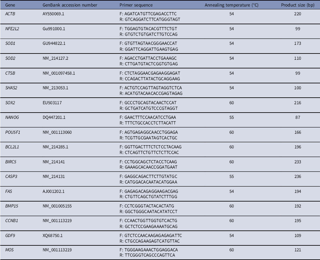
Real-time RT-PCR was performed as described previously (Lee et al., Reference Lee, Bonneau, Takacs, Bazzini, DiVito, Fleming and Giraldez2013). mRNA was isolated from groups of 20 in vitro-matured oocytes using a Dynabeads mRNA Direct Kit (Invitrogen, Carlsbad, CA, USA). cDNA was synthesized from a 2 μg mRNA per sample using an oligo(dT)20 primer and SuperScript III reverse transcriptase (Invitrogen). Real-time RT-PCR was performed using the primer sets listed in Table 2 and a StepOnePlus Real-Time PCR System (Applied Biosystems, Warrington, UK), with a final reaction volume of 20 µl containing SYBR Green PCR Master Mix (Applied Biosystems). The PCR conditions were as follows: 10 min at 95°C, followed by 40 cycles of 15 s at 95°C and 60 s at 55°C or 60°C. Samples were then cooled to 12°C. Relative gene expression was analyzed using the 2−ΔΔCt method (Livak and Schmittgen, Reference Livak and Schmittgen2001) after normalization against the mRNA level of ACTB. The experiment was independently repeated 4–6 times.
Western blot analysis
The protocol was basically the same as that described previously (Lee et al., Reference Lee, Sun, Choi, Uhm and Kim2012). In brief, oocytes (20–30 per sample) were solubilized in 20 μl of 1× sodium dodecyl sulfate (SDS) sample buffer [62.5 mM Tris-HCl (pH 6.8), containing 2% (w/v) SDS, 10% (v/v) glycerol, 50 μM dithiothreitol, and 0.01% (w/v) bromophenol blue or phenol red] and heated for 5 min at 95°C. Proteins were resolved on 5–12% Tris SDS-polyacrylamide gel electrophoresis gels for 1.5 h at 80–100 V. Samples were then transferred to Hybond ECL nitrocellulose membranes (Amersham, Buckinghamshire, UK) at 400 mA for 1.5 h in transfer buffer [25 mM Tris (pH 8.5), containing 200 mM glycine and 20% (v/v) methanol]. After blocking with 5% (w/v) nonfat milk prepared in PBS for 1 h, the membranes were incubated for at least 2 h with an anti-p44/42 MAPK or anti-phospho-p44/42 MAPK antibody diluted 1:300 in blocking solution [1× Tris-buffered saline (pH 7.5), containing 0.1% (v/v) Tween-20% and 5% (w/v) nonfat milk]. Thereafter, the membranes were washed three times in TBST [20 mM Tris-HCl (pH 7.5), containing 250 mM NaCl and 0.1% (v/v) Tween-20] and incubated for 1 h with anti-rabbit IgG-horseradish peroxidase diluted 1:2000 in blocking solution. After three washes with TBST, immunoreactive protein bands were visualized using a chemiluminescent reagent (Invitrogen). The experiment was independently repeated four times.
Statistical analysis
Data from all experiments were analyzed using the general linear model procedure within the Statistical Analysis System software (SAS Institute Inc., 2013). The paired Tukey’s multiple range test was used to determine significant differences. P-values <0.05 were considered significant.
Results
ISO enhances the developmental rate of oxidative stress-exposed porcine oocytes during IVM
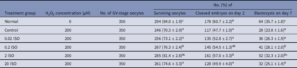
The effects of adding 0.02, 0.2, 2, and 20 μM ISO (0.02, 0.2, 2, and 20 ISO groups, respectively) to IVM medium containing (control group) or lacking (normal group) 200 μM H2O2 on the oocyte maturation efficiency were examined (Table 1). The percentage of surviving oocytes at the MII stage was significantly higher (P < 0.05) in the normal group than in the control, 0.02 ISO, 0.2 ISO, and 20 ISO groups, but did not significantly differ between the normal and 2 ISO groups. The percentage of surviving oocytes at the MII stage was significantly higher (P < 0.05) in the 2 ISO group than in the control, 0.02 ISO, and 20 ISO groups, but did not significantly differ between the normal, 0.2 ISO, and 2 ISO groups (normal, 84.0% ± 1.9%; control, 70.3% ± 2.9%; 0.02 ISO, 73.1% ± 2.2%; 0.2 ISO, 76.3% ± 2.4%; 2 ISO, 81.4% ± 2.8%; and 20 ISO, 74.6% ± 3.3%). After PA, the percentage of cleaved embryos on day 2 was significantly higher (P < 0.05) in the normal, 0.2 ISO, and 2 ISO groups than in the control, 0.02 ISO, and 20 ISO groups, but did not significantly differ between the normal, 0.2 ISO, and 2 ISO groups (normal, 60.7% ± 2.2%; control, 47.7% ± 1.9%; 0.02 ISO, 52.8% ± 2.7%; 0.2 ISO, 54.5% ± 1.3%; 2 ISO, 57.0% ± 3.3%; and 20 ISO, 49.9% ± 4.0%). The percentage of blastocyst formation on day 7 was significantly higher (P < 0.05) in the normal group than in the control, 0.02 ISO, and 20 ISO groups, but did not significantly differ between the normal and 2 ISO groups. The percentage of blastocyst formation on day 7 was significantly higher (P < 0.05) in the 0.2 ISO group than in the control, 0.02 ISO, and 20 ISO groups, but did not significantly differ between the 2 ISO group and the normal and 0.2 ISO groups (normal, 35.7% ± 1.8%; control, 23.8% ± 1.6%; 0.02 ISO, 26.3% ± 1.9%; 0.2 ISO, 28.1% ± 2.0; 2 ISO, 32.3% ± 2.0%; and 20 ISO, 25.1% ± 1.4%). Based on these results, 2 ISO was used in subsequent experiments.
Effect of different concentrations of isorhamnetin on in vitro maturation of porcine oocytes

GV, germinal vesicle; ISO, isorhamnetin.
a–c P < 0.05.
Primers used for quantitative polymerase chain reaction

F, forward; R, reverse.
ISO elicits antioxidative effects during IVM of oxidative stress-exposed porcine oocytes
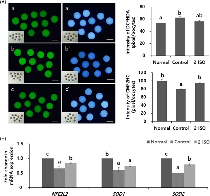
The intracellular ROS and glutathione (GSH) levels were measured to investigate the antioxidative effects of ISO during IVM of H2O2-exposed porcine oocytes (Figure 1A). The fluorescence intensity of the ROS marker dichlorohydrofluorescein diacetate (DCFH-DA) was significantly lower (P < 0.05) in the normal group than in the control group, but did not significantly differ between the normal and 2 ISO groups (normal, 53.4 ± 1.6; control, 62.4 ± 1.7; and 2 ISO, 56.1 ± 2.2). The fluorescence intensity of the GSH marker CellTracker™ Blue 4-chloromethyl-6,8-difluoro-7-hydroxycoumarin (CMF2HC) was significantly higher (P < 0.05) in the normal and 2 ISO groups than in the control group, but did not significantly differ between the normal and 2 ISO groups (normal, 99.5 ± 3.0 pixels/oocyte; control, 79.3 ± 2.6 pixels/oocyte; and 2 ISO, 93.9 ± 3.0 pixels/oocyte). We investigated the effects of ISO on mRNA expression of the antioxidant-related genes nuclear factor erythroid 2-like 2 (NFE2L2), superoxide dismutase 1 (SOD1), and superoxide dismutase 2 (SOD2). The mRNA levels were normalized against those in the normal group (Figure 1B). mRNA expression levels of NFE2L2 and SOD2 were significantly higher (P < 0.05) in the normal group than in the control and 2 ISO groups, and was significantly higher (P < 0.05) in the 2 ISO group than in the control group. mRNA expression of SOD1 was significantly higher (P < 0.05) in the normal group than in the control and 2 ISO groups, but did not significantly differ between the control and 2 ISO groups.
Effect of ISO on the level of oxidative stress during IVM of porcine oocytes. H2O2 was used to induce oxidative stress. (A) Images of oocytes stained with DCFH-DA (green) CellTrackerTM Blue CMF2HC (blue), and quantification of the fluorescence intensities of DCFH-DA and CellTrackerTM Blue CMF2HC. (a–c) ROS staining. (a'–c') GSH staining. (a, a') Normal group. (b, b') Control group. (c, c') 2 ISO group. Scale bars, 100 µm. (B) Relative mRNA expression of antioxidant-related genes (NFE2L2, SOD1, and SOD2). ACTB was used as an internal standard. Data were normalized against the corresponding levels in the normal group. Values are presented as mean ± SEM of X independent experiments (a–c P < 0.05).

ISO enhances cumulus cell expansion, prevents chromosomal misalignment, and upregulates molecular maturation factors during IVM of oxidative stress-exposed porcine oocytes
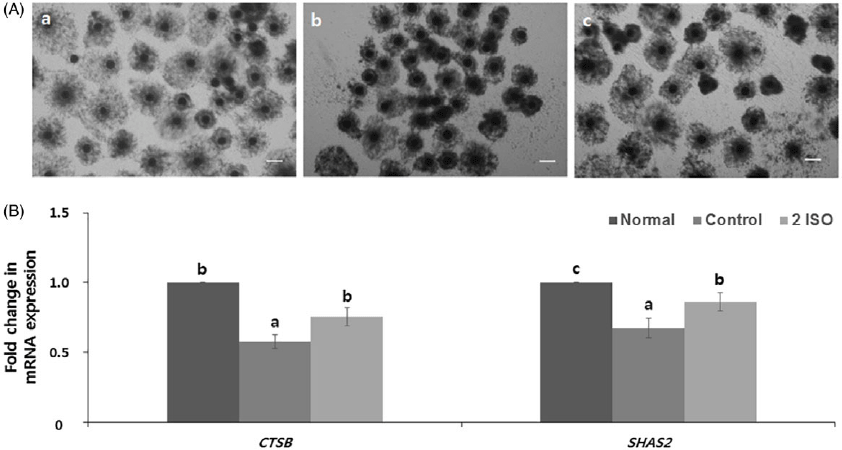
We investigated the effect of ISO on cumulus cell expansion during IVM of H2O2-exposed porcine oocytes at the MII stage (Figure 2). Microscopic analyses indicated that cumulus cell expansion was best in the normal group, and was better in the 2 ISO group than in the control group (Figure 2A). We examined the effects of ISO on mRNA expression of the cumulus cell expansion-related genes cathepsin B (CTSB) and hyaluronan synthase 2 (SHAS2) at the MII stage. mRNA expression of CTSB was significantly higher (P < 0.05) in the normal and 2 ISO groups than in the control group, but did not significantly differ between the normal and 2 ISO groups. mRNA expression of SHAS2 was significantly higher (P < 0.05) in the normal group than in the control and 2 ISO groups, and was significantly higher (P < 0.05) in the 2 ISO group than in the control group (Figure 2B).
Effect of ISO on cumulus cell expansion during IVM of porcine oocytes. (A) Degree of cumulus cell expansion after 44 h of IVM. (a) Normal group. (b) Control group. (c) 2 ISO group. Scale bars, 250 µm. (B) Relative mRNA expression of cumulus cell expansion-related genes (CTSB and SHAS2). ACTB was used as an internal standard. Data were normalized against the corresponding levels in the normal group. Values are presented as the mean ± SEM of five independent experiments (a–c P < 0.05).

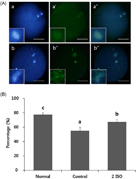
We next evaluated the effect of ISO on chromosomal alignment and spindle organization (Figure 3). Oocytes were classified as abnormal or normal as described previously (Lenie et al., Reference Lenie, Cortvrindt, Eichenlaub-Ritter and Smitz2008) (Figure 3A). The percentage of oocytes with normal chromosomal alignment and spindle organization was significantly higher (P < 0.05) in the normal group than in the control and 2 ISO groups, and was significantly higher (P < 0.05) in the 2 ISO group than in the control group (normal, 78.2% ± 2.3%; control, 55.3% ± 3.4%; and 2 ISO, 68.4% ± 3.1%; Figure 3B).
Effect of ISO on spindle morphology during IVM of porcine oocytes. (A) Normal and abnormal chromosomal alignment and meiotic spindle formation in oocytes. (a, b) Nuclei of oocytes stained with Hoechst 33342. (a', b') Spindles of oocytes stained with an anti--tubulin antibody. (a'', b'') Merged images. (a–a'') Normal. (b–b'') Abnormal. Scale bars, 50 µm. (B) Percentage of oocytes with normal chromosomal alignment and meiotic spindle organization. Values and presented as mean ± SEM of five independent experiments (a–cP < 0.05).

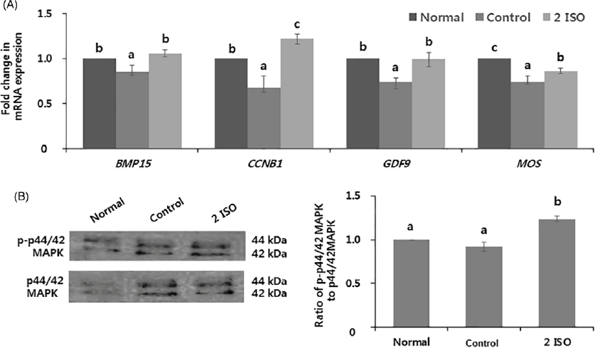
To examine the effect of ISO on oocyte cytoplasmic maturation, we measured maternal gene expression and p44/42 mitogen-activated protein kinase (MAPK) activity (Figure 4). mRNA expression levels of the maternal genes bone morphogenetic protein 15 (BMP15), cyclin B1 (CCNB1), growth differentiation factor-9 (GDF9), and serine/threonine kinase (MOS) were determined. mRNA expression levels of BMP15 and GDF9 were significantly higher (P < 0.05) in the normal and 2 ISO groups than in the control group, but did not differ significantly between the normal and 2 ISO groups. mRNA expression of CCNB1 was significantly higher (P < 0.05) in the 2 ISO group than in the normal and control groups, and was significantly higher (P < 0.05) in the normal group than in the control group (Figure 4A). Western blot analysis revealed that phospho-p44/42 MAPK, the active form of this kinase, migrated as a doublet in lysates of porcine oocytes (Figure 4B). The ratio of phospho-44/42 MAPK to p44/42 MAPK was determined and normalized against that in the normal group. This ratio was significantly higher (P < 0.05) in the 2 ISO group than in the normal and control groups (normal, 1.00 ± 0.00; control, 0.92 ± 0.05; and 2 ISO, 1.24 ± 0.03; Figure 4B).
Effect of ISO on cytoplasmic maturation during IVM of porcine oocytes. (A) Relative mRNA expression of maternal genes (BMP15, CCNB1, GDF9, and MOS). ACTB was uses as an internal standard. Data were normalized against the corresponding levels in the normal group. (B) MAPK activity determined by western blot analysis and the ratio of phospho-p44/42 MAPK to p44/42 MAPK. Values are presented as mean ± SEM of four independent experiments (a–c P < 0.05).

ISO improves the developmental capacity and quality of embryos derived from oxidative stress-exposed porcine oocytes
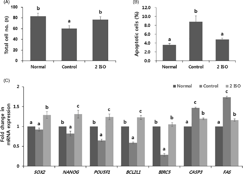
We studied the effect of ISO treatment of H2O2-exposed oocytes on blastocyst formation at day 7. The total cell numbers per blastocyst were significantly higher (P < 0.05) in the normal and 2 ISO groups than in the control group, but did not significantly differ between the normal and 2 ISO groups (normal, 82.9 ± 6.0; control, 60.2 ± 5.7; and 2 ISO, 76.6 ± 5.7; Figure 5A). The percentages of apoptotic cells in blastocysts were significantly lower (P < 0.05) in the normal and 2 ISO groups than in the control group, but did not differ significantly between the normal and 2 ISO groups (normal, 3.6% ± 0.3%; control, 8.9% ± 1.3%; and 2 ISO, 4.9% ± 0.2%; Figure 5B). We measured mRNA expression levels of the development-related genes sex-determining region Y-box 2 (SOX2), homeobox protein NANOG (NANOG), and POU domain, class 5, transcription factor 1 (POU5F1). mRNA expression of SOX2 was significantly higher (P < 0.05) in the 2 ISO group than in the normal and control groups, but did not significantly differ between the normal and control groups. mRNA expression levels of NANOG and POU5F1 were significantly higher (P < 0.05) in the 2 ISO group than in the normal and control groups, and were significantly higher (P < 0.05) in the normal group than in the control group. We measured mRNA expression of the anti-apoptotic genes B-cell lymphoma 2-like 1 (BCL2L1) and baculoviral IAP repeat-containing 5 (BIRC5). mRNA expression of BCL2L1 was significantly higher (P < 0.05) in the 2 ISO group than in the normal and control groups, and was significantly higher (P < 0.05) in the normal group than in the control group. mRNA expression of BIRC5 was significantly higher (P < 0.05) in the normal and 2 ISO groups than in the control group, but did not differ significantly between the normal and 2 ISO groups. We measured mRNA expression levels of the pro-apoptotic genes cysteine-aspartic acid protease 3 (CASP3) and Fas cell surface death receptor (FAS). mRNA expression levels of CASP3 and FAS were significantly lower (P < 0.05) in the normal group than in the control and 2 ISO groups, and wee significantly lower (P < 0.05) in the 2 ISO group than in the control group.
Effect of ISO treatment during IVM of porcine oocytes on subsequent embryo development . (A) Total cell number per blastocyst. (B) Percentage of apoptotic cells in blastocysts. (C) Relative mRNA expression of development-related (SOX2, NANOG, and POU5F1), anti-apoptotic (BCL2L1 and BIRC5), and pro-apoptotic (CASP3 and FAS) genes. ACTB was used as an internal standard. Data were normalized against the corresponding levels in the normal group. Values are presented as mean ± SEM of seven independent experiments (a–cP < 0.05).

Discussion
This study investigated the effect of ISO on IVM of oxidative stress-exposed porcine oocytes and the developmental capacity of embryos derived from these oocytes. ISO increased the GSH level, enhanced cumulus cell expansion, prevented chromosomal misalignment, activated MAPK in porcine oocytes, and increased the total cell number and decreased the percentage of apoptotic cells in blastocysts derived from these oocytes.
Porcine oocytes were matured in vitro for 44 h in the presence of 200 μM H2O2 and 0, 0.02, 0.2, 2, or 20 ISO, or in the absence of ISO and H2O2. However, in vitro-matured oocytes have a variable lack compared with in vivo matured oocytes because the oxygen concentration is higher in vitro than in vivo (Tatemoto et al., Reference Tatemoto, Sakurai and Muto2000). Great efforts have been made to improve IVM efficiency (Yoshida et al., Reference Yoshida, Ishigaki and Pursel1992) that include changing the culture medium composition (Marques et al., Reference Marques, Nicacio, de Oliveira, Nascimento, Caetano, Mendes, Mello, Milazzotto, Assumpção and Visintin2007), altering the atmospheric composition (Kang et al., Reference Kang, Atikuzzaman, Kwon, Park, Kim, Moon, Koo, Jang and Lee2012), supplying hormones (Silvestre et al., Reference Silvestre, Alfonso, García-Mengual, Salvador, Duque and Molina2007), and adding antioxidants (Hennings et al., Reference Hennings, Zimmer, Nabli, Davis, Sutovsky, Sutovsky and Sharpe-Timms2016). Addition of antioxidants to culture medium reduces oxidative stress (Tatemoto et al., Reference Tatemoto, Ootaki, Shigeta and Muto2001). We demonstrated that the percentages of surviving oocytes, cleaved embryos, and blastocysts formation were increased in the 2 ISO group (Table 1). ISO has a high DPPH-scavenging ability dependent on its phenolic hydroxyl group (Zuo et al., Reference Zuo, Yanying, Li, Binbin, Xiongying, Yan and Shuwen2011), and it improves the viability of oxidative stress-exposed retinal pigment epithelial cells by activating PI3K/Akt. The PI3K/Akt signalling pathway is fundamental in animals, and functions in cell growth, proliferation, survival, migration, metabolism, and apoptosis (Knowles et al., Reference Knowles, Platt, Ross and Hurst2009). Therefore, we demonstrated that ISO enhances the development rate of oxidative stress-exposed porcine oocytes, and use 2 ISO in subsequent experiments.
Intracellular oxidative and antioxidative activities are primarily determined by ROS and GSH, respectively. ROS naturally form during metabolic processes (Storz and Imlayt, Reference Storz and Imlayt1999), but external oxygen and an inefficient antioxidant system increase ROS generation in vitro (Armand et al., Reference Armand, Andriamihaja, Gellenoncourt, Bitane, Lan and Blachier2019). An excessively high ROS level directly or indirectly interferes with mitochondrial function (Zhang and Liu, Reference Zhang and Liu2002). Conversely, GSH is a cofactor of glutathione peroxidase and glutathione-S-transferase, and plays an important role in protecting cellular lipids, proteins, and nucleic acids against oxidative stress (Gérard-Monnier and Chaudiere, Reference Gérard-Monnier and Chaudiere1996). This study showed that treatment with 2 ISO altered the ROS and GSH levels (Figure 1A), and increased mRNA expression of the antioxidant-related genes NFE2L2 and SOD2 (Figure 1B). NFE2L maintains mitochondrial redox homeostasis by upregulating antioxidant genes (Ryoo and Kwak, Reference Ryoo and Kwak2018) and plays an important role in defence against oxidative stress (Zelko et al., Reference Zelko, Mariani and Folz2002). Activated NFE2L promotes expression of the antioxidants SOD1 and SOD2 (Dong et al., Reference Dong, Sulik and Chen2008). SOD is the first line of defence against superoxide produced as a by-product of oxidative phosphorylation (Li et al., Reference Li, Huang, Carlson, Melov, Ursell, Olson, Noble, Yoshimura, Berger, Chan, Wallace and Epstein1995). ISO increases the total level of NFE2L2 in mouse-derived C2C12 myoblasts by activating the Nrf2/HO-1 pathway, which involves activation of the ERK pathway (Choi, Reference Choi2016), and it enhances SOD activity in the mouse hippocampus and prefrontal cortex (Ishola et al., Reference Ishola, Osele, Chijioke and Adeyemi2019). ISO acts as an antioxidant by scavenging DPPH, donating electrons to reactive free radicals, and chelating iron (Pengfei et al., Reference Pengfei, Tiansheng, Xianglin and Jianguo2009). It functions as an antioxidant in human cells by activating p38-MAPK (Bao and Lou, Reference Bao and Lou2006). In summary, ISO protects cells against oxidative stress by acting as an antioxidant, thereby reducing the ROS level and increasing the GSH level.
This study investigated the effect of ISO on cumulus cell expansion, chromosomal alignment, and molecular maturation factors in porcine oocytes. Cumulus cells surround the oocyte and play a important role in processes of oocyte maturation and fertilization. These cells expand and surround oocytes after ovulation (Downs et al., Reference Downs, Daniel, Bornslaeger, Hoppe and Eppig1989). At the time of ovulation, cumulus cells have progressed to MII (Gérard-Monnier and Chaudiere, Reference Gérard-Monnier and Chaudiere1996). During cumulus cell expansion, an extracellular matrix consisting of hyaluronic acid (HA), proteoglycans, and proteins accumulates in the intercellular space and induces oocyte maturation (Russell and Salustri, Reference Russell and Salustri2006). In the present study, treatment with 2 ISO enhanced cumulus cell expansion and expression of the cumulus expansion-related gene SHAS2 (Figure 2). SHAS2 stimulates production and extrusion of HA (Itano et al., Reference Itano, Sawai, Yoshida, Lenas, Yamada, Imagawa, Shinomura, Hamaguchi, Yoshida, Ohnuki, Miyauchi, Spicer, McDonald and Kimata1999), and its expression increases during cumulus cell expansion (Fülöp et al., Reference Fülöp, Salustri and Hascall1997). The cumulus cell expansion-related gene CTSB plays a regulatory role in cell death via caspase-3 stimulation in oocytes (Eykelbosh and Van Der Kraak, Reference Eykelbosh and Van Der Kraak2010), and its upregulation in cumulus cells indicates that oocytes have a low developmental competence (Bettegowda et al., Reference Bettegowda, Patel, Lee, Park, Salem, Yao, Ireland and Smith2008). In summary, ISO protects cumulus cells against oxidative stress and improves the quality of cumulus–porcine oocyte complexes under oxidative stress. Interestingly, the present study showed that ISO increased the percentage of oocytes with normal chromosomal alignment and spindle organization (Figure 3B), elevated MAPK activity, and upregulated maternal gene expression (Figure 4). The meiotic spindle consists of microtubules and is crucial for normal chromosomal alignment and separation of maternal chromosomes during MI and MII in oocytes (Liu et al., Reference Liu, Sun, Li, Jiao and Wang2003). It is essential for the maintenance of chromosomal organization and formation of the second polar body (Schatten et al., Reference Schatten, Simerly and Schatten1985). Oxidative stress increases abnormal spindle alignment (Choi et al., Reference Choi, Banerjee, Falcone, Bena, Agarwal and Sharma2007), and spindle disruption promotes chromosomal misalignment (Eroglu et al., Reference Eroglu, Toth and Toner1998). MAPK, which regulates cell cycle progression by modulating microtubules and actin filaments, is an essential regulator of oocyte maturation (Sun et al., Reference Sun, Wu, Lai, Bonk, Cabot, Park, Day, Prather and Schatten2002), but its activity decreases upon oxidative stress (Inoue et al., Reference Inoue, Hisamoto, An, Oliveira, Nishida, Blackwell and Matsumoto2005). MAPK is activated at the germinal vesicle breakdown stage, localizes to the cytoplasm and around chromosomes from MI to MII, and is essential for resuming meiosis in MII and maintaining arrest (Villa-Diaz and Miyano, Reference Villa-Diaz and Miyano2004). MAPK is activated by MOS protein, an active component of cytostatic factor, which is responsible for meiotic arrest at MII (Newman and Dai, Reference Newman and Dai1996). GDF9 and BMP15 play an important role in the regulation of fertility (Juengel et al., Reference Juengel, Bodensteiner, Heath, Hudson, Moeller, Smith, Galloway, Davis, Sawyer and McNatty2004) and regulate oogenesis by interacting with each other (Hussein et al., Reference Hussein, Thompson and Gilchrist2006). CCNB is an important meiotic regulator and forms a complex with cyclin-dependent kinase 1, which permits transition from GII to M phase (Robert et al., Reference Robert, Hue, McGraw, Gagné and Sirard2002). Therefore, upregulation of these genes is beneficial in oxidative stress-exposed porcine oocytes. A recent study reported that ISO inhibits the proliferation of cells arrested at the GII/M phase and promotes the formation of cytoplasmic vacuoles, which are indicative of apoptotic cell death mediated by ROS and the ERK signalling pathway (Chen et al., Reference Chen, Song, Wang, Shen, Xie, Li, Jiang, Zhao, Feng, Zhou, Zhou, Zeng, Ji and Chen2021). In summary, we demonstrate that ISO protects the nucleus and regulates maturation factors in oxidative stress-exposed porcine oocytes. Collectively, these results indicate that ISO improves porcine oocyte maturation under oxidative conditions.
To further investigate whether ISO affects subsequent embryo development, oocytes were parthenogenetically activated and cultured in vitro for 7 days. Treatment with 2 ISO increased the total cell number per blastocyst and reduced the percentage of apoptotic cells (Figure 5A,B), and regulated development-related and apoptosis-related genes (Figure 5C). Apoptosis is associated with cellular stress and cell death (Mizushima, Reference Mizushima2007). Apoptosis supports embryo survival under healthy conditions by selectively eliminating abnormal cells with nuclear and chromosomal abnormalities, which may lead to the loss of the whole embryo during early embryonic development (Choi et al., Reference Choi, Park, Lee, Kim, Jeong, Lee, Park, Kim, Hossein, Jeong, Kim, Hyun and Hwang2008). It is important that the tendency for the apoptosis is consistently balanced, because high percentages of apoptotic cells inhibit embryonic development (Kwak et al., Reference Kwak, Cheong, Jeon, Lee, Choi, Jeung and Hyun2012). In a recent study, resveratrol supplementation downregulated apoptosis-related genes and demonstrated better developmental competence in porcine oocytes (Kwak et al., Reference Kwak, Cheong, Jeon, Lee, Choi, Jeung and Hyun2012). Another study found that BME modulated GSH levels and raised total cell number in blastocysts derived from matured porcine oocytes (Abeydeera et al., Reference Abeydeera, Wang, Cantley, Prather and Day1998). Cell numbers have been used to determine embryo viability and increased cell numbers have been associated with the advanced development of the embryo (Papaioannou and Ebert, Reference Papaioannou and Ebert1988). We examined the expression of development-related (SOX2, NANOG, and POU5F1) and apoptosis-related (BCL2L1, BIRC5, CASP3, and FAS) genes. Recent studies have shown that SOX2, NANOG, and POU5F1 contribute to activation of zygotic genes during the maternal-to-zygote transition (Lee et al., Reference Lee, Bonneau, Takacs, Bazzini, DiVito, Fleming and Giraldez2013), whereas POU5F1 and SOX2 function in all the main embryonic regulatory pathways (Leichsenring et al., Reference Leichsenring, Maes, Mössner, Driever and Onichtchouk2013). Anti-apoptotic members of the BLC-2 family regulate pro-apoptotic proteins (Marques et al., Reference Marques, Nicacio, de Oliveira, Nascimento, Caetano, Mendes, Mello, Milazzotto, Assumpção and Visintin2007). BIRC5 belongs to the inhibitor of apoptosis family and is involved in the regulation of the mitotic spindle, especially at the G2/M phase. BIRC5 protein is expressed during embryonic and fetal development (Ambrosini et al., Reference Ambrosini, Adida and Altieri1997). FAS activates caspase-3 by stimulating denitrosylation of thiol in its active site (Mannick et al., Reference Mannick, Hausladen, Liu, Hess, Zeng, Miao, Kane, Gow and Stamler1999). Our results showed that ISO improves the developmental capacity and quality of embryos under oxidative conditions.
In conclusion, this study demonstrates that the antioxidant ISO effectively protects oocytes against oxidative stress by reducing the level of ROS and thereby minimizes the deterioration in oocyte quality, leading to subsequent good-quality embryonic development. Therefore, ISO may be a good supplement during porcine oocyte maturation to improve the efficiencies of in vitro fertilization, intracytoplasmic sperm injection, and somatic cell nuclear transfer.
Acknowledgements
This work was supported by a grant (715003-07) from the Research Center for Production Management and Technical Development for High Quality Livestock Products through Agriculture, Food and Rural Affairs Convergence Technologies Programme for Educating Creative Global Leader, Ministry of Agriculture, Food and Rural Affairs. Also this work was supported by a grant from Jeollanam-do Science and Technology R&D Project (Development of stem cell-derived new drug), funded by the Jeollanam-do, Korea.
Conflict of interest
The authors do not have any conflicts of interest to declare.