Ketosis and fatty liver are common metabolic diseases in perinatal dairy cows (Raboisson et al., Reference Raboisson, Mounie and Maigne2014; Itle et al., Reference Itle, Huzzey, Weary and von Keyserlingk2015) and are associated with decreased health status and reproductive performance. Clinical investigations demonstrated that dairy cows with ketosis or fatty liver display high blood levels of insulin and massive hepatic lipid accumulation (Mann et al., Reference Mann, Leal Yepes, Duplessis, Wakshlag, Overton, Cummings and Nydam2016). Insulin can increase lipid synthesis and decrease lipid oxidation through the AMPK signaling pathway, thereby inducing TG accumulation (Li et al., Reference Li, Li, Ding, Dong, Zhang, Huang, Lei, Wang, Liu and Li2018), whereas impairment of the insulin signaling pathway is associated with the development of fatty liver and type II diabetes. Hepatic insulin signaling pathway is influenced by several factors, one negative regulatory factor being phosphatase and tension homolog (PTEN), a ubiquitously expressed tumor suppressor gene (Jayakumar et al., Reference Jayakumar, Lanjewar and Axiotis2018; Jia et al., Reference Jia, Zhan, Li, Li, Li and Qian2018). In addition, PTEN is a protein tyrosine phosphatase, and can suppress the activation of phosphoinositide 3-kinase (PI3K) by catalyzing PIP3 to PIP2, and thereby negatively influence the PI3K/AKT signaling pathway (Worby and Dixon, Reference Worby and Dixon2014). Gao et al. indicated that the over-expression of PTEN enhanced the impairment of insulin signaling in mice (Gao et al., Reference Gao, Su, Wang, Zhu, Chen, Liu and He2013). These studies indicate that PTEN plays an important role in the regulation of hepatic glucose and lipid metabolism.
The glucose and lipid metabolism of dairy cows is different from that of monogastric animals such as humans and mice, and the role of PTEN in regulating insulin pathway is poorly understood in dairy cows. Therefore, we investigated the regulatory mechanism by which PTEN affects the insulin pathway-mediated glucose and lipid metabolism in dairy cows.
Materials and methods
Detailed materials and methods are given in the online Supplementary File.
Cell culture
Hepatocytes were isolated by a modified two-step collagenase perfusion method from a hepatic caudate lobe obtained post mortem from a newborn calf (Zhang et al., Reference Zhang, Li, Gao, Liu, Kong, Li, Wang, Zhang, Wang and Zhang2012; Deng et al., Reference Deng, Li, Fu, Yin, Zhang, Wang, Wang, Liu, Yuan, Sun, Wang, Liu and Li2014). After digestion, we used 100 ml RPMI-1640 culture medium containing 0.2% bovine serum albumin with pre-cooling to terminate the digestion. The hepatocyte suspension was filtered sequentially with 100 mesh (150 µm), and 200 mesh (75 µm) cell sieves. Cell density was adjusted to 2 × 10⁶ cells/ml with adherent 1640 culture medium. The hepatocytes were seeded into a 6-well tissue culture plate and incubated at 37 °C in 5% CO2.
AD-PTEN, AD-shPTEN and NC transfected hepatocytes
PTEN over-expression adenovirus labeled with GFP (green fluorescent protein) (AD-PTEN), PTEN low-expression adenovirus labeled with GFP (AD-shPTEN), and adenovirus labeled with GFP (NC: negative control) were constructed (Fu et al., Reference Fu, Deng, Yang, Ding, Wang, Li, Li, Wang, Li and Liu2012). The recombinant adenoviruses were transfected into cow hepatocytes with the multiplicity of infection (MOI) of 100 at 48 h after the hepatocytes were seeded for 72 h. At the same time, control group (non-transfected) was incubated.
Quantitative real-time PCR
The mRNA expression levels were normalized to the house-keeping gene—β-actin. Real-time PCR was conducted under the following conditions: initial denaturation at 95 °C for 3 min, 45 cycles of amplification (denaturation at 95 °C for 15 s, annealing at 60 °C for 1 min). The relative expression of genes was calculated by the 2−ΔΔCt method and was normalized to abundance of β-actin.
Protein extraction and western blot analysis
Hepatocytes were lysed with lysis buffer, and the protein concentration was measured using the BCA protein assay kit (Pierce, Rockford, IL, USA). Prepared proteins were obtained by standard SDS-PAGE with 10% (w/v) polyacrylamide gels (0.1% SDS), and electrotransferred onto PVDF membranes (Roche, Basel, Switzerland). The resulting bands were detected by ECL kit (Millipore, Boston, Massachusetts, USA). The relative expression levels of the proteins were normalized to β-actin level.
Statistics
All results are expressed as the means ± standard error (se). Statistical analysis was carried out using the statistical analysis program SPSS 13.0 (SSPS, Chicago, IL, USA). P < 0.05 was considered to be significant.
Results
Low and over-expression of PTEN in bovine hepatocytes
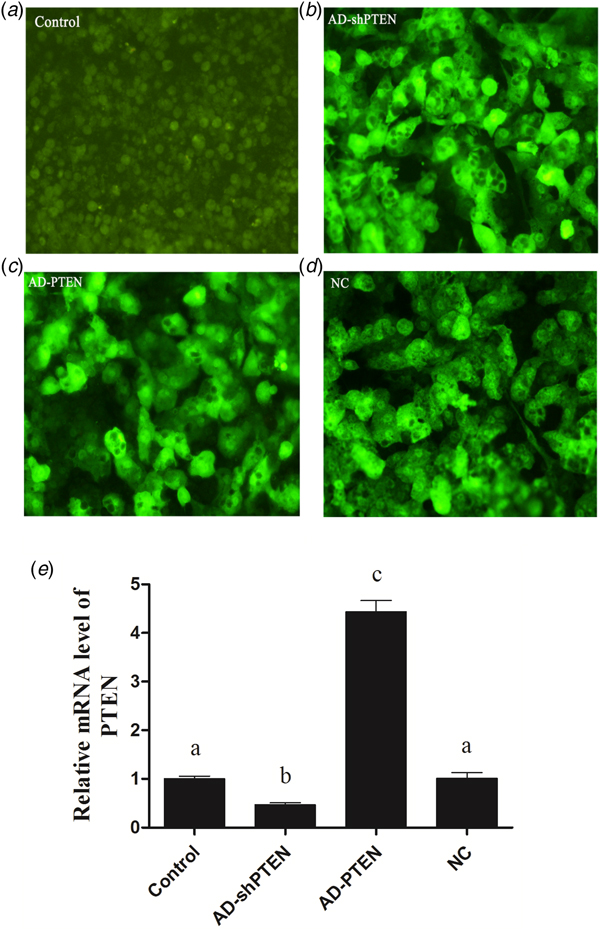
Hepatocytes were transfected with AD-shPTEN, AD-PTEN and NC. The transfection efficiency as observed by confocal laser scanning microscopy exceeded 95% (Fig. 1). NC was used as a negative control that didn't influence the PTEN mRNA expression. Compared with control and NC groups, AD-shPTEN significantly decreased the mRNA and protein expression levels of PTEN in cow hepatocytes, while AD-PTEN significantly increased the mRNA and protein expression levels of PTEN (Fig. 2; P < 0.05).
Results of fluorescence confocal microscopy and the relative mRNA level of PTEN in hepatocytes. (a) Control (no infection) (100×). (b) Hepatocytes infected with AD-shPTEN (the low expression adenovirus vector) (200×). (c) Hepatocytes infected with AD-PTEN (the over expression adenovirus vector) (200×). (d) Hepatocytes infected with NC (negative control) (200×). (e) The relative mRNA level of PTEN was examined by qRT-PCR in hepatocytes after infecting adenovirus vectors for 48 h. Control (no treatment), AD-shPTEN (the low expression adenovirus vector), AD-PTEN (the over expression adenovirus vector), NC (negative control). Values are the means ± se (n = 6), different letters are statistically different, P < 0.05.

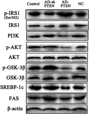
The protein expression levels of p-IRS1 (ser302), IRS1, PI3K, p-AKT, AKT, p-GSK-3β, GSK-3β, SREBP-1c, FAS and β-actin. The protein expression level was carried out according to a western blot after infecting with control (no infection), AD-shPTEN (the low expression adenovirus vector), AD-PTEN (the over expression adenovirus vector) and NC (negative control).

The effect of PTEN on insulin signaling pathway in cow hepatocytes
To investigate the role of PTEN on insulin pathway in calf hepatocytes, we determined the protein levels of phospho-IRS1 (Ser302), IRS1, PI3K, p-AKT, and AKT using western blotting. The protein level of PI3K and phosphorylation level of AKT were significantly increased in AD-shPTEN group compared with control group, while phosphorylation level of IRS1 was significantly decreased (Fig. 3). Conversely, PTEN over-expression markedly inhibited the PI3K/AKT pathway. Collectively, PTEN expression negatively regulated the activation of PI3K/AKT pathway in cow hepatocytes (Fig. 3).
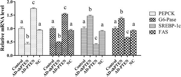
The relative mRNA levels of PEPCK, G6-Pase, SREBP-1c and FAS. Control (no treatment), AD-shPTEN (the low expression adenovirus vector), AD-PTEN (the over expression adenovirus vector), NC (negative control). The relative mRNA level of PTEN was examined by qRT-PCR in hepatocytes after infecting adenovirus vectors for 48 h. Values are the means ± se (n = 6), different letters are statistically different, P < 0.05.

The mRNA levels of PEPCK, G6-Pase, ACC1 and FAS in hepatocytes
We determined the phosphorylation level of GSK-3β and the expressions of SREBP-1c, PEPCK, G6-Pase, ACC1 and FAS. The phosphorylation level of GSK-3β was significantly increased in the AD-shPTEN group and decreased in the AD-PTEN group, indicating that PTEN silencing could inhibit the GSK-3β activity and PTEN over-expression could increase the GSK-3β activity (Fig. 3). Furthermore, the mRNA expression levels of PEPCK and G6-Pase were significantly decreased in the AD-shPTEN group. The protein and mRNA levels of SREBP-1c and its target genes ACC and FAS were significantly increased in the AD-shPTEN group and decreased in the AD-PTEN group.
Discussion
Dairy cows with fatty liver or ketosis exhibit insulin resistance and decreased insulin sensitivity (Li et al., Reference Li, Ding, Wang, Liu, Huang, Zhang, Guo, Wang, Li, Liu, Wu and Li2016). Furthermore, defects in the insulin receptor substrate (IRS)/PI3K/AKT signaling pathway are associated closely with liver steatosis and insulin resistance, and then are involved in the development of ketosis or fatty liver in dairy cows with NEB (LeBlanc, Reference LeBlanc2010; Wang et al., Reference Wang, Hou, Qu, Wang, Gao and Li2014). Hence, the regulation of insulin signaling pathway (PI3K/AKT) is an effective measure for the prevention and treatment of ketosis or fatty liver. PTEN is a major negative regulator for the PI3K/AKT pathway. Importantly, PTEN plays a significant role in the hepatic glucose and lipid metabolism (Chen et al., Reference Chen, Chen, He and Stiles2018). Liver-specific deletion of PTEN in mice induces steatosis, steatohepatitis and enhanced peripheral glucose metabolism (Peyrou et al., Reference Peyrou, Bourgoin, Poher, Altirriba, Maeder, Caillon, Fournier, Montet, Rohner-Jeanrenaud and Foti2015). Loss of PTEN function in hepatocytes could, thus, represent an important mechanism triggering the development of steatosis and affecting insulin sensitivity. Low PTEN expression or loss of PTEN function was observed in human liver cancer and non-alcoholic fatty liver disease (NAFLD) (Peyrou et al., Reference Peyrou, Bourgoin and Foti2010).
The hepatic glucose and lipid metabolism in dairy cows is different from that of monogastric animals such as humans and mice. Unfortunately, the regulation mechanisms of PTEN on the hepatic glucose and lipid metabolism of dairy cows are not well known. We recombined adenovirus that induced over-expression and silencing of PTEN, respectively, to investigate the regulation role of PTEN on the insulin signaling pathway.
Insulin receptor substrate 1 (IRS1) is a key mediator of the signaling events that lie downstream of the insulin receptor. IRS1-deficient mice showed normal glucose tolerance and peripheral insulin resistance (Saltiel and Kahn, Reference Saltiel and Kahn2001). Nakamura et al. (Reference Nakamura, Tajima, Zolzaya, Sato, Inoue, Yoneda, Fujita, Nozaki, Kubota, Haga, Kubota, Nagashima, Nakajima, Maeda, Kadowaki and Terauchi2012) reported that IRS1−/− mice fed a high fat diet were protected against hepatic steatosis despite exhibiting severe hyperinsulinemia, as compared with the wild type mice fed the same diet (Nakamura et al., Reference Nakamura, Tajima, Zolzaya, Sato, Inoue, Yoneda, Fujita, Nozaki, Kubota, Haga, Kubota, Nagashima, Nakajima, Maeda, Kadowaki and Terauchi2012). We found that PTEN silencing by AD-shPTEN decreased the phosphorylation of IRS1, while PTEN over-expression significantly increased the IRS1 phosphorylation. The PI3K/AKT pathway is a key component of the insulin signaling cascade and is considered necessary for hepatic glucose and lipid metabolism (Wang et al., Reference Wang, Sun, Li, Dong, Li and Zhao2015). Knockouts of the regulatory subunit of PI3K, including a heterozygous deletion of p85α or p85β, display increased insulin sensitivity (Terauchi et al., Reference Terauchi, Tsuji, Satoh, Minoura, Murakami, Okuno, Inukai, Asano, Kaburagi, Ueki, Nakajima, Hanafusa, Matsuzawa, Sekihara, Yin, Barrett, Oda, Ishikawa, Akanuma, Komuro, Suzuki, Yamamura, Kodama, Suzuki, Yamamura, Kodama, Suzuki, Koyasu, Aizawa, Tobe, Fukui, Yazaki and Kadowaki1999; Ueki et al., Reference Ueki, Yballe, Brachmann, Vicent, Watt, Kahn and Cantley2002). Previously, studies demonstrated that liver-specific PRSS8 knockout mice decreased the phosphorylation of AKT in liver and developed insulin resistance (Uchimura et al., Reference Uchimura, Hayata, Mizumoto, Miyasato, Kakizoe, Morinaga, Onoue, Yamazoe, Ueda, Adachi, Miyoshi, Shiraishi, Ogawa, Fukuda, Kondo, Matsumura, Araki, Tomita and Kitamura2014). We found that the expression of PI3K and the phosphorylation of AKT increased in AD-shPTEN groups but decreased in AD-PTEN groups (see Fig. 3). These results demonstrate that PTEN could negatively regulate activation of the PI3K/AKT signaling pathway.
GSK-3β is a key enzyme mediating insulin signaling via inhibition of glycogen synthase (GS), leading to a decrease in glycogen synthesis in the liver (Rayasam et al., Reference Rayasam, Tulasi, Sodhi, Davis and Ray2009). PEPCK and G6-Pase are the key enzymes in the process of gluconeogenesis. AKT activation in liver can contribute to the insulin-mediated suppression of gluconeogenesis with an associated down-regulation of PEPCK and G6-Pase in mice (Kuo et al., Reference Kuo, Lin and Shih2014). We found that PTEN silencing significantly increased the phosphorylation of GSK-3β and decreased the mRNA expressions of PEPCK and G6-Pase, while PTEN over-expression displayed the reverse. This indicates that PTEN regulates the key enzymes in hepatic gluconeogenesis through the PI3K/AKT pathway.
SREBP-1c, an AKT downstream molecule, is the most important inducer of the de novo hepatic lipogenesis by modulating lipogenic enzymes, such as FAS (Tappy and Le, Reference Tappy and Le2010). Insulin-mediated activation of the PI3K-PKB/AKT pathway increases SREBP-1c expression in cultured rat hepatocytes. We found that the expressions of SREBP-1c and FAS were markedly increased in AD-shPTEN group and significantly decreased in AD-PTEN group. Horie et al. reported that down-regulation of PTEN resulted in steatohepatitis (Horie et al., Reference Horie, Suzuki, Kataoka, Sasaki, Hamada, Sasaki, Mizuno, Hasegawa, Kishimoto, Iizuka, Naito, Enomoto, Watanabe, Mak and Nakano2004). Our data further confirmed this observation, because PTEN silencing significantly increased the lipid synthesis gene expression. Dairy cows with ketosis or fatty liver exhibit decreased insulin sensitivity. Our data demonstrated that PTEN is an effective negative regulator for PI3K/AKT signaling pathway. Accordingly, PTEN is a target molecule to improve insulin resistance and reduce ketosis or fatty liver.
In conclusion, we have shown that PTEN can negatively regulate insulin pathway PI3K/AKT inhibiting key enzymes in gluconeogenesis and lipid synthesis by down-regulating the expressions of PEPCK, G6-Pase, SREBP-1c and FAS in bovine hepatocytes.
Supplementary material
The supplementary material for this article can be found at https://doi.org/10.1017/S0022029919000128.
Acknowledgements
This work was supported by the National Natural Science Foundation of China (Beijing, China; grant no. 31602121 and 31460681), Dr start-up fund in Inner Mongolia University for Nationalities (BS361).
Financial support
None.