Introduction
Mammalian sperm undergo many physiological and biochemical changes in the oviduct before fertilization and, collectively, these changes are called capacitation (Austin, Reference Austin1952). The changes include the acrosome reaction, hyperactivation (Yanagimachi, Reference Yanagimachi, Knobil and Neil1994), and tyrosine phosphorylation of flagellar proteins (Visconti et al., Reference Visconti, Bailey, Moore, Pan, Olds-Clarke and Kopf1995). As a capacitation-associated change, hyperpolarization of the membrane potential has been reported previously in mouse, bull (Zeng et al., Reference Zeng, Clark and Florman1995), horse (McPartlin et al., Reference McPartlin, Visconti and Bedford-Guaus2011) and human (Brewis et al., Reference Brewis, Morton, Mohammad, Browes and Moor2000). The mechanisms of capacitation-associated hyperpolarization of membrane potential have been investigated intensively, especially in mouse models. It has been proposed that capacitation-associated hyperpolarization in mouse sperm is caused by alkalization-induced activation of the Ksper channel (Navarro et al., Reference Navarro, Kirichok and Clapham2007). Further studies have shown that in mice the molecular determinant of the Ksper channel is the Slo3 K+ channel (Schreiber et al., Reference Schreiber, Wei, Yuan, Gaut, Saito and Salkoff1998; Martínez-López et al., Reference Martínez-López, Santi, Treviño, Ocampo-Gutiérrez, Acevedo, Alisio, Salkoff and Darszon2009; Santi et al., Reference Santi, Martínez-López, de la Beltrán, Butler, Alisio, Darszon and Salkoff2010; Zeng et al., Reference Zeng, Yang, Kim, Lingle and Xia2011; Chávez et al., Reference Chávez, de la Vega-Beltrán, Escoffier, Visconti, Treviño, Darszon, Salkoff and Santi2013; Escoffier et al., Reference Escoffier, Navarette, Haddad, Santi, Darszon and Visconti2015), and is Slo1 or Slo3 in humans (Mannowetz et al., Reference Mannowetz, Naidoo, Choo, Smith and Lishko2013; Brenker et al., Reference Brenker, Zhou, Müller, Echeverry, Trötschel, Poetsch, Xia, Bönigk, Lingle, Kaupp and Strünker2014). Other studies have reported that inhibition of the epithelial Na+ channel (ENaC), and the subsequent reduction in intracellular Na+ concentration (Hernández-González et al., Reference Hernández-González, Sosnik, Edwards, Acevedo, Mendoza-Lujambio, López-González, Demarco, Wertheimer, Darszon and Visconti2006; Martínez-López et al., Reference Martínez-López, Santi, Treviño, Ocampo-Gutiérrez, Acevedo, Alisio, Salkoff and Darszon2009; Escoffier et al., Reference Escoffier, Krapf, Navarrete, Darszon and Visconti2011; de la Vega-Beltrán et al., Reference de la Vega-Beltrán, Sánchez-Cárdenas, Krapf, Hernández-González, Wertheimer, Treviño, Visconti and Darszon2012) and increase in Cl– flux through cystic fibrosis transmembrane conductance regulator chloride channel (CFTR), are involved in the regulation of mouse sperm membrane potential hyperpolarization (Hernández-González et al., Reference Hernández-González, Treviño, Castellano, de la Vega-Beltrán, Ocampo, Wertheimer, Visconti and Darszon2007). Capacitation-associated hyperpolarization is necessary and sufficient to permit a solubilized zona pellucida (sZP)-induced acrosome reaction in mouse sperm (de la Vega-Beltrán et al., Reference de la Vega-Beltrán, Sánchez-Cárdenas, Krapf, Hernández-González, Wertheimer, Treviño, Visconti and Darszon2012).
We reported previously that in hamster the fluid in the oviduct, where fertilization occurs, contains a high K+ concentration (~20 mEq/l; Takei and Fujinoki, Reference Takei and Fujinoki2016). Mouse and human oviduct and uterine fluid were shown to contain 17.8–28.6 mM potassium (Roblero et al., Reference Roblero, Biggers and Lechene1976; Borland et al., Reference Borland, Hazra, Biggers and Lechene1977, Reference Borland, Biggers, Lechene and Taymor1980), although there are conflicting studies that report lower K+ concentrations in human oviduct fluid (Lopata et al., Reference Lopata, Patullo, Chang and James1976). Rat uterine fluid also contains a high K+ concentration (37.4–37.5 mEq/l; Howard and De Feo, Reference Howard and De Feo1959).
As K+ high concentrations depolarize the membrane potential, high K+ content in the oviduct appears to be harmful for fertilization in vivo, by preventing capacitation-associated hyperpolarization and therefore the acrosome reaction. Conversely, some studies have reported that high K+ concentration in the oviduct facilitated an acrosome reaction in humans and improved embryo development in mice (Roblero and Riffo, Reference Roblero and Riffo1986; Roblero et al., Reference Roblero, Guadarrama, Ortiz, Fernández and Zegers-Hochschild1988). In addition, in rat sperm, a high ratio of K/Na ions shortened the latency of sperm penetration by accelerating capacitation (Toyoda and Chang, Reference Toyoda and Chang1974). Although it has been shown that an increase in Na+ concentration in the medium delayed hamster sperm hyperactivation (Takei and Fujinoki, Reference Takei and Fujinoki2016), it is still unclear whether high K+ concentration or the ratio of K/Na affected hamster sperm capacitation and fertilization.
Therefore, the present study investigated the effect of a high K+ concentration in combination with Na+ on capacitation-associated processes (hyperpolarization, hyperactivation, acrosome reaction) and in vitro fertilization (IVF) using hamsters as a model.
Materials and methods
Reagents and solutions
HEPES was obtained from Dojindo (Kumamoto, Japan). Bovine serum albumin (BSA) fraction V was purchased from Merck KGaA (Darmstadt, Germany). DiSC3(5) (3,3′-dipropylthiadicarbocyanine iodide) was purchased from Tokyo Chemical Industry Co., Ltd (Tokyo, Japan). All other chemicals were purchased from Wako Pure Chemical (Osaka, Japan) unless otherwise noted.
Modified Tyrode’s albumin lactate pyruvate medium (mTALP) consisted of 75–150 mM NaCl, 2.68–20 mM KCl, 0.36 mM NaH2PO4, 1.8 mM CaCl2, 0.49 mM MgCl2, 35.7 mM NaHCO3, 4.5 mM glucose, 1 mM sodium pyruvate, 9.0 mM lactic acid, 0.5 mM hypotaurine, 0.05 mM (−)epinephrine, 0.2 mM sodium taurocholate, 5.26 μM sodium metasulfate, 0.1 mM EDTA, 0.05% (w/v) penicillin G, 0.05% (w/v) streptomycin sulfate and 15 mg/ml BSA (Maleszewski et al., Reference Maleszewski, Kline and Yanagimachi1995). Osmotic pressure of the medium was not compensated for as it had been previously shown that osmotic pressure within the range tested in the present study did not affect either percentage of motile sperm, hyperactivation or viability (Takei and Fujinoki, Reference Takei and Fujinoki2016).
Coomassie brilliant blue (CBB) solution consisted of 0.22% (w/v) CBB G-250, 50% (v/v) methanol, 10% (v/v) glacial acetic acid and 40% (v/v) pure water.
Animals and collection of sperm
Sperm were obtained from the cauda epididymis of sexually matured male golden hamsters (Mesocricetus auratus). Hamsters were euthanized by an overdose of isoflurane or CO2. The cauda epididymis was isolated and punctured with a needle, and sperm were gently squeezed out from the cauda epididymis. Squeezed sperm were used in subsequent experiments. All animals used in the experiments were kept and used in accordance with the Guidelines for Animal Experimentation of Dokkyo Medical University. This study was approved by the Animal Care and Use Committee of Dokkyo Medical University (experiment permission number: 0996).
Preparation of hyperactivation and analysis of sperm motility
Hyperactivated sperm were prepared and analyzed in accordance with methods described previously (Takei and Fujinoki, Reference Takei and Fujinoki2016). Briefly, drops of epididymal sperm were suspended in 3 ml mTALP in 3.5 cm dishes (Iwaki). The suspended sperm were incubated in a humid CO2 incubator (37°C, 5% CO2 and 95% air) for up to 4 h. Sperm motility was observed using a charge coupled device (CCD) camera (Progressive 3CCD, Sony, Tokyo, Japan) mounted on an inverted phase-contrast microscope (IX70, Olympus, Tokyo, Japan) equipped with a small CO2 incubator (MI-IBC, Olympus, Tokyo, Japan), and recorded every 30 min on videotape using a video recorder (NV-SX10, Panasonic, Osaka, Japan). Alternatively, sperm motility was observed using a CCD camera (C2400, Hamamatsu Photonics, Hamamatsu, Japan) mounted on a phase-contrast microscope (CX-43, Olympus) equipped with a microwarm plate (MP-1000, Kitazato Science Co., Ltd, Fuji, Japan), and recorded using a Blue Ray recorder (2B-C10BT1, SHARP. Osaka, Japan).
The percentages of motile and hyperactivated sperm were analyzed by counting the total sperm, motile sperm and hyperactivated sperm during slow-motion playback of the video recordings. Hyperactivation was defined as sperm motility with asymmetric and whiplash flagellar motions and a circular and/or octagonal swimming locus (see movies in Fujinoki et al., Reference Fujinoki, Takei and Kon2016). All analyses were performed using a blinded method.
Measurement of membrane potential
Measurement of the sperm membrane potential was performed as reported previously with modifications (Takei and Fujinoki, Reference Takei and Fujinoki2016). Briefly, sperm were suspended in 1.2 ml mTALP at 5 × 10⁵ to 1 × 10⁶ cells/ml in a 1.5-ml plastic tube, with or without incubation at 37°C for 170 min. After incubation, 5 μM DisC3(5) (final) was added and sperm incubated for an additional 8 min. Subsequently, 1 μM CCCP (final) was added to cancel the mitochondrion potential, and sperm incubated for a further 2 min at 37°C. After incubation, fluorescence was measured at an excitation/emission wavelength of 620 nm/670 nm using a fluorescence spectrophotometer (F-4010, Hitachi, Japan). After fluorescence determination, a calibration of the membrane potential was performed as described previously (Espinosa and Darszon, Reference Espinosa and Darszon1995) by adding 1 μM valinomycin and then increasing the K+ concentration using KCl in a stepwise manner. When the initial KCl concentration in mTALP was 2.68 mM, the K+ concentration was sequentially increased to 2.68, 8, 12, 20 or 36 mM (corresponding to the membrane potential of −93.8, −64.6, −53.8, −40.2 and −24.5 mV, respectively, in accordance with the Nernst equation). When the initial KCl concentration was 10 mM, the K+ concentration was sequentially increased to 10, 20, 36 and 52 mM (corresponding to −58.7, −40.2, −24.5 and −14.6 mV, respectively). When the initial KCl concentration was 20 mM, the K+ concentration was sequentially increased to 20, 36, 52 and 68 mM (corresponding to −40.2, −24.5, −14.6 and −7.5 mV, respectively). Intracellular K+ concentration used to determine the membrane potential was estimated to be 90 mM, as reported previously (Zeng et al., Reference Zeng, Clark and Florman1995).
Acrosome reaction assay
The acrosome reaction was assessed using a method reported previously, with some modifications (Bendahmanel et al., Reference Bendahmanel, Zeng and Tulsiani2002). Briefly, epididymal sperm were suspended in 3 ml mTALP in 3.5 cm dishes and incubated at 37°C for the time indicated. After incubation for the indicated time, a 100 μl sperm suspension was collected and a one-half volume of 12% (w/v) paraformaldehyde in 50 mM phosphate buffer (pH 7.4) was added to the sperm suspension, and left for 15 min to fix the sperm. After fixation, the sperm suspension was centrifuged at 500 g for 5 min at room temperature. The pelleted sperm were washed two times with 250 μl 100 mM ammonium acetate (pH 9.0), and resuspended in 200 μl ammonium acetate solution. Aliquots (10 μl) of the sperm suspension were put into the wells of non-coat Glass slides printed with water-repellent coating material (Matsunami Glass Ind., Ltd, Osaka, Japan) and air dried. The dried samples were stained with CBB solution for 2 min at room temperature, and washed twice with distilled water. The washed slides were air dried, mounted in Permount (Fisher Scientific Inc., Waltham, USA), and observed using a phase-contrast microscope (CX41, Olympus, Tokyo, Japan). The acrosomal region that stained blue was considered to be acrosome intact, and the acrosomal region without such staining was considered to be acrosome reacted. At least 200 sperm were analyzed for each experiment, and the percentages of acrosome reacted sperm were calculated.
IVF experiments
IVF was performed as reported previously with some modifications (Bavister, Reference Bavister1989). Briefly, sexually mature female hamsters were superovulated by intraperitoneal administration of 30 IU pregnant mare serum gonadotropin (PMSG) and subsequent intraperitoneal injection of 30 IU human chorionic gonadotropin (hCG) 53.5 h after injection of PMSG. Cumulus–oocyte complexes (COC) were collected from the ampulla of the oviduct at 15.5 h after injection of hCG. Collected COCs were placed in 300-μl droplets of 101 mM NaCl mTALP containing 2.68 or 20 mM KCl under mineral oil in a 3.5-cm dish. Sperm were preincubated in the corresponding mTALP for 3 h to achieve capacitation before insemination, and then inseminated at a final concentration of 106 sperm/ml. The sperm–COC suspension was then incubated for 4 h at 37°C in 5% CO2 in air. After incubation, oocytes were washed in fresh mTALP and fertilization was examined using an inverted microscopy (DM IRB, Leica Microsystems, Wetzlar, Germany) equipped with Hoffman modulation contrast optics. Fertilization was confirmed by the criteria of whether oocytes had: (i) two pronuclear, (ii) two polar body, and (iii) existence of penetrated sperm. Oocytes with two pronuclear and at least one of above criteria were considered to be fertilized (Bavister, Reference Bavister1989; Visconti et al., Reference Visconti, Bailey, Moore, Pan, Olds-Clarke and Kopf1995).
Statistical analysis
Statistical analysis was conducted using one-way analysis of variance (ANOVA) and Tukey–Kramer’s post hoc test. For the IVF experiment, the statistical differences of the values were analyzed using Student’s t-test. P-values < 0.05 were considered to be statistically significant.
Results
Effect of extracellular K+ on the membrane potential of hamster sperm
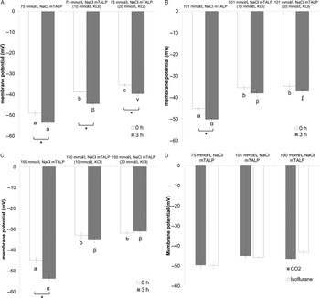
We first examined the effect of high K+ concentration in combination with Na+ on capacitation-associated hyperpolarization of membrane potential using fluorescent dye DiSC3(5) (Fig. 1). In 75 mM NaCl mTALP, an increase in KCl concentration to 10 or 20 mM significantly depolarized (increased) the membrane potential compared with that of the normal KCl concentration (2.68 mM) at 0 h incubation (uncapacitated; Fig. 1 A). When incubated for 3 h (capacitated), the membrane potentials were significantly hyperpolarized (decreased), compared with those at 0 h, at all the KCl concentrations tested (Fig. 1 A). When compared within the 3 h incubation groups, an increase in KCl concentration to 10 or 20 mM significantly depolarized the membrane potential, compared with that found with 2.68 mM KCl (Fig. 1 A). The membrane potential of sperm capacitated in the presence of 10 and 20 mM KCl at 3 h of incubation was significantly higher (depolarized; P < 0.05) compared with the membrane potential of uncapacitated sperm in the presence of 2.68 mM KCl at 0 h of incubation.
Effect of the K+ concentration on membrane potential. Sperm were suspended in 75 mM NaCl mTALP (A), 101 mM NaCl mTALP (B) or 150 mM NaCl mTALP (C) containing 2.68–20 mM KCl, and the effect of K+ concentration on the hamster sperm membrane potential was investigated using fluorescent dye DiSC3 (5) at 0 h and 3 h incubation. (D) Effect of the euthanizing method (CO2 or isoflurane) on hamster sperm membrane potential was investigated at 0 h in 75, 101 or 150 mM NaCl mTALP. Data were expressed as mean ± SEM. Different superscripts using the roman alphabet indicate significant differences in the 0 h group (P < 0.05). Different superscripts in Greek characters indicate significant differences in the 3 h group (P < 0.05). Asterisks indicate significant differences between the 0 h and 3 h of incubation (P < 0.05). (A–C) n = 6; (D) n = 4.

Similar results were obtained when the NaCl concentration was raised to 101 mM (Fig. 1 B). An increase in KCl concentration to 10 or 20 mM significantly depolarized the membrane potential at both 0 h and 3 h of incubation (Fig. 1 B). Capacitation-associated hyperpolarization was observed in standard 101 mM NaCl mTALP containing 2.68 mM KCl (Fig. 1 B). Significant hyperpolarization was not observed when KCl concentration was raised to 10 or 20 mM, although there was a tendency to show hyperpolarization in capacitated sperm. The membrane potential of capacitated sperm in the presence of 10–20 mM KCl was significantly higher (P < 0.05) compared with that of uncapacitated sperm (0 h of incubation) in mTALP containing 2.68 mM KCl (Fig. 1 B).
Similar results were obtained when the NaCl concentration was raised further to 150 mM, (Fig. 1 C). An increase in KCl concentration to 10 or 20 mM significantly depolarized the membrane potential at both 0 h and 3 h of incubation (Fig. 1 C). Significant hyperpolarization upon capacitation was also observed in 150 mM NaCl mTALP containing 2.68 mM KCl. Significant hyperpolarization was not observed when the KCl concentration was increased to 10 or 20 mM, as was the case for 101 mM NaCl mTALP (Fig. 1 C). Capacitated membrane potential in the presence of 10 or 20 mM KCl was significantly higher compared with the uncapacitated membrane potential in mTALP containing 2.68 mM KCl (Fig. 1 C; P < 0.01). Taken together, these results showed that capacitation-associated hyperpolarization also occurred in hamster sperm, regardless of Na+ concentration as long as K+ concentration was low (2.68 mM).
Significant depolarization of membrane potential of sperm suspended in 150 mM NaCl mTALP (2.68 mM KCl) compared with sperm in 75 mM NaCl mTALP (2.68 mM KCl) was observed at 0 h (Fig. 1 A–C, bar at the left side).
To eliminate the possibility that the euthanizing method (isoflurane) affected the membrane potential, we measured the membrane potential of sperm from hamsters euthanized by CO2 in 75–150 mM NaCl mTALP containing 2.68 mM KCl at 0 h (Fig. 1 D). There was no significant difference in membrane potential between hamsters euthanized with isoflurane or with CO2 at any NaCl concentration tested (Fig. 1 D).
Effect of extracellular K+ on percentage of motile sperm in hamster
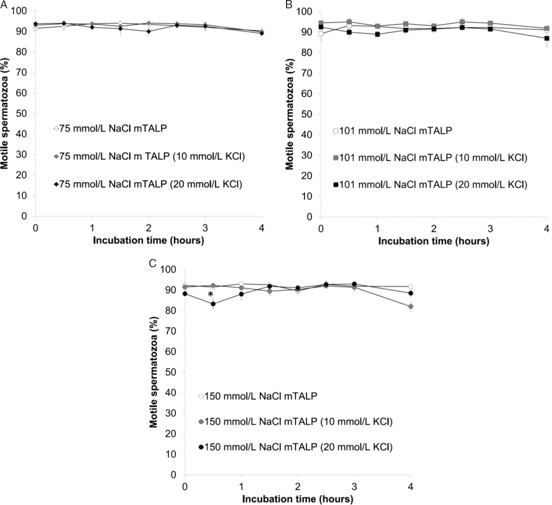
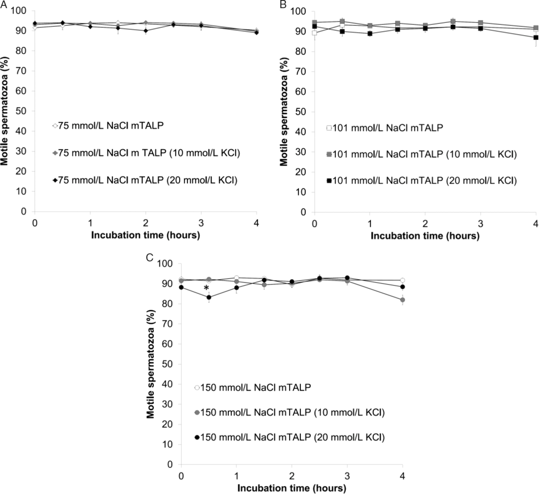
Next, we investigated the effect of a high K+ concentration in combination with Na+ on the percentage of motile sperm (Fig. 2). In 75 mM NaCl mTALP and 101 mM NaCl mTALP, an increase in K+ concentration from 2.68 to 20 mM had no effect on the percentage of motile sperm (Fig. 2 A, B). An increase in the K+ concentration to 20 mM in 150 mM NaCl mTALP transiently reduced the percentage of motile sperm at 0.5 h of incubation (P < 0.05), but no significant effect due to K+ concentration was seen at 1 h of incubation and thereafter (Fig. 2 C).
Effect of K+ concentration on percentage of motile sperm. Sperm were suspended in mTALP containing 2.68–20 mM KCl, and the effect of the K+ concentration on the percentage of motile sperm was evaluated. Data are expressed as mean ± SEM. Asterisks indicate significant differences compared with the normal mTALP containing 2.68 mM KCl (P < 0.05); n = 4.
Effect of extracellular K+ on hyperactivation in hamster sperm
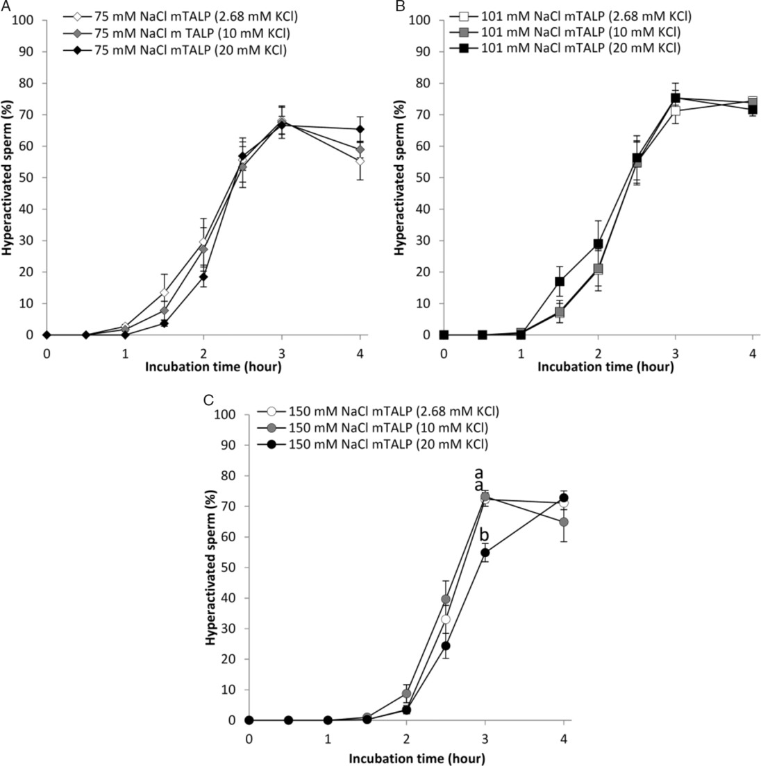
In the next step, we investigated the effect of a high K+ concentration in combination with Na+ on hyperactivation (Fig. 3). In 75 mM and 101 mM NaCl mTALP, the percentage of hyperactivated sperm was not significantly affected by 2.68–20 mM KCl throughout the incubation time periods (Fig. 3 A, B).
Effect of K+ concentration on hyperactivation. Sperm were suspended in mTALP containing 2.68–20 mM KCl, and the effect of the K+ concentration on hamster sperm hyperactivation was evaluated. Data were expressed as mean ± SEM. Different superscripts in the roman alphabet indicate significant differences between the data (P < 0.01); n = 4 at 0–1.5 h time points and n = 8 at 2–4 h time points.
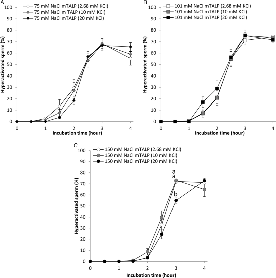
In 150 mM NaCl mTALP, 20 mM KCl significantly delayed hyperactivation, that is 20 mM KCl significantly suppressed hyperactivation at 3 h compared with normal 150 mM NaCl mTALP, but no significant difference was observed at 4 h of incubation (Fig. 3 C). There was no significant variation in the peak hyperactivation value (70–80%) associated with 150 mM NaCl mTALP, 150 mM NaCl mTALP (10 mM KCl) and 150 mM NaCl mTALP (20 mM KCl).
Effect of extracellular K+ on acrosome reaction in hamster sperm
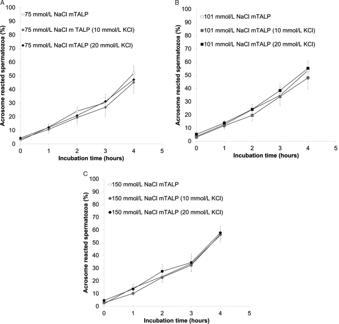
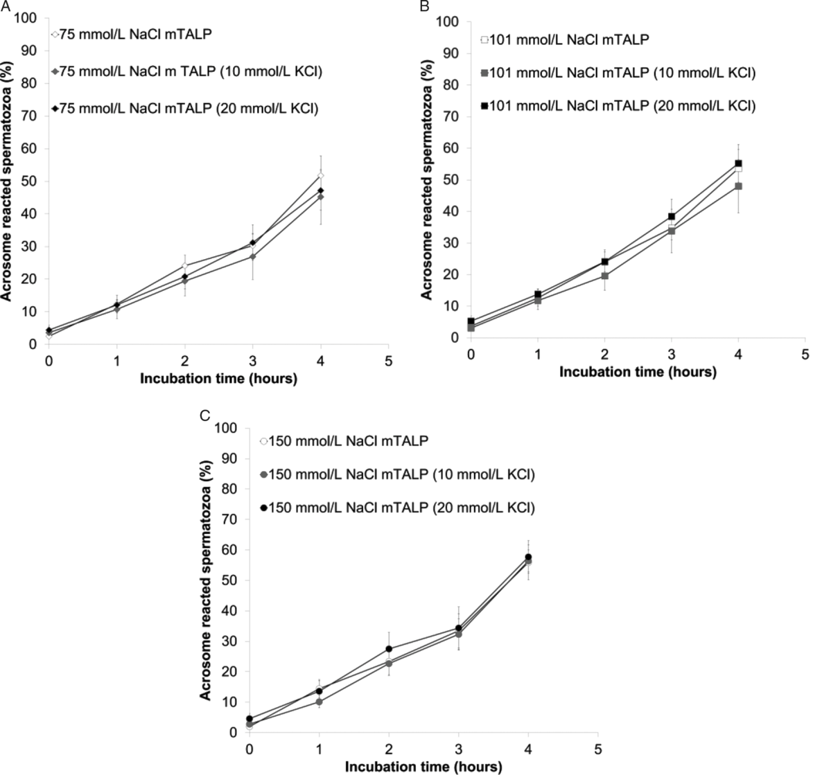
Next, we investigated the effect of high K+ concentration in combination with Na+ on acrosome reaction (Fig. 4). Changing the KCl concentration in the range 2.68–20 mM did not have any effect on the spontaneous acrosome reaction at any of the following NaCl concentrations in mTALP (75 mM NaCl, 101 mM NaCl or 150 mM NaCl; Fig. 4).
Effect of the K+ concentration on the acrosome reaction. Sperm were suspended in mTALP containing 2.68–20 mM KCl, and the effect of K+ concentration on hamster sperm acrosome reaction was determined. Acrosomal status was evaluated by CBB staining. Data are expressed as mean ± SEM. No significant difference was observed in any of the conditions tested (P < 0.05); n = 5.
Effect of extracellular K+ on IVF
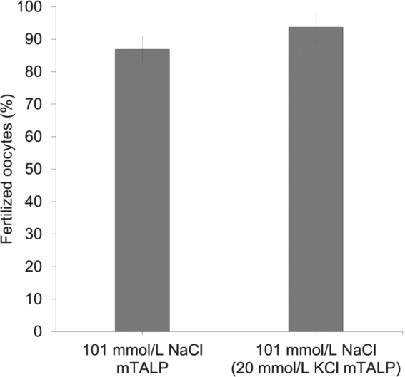
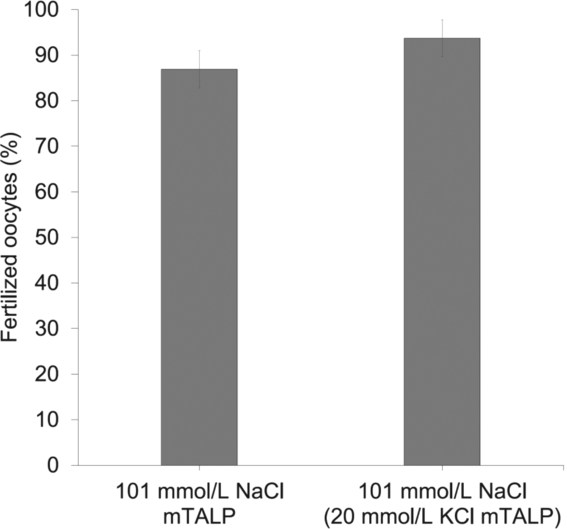
Finally, we tested the effect of change of extracellular K+ from 2.68 to 20 mM in 101 mM mTALP on IVF (Fig. 5). Sperm were preincubated in 101 mM NaCl mTALP containing 2.68 or 20 mM KCl, and inseminated in the same medium containing oocytes. In total, 55 oocytes for 101 mM NaCl mTALP containing 2.68 and 56 oocytes for 101 mM NaCl mTALP, 20 mM KCl from seven females were examined for three males. IVF was repeated three times. The results showed that increase in KCl concentration to 20 mM in 101 mM NaCl mTALP compared with 2.68 mM KCl did not significantly affect fertilization (Fig. 5; P = 0.166).
Effect of the K+ concentration in 101 mM NaCl mTALP on IVF. Hamster sperm suspended in 101 mM NaCl mTALP containing 2.68 or 20 mM KCl were incubated for 3 h, and subsequently co-incubated with oocytes in corresponding mTALP for 4 h. Then oocytes were washed and the number of fertilized oocytes were determined. The percentages of fertilized oocytes were expressed as mean ± SEM. No significant difference was observed (P = 0.166). In total, 55 oocytes for 101 mM NaCl mTALP containing 2.68 and 56 oocytes for 101 mM NaCl mTALP 20 mM KCl from seven females were examined with three males. IVF was repeated three times.
Discussion
In the present study, we investigated whether or not the oviduct K+ concentration (~20 mM) affected capacitation-associated events and fertilization. The results showed that increasing the KCl concentration had no significant effect on fertilization, and only a minor effect on sperm capacitation-associated events. These results indicated that there were not any positive or negative effects on hamster fertilization associated with physiological K+ concentration. It seems quite natural that oviduct K+ concentration would not have negative effects on fertilization, however as there have been conflicting studies regarding the effects of high K+ concentrations (≥20 mM) on sperm capacitation, fertilization and development (Toyoda and Chang, Reference Toyoda and Chang1974; Roblero and Riffo, Reference Roblero and Riffo1986; Roblero et al., Reference Roblero, Guadarrama, Ortiz, Fernández and Zegers-Hochschild1988; de la Vega-Beltrán et al., Reference de la Vega-Beltrán, Sánchez-Cárdenas, Krapf, Hernández-González, Wertheimer, Treviño, Visconti and Darszon2012), there was a need to confirm the effect of oviduct K+ concentration on capacitation/fertilization. This study clearly showed that, in the hamster oviduct, high K+ concentrations did not have a negative effect on fertilization.
When focused on sperm capacitation-associated hyperpolarization, our results indicated that hyperpolarization of membrane potential during capacitation was a general phenomenon in vitro among mammals including hamsters. However, 20 mM KCl (equivalent to the oviduct K+ concentration) suppressed capacitation-associated hyperpolarization when in the presence of >101 mM NaCl, and the membrane potential of the capacitated spermatozoa was depolarized by 20 mM KCl or significantly higher compared with that of uncapacitated sperm in mTALP containing 2.68 mM KCl (Fig. 1). Conversely, the spontaneous acrosome reaction was unaffected by 20 mM KCl (Fig. 4). These results indicated that there was no correlation between membrane potential status and spontaneous acrosome reaction, which seemed to contradict results from previous studies using mouse and bull sperm (Zeng et al., Reference Zeng, Clark and Florman1995; de la Vega-Beltrán et al., Reference de la Vega-Beltrán, Sánchez-Cárdenas, Krapf, Hernández-González, Wertheimer, Treviño, Visconti and Darszon2012). This discrepancy could be due to differences in the methods used to evaluate the acrosome reaction; the previous studies evaluated sZP-induced acrosome reactions, whereas the present study evaluated the spontaneous acrosome reaction. In the previous study, spontaneous acrosome reactions remained unchanged using K+ concentrations that were high enough in a previous study to prevent hyperpolarization (de la Vega-Beltrán et al., Reference de la Vega-Beltrán, Sánchez-Cárdenas, Krapf, Hernández-González, Wertheimer, Treviño, Visconti and Darszon2012). In the present study, we evaluated the spontaneous acrosome reaction but not the trigger-induced acrosome reaction for following reasons. First, occurrence of spontaneous acrosome reaction indicated completion of the capacitation process (Hirohashi and Yanagimachi, Reference Hirohashi and Yanagimachi2018). Second, recent in vivo observations utilizing sperm from transgenic mice expressing enhanced green fluorescent protein in their acrosome presented evidence that suggested that the ZP-induced acrosome reaction was not necessary for fertilization success in vivo and was not physiologically relevant (Jin et al., Reference Jin, Fujiwara, Kakiuchi, Okabe, Satouh, Baba, Chiba and Hirohashi2011; Hino et al., Reference Hino, Muro, Tamura-Nakano, Okabe, Tateno and Yanagimachi2016; Muro et al., Reference Muro, Hasuwa, Isotani, Miyata, Yamagata, Ikawa, Yanagimachi and Okabe2016, Hirohashi and Yanagimachi, Reference Hirohashi and Yanagimachi2018). Further studies to investigate the need for a physiological trigger to induce the acrosome reaction for fertilization in vivo, and to identify the ‘trigger’ molecule are needed to determine the relationship between the acrosome reaction and membrane potential. However, those experiments were beyond the scope of this study.
The present study raised the possibility that capacitation-associated hyperpolarization was not necessary for the acrosome reaction. As capacitation-associated hyperpolarization of membrane potential in vitro is a universal phenomenon among mammalian species, capacitation-associated hyperpolarization should have some physiological implications. Therefore, we proposed the hypothesis that changes in channel properties upon capacitation, such as activation of Slo3 (Santi et al., Reference Santi, Martínez-López, de la Beltrán, Butler, Alisio, Darszon and Salkoff2010; Zeng et al., Reference Zeng, Yang, Kim, Lingle and Xia2011; Chávez et al., Reference Chávez, de la Vega-Beltrán, Escoffier, Visconti, Treviño, Darszon, Salkoff and Santi2013; Escoffier et al., Reference Escoffier, Navarette, Haddad, Santi, Darszon and Visconti2015), inactivation of ENaC (Hernández-González et al., Reference Hernández-González, Sosnik, Edwards, Acevedo, Mendoza-Lujambio, López-González, Demarco, Wertheimer, Darszon and Visconti2006), or resultant changes in intracellular ionic conditions upon capacitation (such as decrease in intracellular Na+ concentration; Escoffier et al., Reference Escoffier, Krapf, Navarrete, Darszon and Visconti2011) that led the sperm membrane potential to hyperpolarize upon in vitro capacitation, are physiologically relevant, rather than the membrane potential itself. Further studies are needed to determine if hyperpolarization is necessary or not for the acrosome reaction. There remains the possibility that the hyperpolarization of membrane potential of individual cells is masked by the majority of the sperm for which membrane potential has been depolarized by K+, as the present study measured mass sperm membrane potential. Also, it was shown that only a subpopulation of sperm underwent membrane potential hyperpolarization during capacitation (Escoffier et al., Reference Escoffier, Navarette, Haddad, Santi, Darszon and Visconti2015). Measurements of membrane potential of individual cells such as by using electrodes are necessary to determine if hyperpolarization is needed for the acrosome reaction.
We previously reported that the hamster sperm resting potential was around −80 mV (Takei and Fujinoki, Reference Takei and Fujinoki2016). In the present study, however, resting potentials in mTALPs containing a normal K+ concentration were −45 to 50 mV (Fig. 1). Moreover, the present study showed that an increase in NaCl concentration from 75 to 150 mM significantly depolarized the hamster sperm membrane potential at 0 h (Fig. 1); this finding conflicted with findings in our previous study (Takei and Fujinoki, Reference Takei and Fujinoki2016). These differences were caused by modification of the employed methods, e.g. cell concentration was reduced to 5 × 10⁵ to 1 × 10⁶, dye concentration was raised to 5 μM, pre-incubation time with dye was prolonged to 8 min and the sequence of experimental procedure was modified. As the results obtained in the present study using the modified method agreed with other previous studies on sperm membrane potential in mouse, bovine and horse (Zeng et al., Reference Zeng, Clark and Florman1995; McPartlin et al., Reference McPartlin, Visconti and Bedford-Guaus2011), the present results seem to be more significant and therefore we have had to modify our previous conclusion that extracellular Na+ had no effect on membrane potential (Takei and Fujinoki, Reference Takei and Fujinoki2016). However, delay of hyperactivation by extracellular Na+ observed in the previous study seemed not cause a change in membrane potential, because in the present study the increase in extracellular K+ with 75 and 101 mM NaCl mTALP did not delay hyperactivation (Fig. 3 B).
When focused on capacitation-associated events other than hyperpolarization, this study showed that hyperactivation was transiently suppressed by increasing the KCl concentration in 150 mM NaCl mTALP, but peak hyperactivation values were not affected by KCl concentration (Fig. 3). Acrosome reaction was not affected by any increase in KCl concentration (Fig. 4). In addition, there was no correlation with high K/Na ratio [such as 75 mM NaCl mTALP (20 mM KCl)] and capacitation-associated events (hyperactivation and acrosome reaction). These results disagreed with previous studies that showed that in human sperm high K+ concentration (25 mM) improved the acrosome reaction (Roblero et al., Reference Roblero, Guadarrama, Ortiz, Fernández and Zegers-Hochschild1988), and which showed that in rat sperm a high K/Na ratio facilitated sperm penetration (Toyoda and Chang, Reference Toyoda and Chang1974). These facts suggested variation in the regulatory mechanisms of capacitation among species (rat, human and hamster), even though rat and hamster are both rodents.
In the present study, the results indicated that increasing the KCl concentration of the medium to oviduct levels had a suppressive effect on hamster sperm capacitation: hyperactivation in 150 mM mTALP was significantly delayed by 20 mM KCl at 3 h incubation (Fig. 3 A, C). In addition, membrane potential hyperpolarization was also suppressed by 20 mM KCl in 101 and 150 mM NaCl mTALP (Fig. 1). These results suggested that in hamsters the oviduct environment was not necessarily optimum for sperm and may therefore serve as a selective pressure. Conversely, a previous study suggested that oviduct K+ concentration had some beneficial effect on embryo development, namely that the oviduct environment was desirable for the oocyte/embryo (Roblero and Riffo, Reference Roblero and Riffo1986). Therefore, further studies are needed on the effect of high K+ concentrations on development that would lead to the production of a more efficient medium for in vitro fertilization. The elucidation of these problems should lead to a better understanding of fertilization, and the development of assisted reproductive technologies.
Acknowledgements
We would like to express my thanks to Professor Dr Hiroyuki Sugimoto of the Department of Biochemistry, Dokkyo Medical University for providing us with the spectrophotometer used in the study.
Financial support
This work was supported by JSPS KAKENHI (GLT, Grant Number JP15K21323) and (HK, Grant Number JP15K21324).
Conflicts of interest
None.
Ethical standards
The authors assert that all procedures contributing to this work comply with the ethical standards of the relevant national and institutional committees on human experimentation and with the Helsinki Declaration of 1975, as revised in 2008. The authors assert that all procedures contributing to this work comply with the ethical standards of the relevant national and institutional guides on the care and use of laboratory animals.