A combination of aortic valve atresia and an interrupted aortic arch is a rare form of congenital heart disease that is incompatible with life unless blood supply to the myocardium and brain is provided through one of the different mechanisms. Prompt identification of this entity is important because these patients may need interventions in order to maintain a reliable source of brain and myocardial perfusion.
Case report
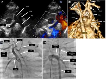
A term female newborn baby was admitted at 2 hours of life to the neonatal intensive care unit with low saturation. Echocardiogram showed double inlet left ventricle, non-restrictive ventricular septal defect, hypoplastic sub-aortic right ventricle, type B interrupted aortic arch (arch interrupted between the left common carotid and the left subclavian artery), large non-restrictive patent ductus arteriosus continue as descending aorta, and normal origin of the right subclavian artery. The aortic valve was atretic with a small (3 mm) ascending aorta and right-sided arterial ductus arising from the right pulmonary artery was supplying the brachiocephalic artery which in turn retrogradely perfusing the coronary arteries (Fig 1a). The baby was started on prostaglandin in order to maintain the ductal patency. Computed tomography (CT) angiogram showed aortic atresia, type B interrupted aortic arch and large arterial ductus from the left pulmonary artery continues as descending aorta, right-sided arterial ductus from the right pulmonary artery supplying the head and neck vessels, ascending aorta, and coronaries (Fig 1b). Management options we had for the baby were either do a Damus–Kaye–Stensel operation with an aortic arch repair or a hybrid procedure which include placing stents in both the ducti and bilateral pulmonary artery banding. Considering the high-risk status with primary surgical repair, it was decided to proceed with the later.
( a ) Echocardiographic sagittal image shows RBCA=right brachiocephalic artery; LCCA=left common carotid artery; RDA=right ductus arteriosus; AAO=ascending aorta. ( b ) CT angiogram 3DVR images (looking from behind) shows interrupted aortic arch type B with left arterial ductus (*) continues as descending aorta (DAO); LCCA=left common carotid artery; RCCA=right common carotid artery; RBCA=right brachiocephalic artery; RSCA=right subclavian artery; LSCA=left subclavian artery; RDA=right ductus arteriosus; RPA=right pulmonary artery; AAO=ascending aorta. ( c ) Angiogram shows RSCA=right subclavian artery; RCCA=right common carotid artery; LCCA=left common carotid artery; AAO=ascending aorta; RDA=right ductus arteriosus with 4-mm stent deployed; RPA=right pulmonary artery. ( d ) Angiogram shows stented LDA – left ductus arteriosus; LSCA=left subclavian artery from DAO=descending aorta; LPA=left pulmonary artery.

Cardiac catheterisation showed no forward flow the ventricle to the aorta. Blood supply to the ascending aorta and coronary arteries were provided by a right arterial duct which took origin from the right pulmonary artery. There was type B interrupted aortic arch. The descending aorta was perfused by a left arterial duct and the right subclavian was arising normally from the brachiocephalic artery. A 4-mm stent was deployed in the right arterial ductus and an 8-mm stent in the left arterial ductus (Fig 1c and d). The baby tolerated the procedure well; prostaglandin was discontinued, and bilateral pulmonary artery banding was done on the same day. The baby had an uneventful post-operative course, extubated on 2nd post-operative day, and discharged home after 10 days in stable clinical condition with a plan to proceed with single-ventricle palliation by doing a comprehensive stage 2 (Norwood operation and Glenn shunt) in the future.
Discussion
Aortic valve atresia and an interrupted aortic arch is a rare congenital heart disease in which the survival of the patient depends on the blood supply to the ascending aorta and myocardium. Most of the time associated lesions provide an alternate source of blood supply to the brain and heart. Different reported sources of blood supply to the blind ascending aortic segment are (1) aorto-pulmonary (AP) window, (2) double aortic arch, (3) bilateral arterial ductus, and (4) aberrant right subclavian artery. Reference Baker-Smith, Milazzo and Frush1–Reference Lacour-Gayet, Kandachar and Al-Bulushi3 When there are no above-mentioned sources of blood supply is available, it is through the circle of Willis, the blood flow to the brain and myocardium is maintained in a retrograde manner through carotids ascending aorta and then to the coronary arteries. Reference Tannous, Moulick and Jonas4
In the absence of an associated anomaly to perfuse the blind segment, type A interrupted aortic arch with aortic atresia is not compatible with life. However, in cases of type-A interruption and aortic atresia with aberrant right subclavian artery there could be minimal supply to the brain from the descending aorta through the aberrant right subclavian artery and right vertebral artery through circle of Willis. In type-B interruption and aortic atresia minimal supply to the brain and heart is by the left subclavian artery and left vertebral artery through circle of Willis. In above two situations, this blood supply might not be adequate to maintain both cerebral and myocardial perfusion. However, in type C interruption and aortic atresia, brain and coronary perfusion are supported by both the left carotid artery and left vertebral artery which in combination might provide adequate blood supply to the brain and myocardium and could be compatible with life. In the majority of the reported cases, perfusion to the brain and myocardium is provided by bilateral ductus, AP window or double arch. Reference Lacour-Gayet, Kandachar and Al-Bulushi3 Only very few cases had no direct connection to the ascending aorta and all had an aberrant right subclavian artery. Reference Careddu, Oppido and Ialonardi5 In our case, cerebral and coronary perfusion was maintained through a right-sided arterial ductus. Hence it was crucial to keep ductal patency with prostaglandin. Considering the high risk of a Damus–Kaye–Stensel operation with aortic arch repair, we decided to proceed with a hybrid procedure which includes placing stents in both the ducti and bilateral pulmonary artery banding.
Our report adds to the literature like the 3rd reported case of an interrupted aortic arch, aortic atresia with bilateral arterial ductus. Prompt identification of this disease is important to ensure survival by keeping ductal patency using prostaglandin. A hybrid procedure with bilateral ductal stenting and branch pulmonary artery banding is an alternative low-risk initial approach which can be considered if the surgical procedure is found to be high risk.
Acknowledgements
None.
Financial support
This research received no specific grant from any funding agency, commercial or not-for-profit sectors.
Conflicts of interest
None.
Ethical standards
The authors assert that all procedures contributing to this work comply with the ethical standards of the Helsinki Declaration of 1975, as revised in 2008, and has been approved by the institutional review board of Hamad Medical Corporation, Qatar.