Implications
The economic value of a pig carcass depends on its meat content, which is determined after slaughtering. Therefore, the improvement of carcass quality is one of the main breeding goals in pig production, and breeding animals have to be selected for their body composition, aiming for a high meat content. This study evaluated two techniques, both able to predict body composition in live pigs: dual-energy X-ray absorptiometry (quantitative analysis technique) and magnetic resonance imaging (imaging technique). It was concluded that the combination of both techniques is very suitable to predict the carcass value in live pigs.
Introduction
Selection for improved body composition in pig breeding is of major concern in order to achieve genetic improvement of economically important performance traits such as carcass quality. The prediction of the carcass value before slaughtering, however, is still a challenging issue in the field of animal breeding. Different methods were examined for their precision in predicting carcass composition in live animals (Szabo et al., Reference Szabo, Babinszky, Verstegen, Vangen, Jansman and Kanis1999). One method that seems to be very suitable for analyzing the body composition in different species in vivo, especially in pigs, is the dual-energy X-ray absorptiometry (DXA). DXA quickly and accurately provides reliable information about mass and percentage of lean tissue and fat, bone mineral content (BMC), and bone mineral density (BMD; Suster et al., Reference Suster, Leury, Ostrowska, Butler, Kerton, Wark and Dunshea2003; Marcoux et al., Reference Marcoux, Faucitano and Pomar2005; Scholz and Förster, Reference Scholz and Förster2006). A further, non-destructive method to reliably analyze body composition is the use of imaging techniques (Szabo et al., Reference Szabo, Babinszky, Verstegen, Vangen, Jansman and Kanis1999). Whereas computed tomography (CT) is based on ionizing radiation, magnetic resonance imaging (MRI) represents a cross-sectional imaging technique without any exposure to radiation (Szabo et al., Reference Szabo, Babinszky, Verstegen, Vangen, Jansman and Kanis1999; Scholz and Baulain, Reference Scholz and Baulain2009). The technique is based on the magnetic properties of protons, which are part of all soft tissues of the body. The MRI technique uses a strong magnetic field, inside which protons align alongside its direction. Short pulses of targeted radio waves are used to deflect the nuclei from their alignment. By drifting back to their initial position, they emit the absorbed radio waves in a tissue-specific way. According tuners and sophisticated computers are used to remodel digital images by the received electromagnetic signals (Baulain, Reference Baulain1997).
In some sectors of the pork industry, for example in the dry-cured meat production of the Iberian pig, particular carcass regions are of higher economic importance than others. Cerdo Ibérico is an autochthonous, rural pig breed, originating from the Iberian peninsula and very uncommon outside Spain. It is the most meaningful Mediterranean swine type in population and economic importance. The Iberian breed is characterized by its fat-producing ability and perfectly adapted to the ecosystem of the Mediterranean oak forest (Lopez-Bote, Reference Lopez-Bote1998; Daza et al., Reference Daza, Olivares, Rey, Ruiz and López-Bote2008). Even when reared and fattened intensively, meat products of the Iberian pig differ considerably in quality from conventional pig breeds (Serra et al., Reference Serra, Gil, Pérez-Enciso, Oliver, Vázquez, Gispert, Díaz, Moreno, Latorre and Noguera1998). Therefore, the prediction of loin, ham or shoulder yield in vivo could be useful in selecting breeding animals.
In fact, carcass dissection during progeny testing is the most common technique to measure specific carcass portions in order to estimate breeding values for selection decisions. It is, however, a time-consuming, expensive and finally fatal method. The potential of virtual dissections in live animals is provided by different imaging techniques as recently discussed by Scholz and Baulain (Reference Scholz and Baulain2009). A method to quantify exactly the lean meat and fat content of main carcass sections in vivo could provide an important tool in economically oriented pig breeding. The current study aimed at evaluating the use of DXA as a quantitative analysis method of whole-body composition in combination with MRI as a digital imaging technique by comparing live pigs of different genetic background and different genders covering a wide variation of body composition.
Material and methods
The study was performed at the Livestock Center Oberschleissheim of the Veterinary Faculty of the University of Munich. The protocol of the study was approved by the Government of Upper Bavaria with the tracking number 55.2-1-54-2531.2-60-07.
Animals
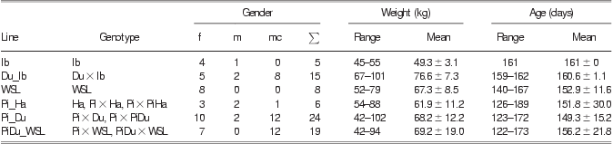
A total number of 77 pigs (7 boars, 33 barrows, 37 gilts) were examined during the study. Only seven intact boars were used in the study, because there was no need for a higher number of potential breeding boars. In addition, on the basis of the possible problems with the taste of pork originating from intact boars, boar fattening is not a standard procedure in Germany yet. The pigs originated from different autochthonous or conventional breeds or crossbreeds: Cerdo Ibérico (Ib; Negro Lampiño or Black Hairless strain, n = 5; Clemente et al., Reference Clemente, Membrillo, Azor, Dorado, Rodero and Molina2006), Duroc (Du) × Ib (Du_Ib; n = 15), German Landrace and German Large White = White Sow Lines (WSL; n = 8), Hampshire (Ha) and Pietrain (Pi) × Ha and Pi × PiHa (Pi_Ha; n = 6), Pi × Du and Pi × PiDu (Pi_Du; n = 24), Pi or PiDu × WSL (PiDu_WSL; n = 19). The pigs ranged from 42 to 102 kg in BW and from 122 to 189 days in age (Table 1).
Animals of the study according to breeding group, age and weight
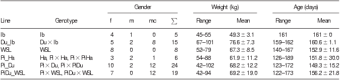
f = female; m = male; mc = male castrated; ∑ = sum; Ib = Cerdo Ibérico; Du = Duroc; WSL = White Sow Lines (German Landrace + German Large White); Pi = Pietrain; Ha = Hampshire.
Management and housing of the animals
The pigs were housed in an outer climate barn in groups of up to 30 pigs within a pen with deep straw. They were fed a fattening diet of pelleted food with 16.8% CP dry matter (DM) and 13.29 MJ metabolizable energy (ME)/kg DM, comprising 22.3% wheat, 31.0% barley, 22.3% corn, 14.4% soy meal, 7.0% soybeans toasted and 3% mineral mix (Mineral E, Fa. Salvana, Wertingen, Germany). Food was provided on a restricted basis depending on an age-related feeding curve through a Double-Fitmix station (Fa. PigTek, Schüttorf, Germany). Alone the Ib pigs and two of the Du_Ib pigs (boars) were housed in a different barn at the Livestock Center. They were kept on straw in one group and were fed a low-protein (Nieto et al., Reference Nieto, Miranda, García and Aguilera2002) and low-energy (12.09 MJ ME/kg DM) diet. The pelleted food contained 6.0% wheat, 48.7% barley, 37.0% oats, 4.8% soybeans toasted and 3.5% mineral mix (Premium-Z, Fa. Salvana).
DXA analysis
Single DXA scans were performed using a GE Lunar DPX-IQ (Lunar software version 4.7e, GE Healthcare, Pittsburgh, PA, USA) scanner. A detailed explanation of the technical details of DXA is given by Pietrobelli et al. (Reference Pietrobelli, Formica, Wang and Heymsfield1996). The whole-body scan mode ‘adult normal’ was used on all animals. Every second day, a calibration scan of a phantom was performed to ensure the maintenance of accurate body composition results. Measurements made by DXA included soft lean tissue mass (DXA-LM; g), which represents the mass of all tissues of the body that are not identified as bone or fat tissue; soft lean tissue percentage (DXA-%LM; %), which represents the percentage of the DXA-LM related to the total tissue mass (TTM); fat tissue mass (DXA-FM; g), which represents the mass of all tissues of the body that are not identified as bone or lean tissue; fat percentage (DXA-%FM; %), which represents the percentage of the DXA-FM related to the TTM; bone mineral content (DXA-BMC; g), representing the mass of bone mineral inside all tissues that are identified as bone; bone mineral percentage (DXA-%BM; %), which represents the percentage of the bone mineral related to the TTM; bone mineral density (DXA-BMD; g/cm2), representing the content of bone mineral per cm2 bone area of the body; and TTM (DXA-TTM; g) as a sum of DXA-LM, DXA-FM and DXA-BMC (Scholz and Förster, Reference Scholz and Förster2006).
MRI scans
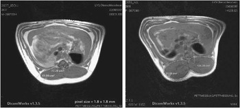
A 0.2 Tesla Magnetom Open combined with a large body coil (Siemens, Erlangen, Germany) was used for MRI scanning. The MRI protocol consisted of a T1 weighted spin echo sequence (TR 380 ms, TE 15 ms, flip angle 90°, 10 slices, number of acquisitions 4), a field of view of 461 × 461 mm with an image matrix of 256 × 256 pixels and a transversal slice thickness of 15 mm with a distance factor of 0.25. The acquisition time for one sequence was 6 min and 32 s. Each animal was scanned once. The MRI scans were performed to compare loin eye muscle area (MRI-LA) and subcutaneous back fat area above the loin eye muscle area (MRI-FA) in the region of the 14th vertebra of both body sides – accordingly to the site of the standard performance testing procedure of German pig breeders. Therefore, after performing a localizer scan, the slices of the specified protocol were positioned on the coronary plane with the first slice in the region of the 13th vertebra, in order to see the region of interest on the second or third slice. The muscle and FAs were calculated using the software Dicom Works 1.3.5 (Inviweb, Lyon, France). The corresponding areas were defined manually after visually distinguishing between muscle and fat by qualified personnel as shown in Figure 1.
Magnetic resonance images of a Duroc–Iberian crossbred pig (left hand; Du_Ib) and a Hampshire pig (right hand; Ha) with delineated areas of loin and above back fat. Both pigs represent different body compositions; Du_Ib as fat and Ha as lean line.

Treatment
Both examinations of the pigs were performed in one session. Before the scans, the pigs were fasted for ∼16 h. Examinations started with weighing the pigs in order to calculate the required amount of anesthetic. The animals were sedated by an intramuscular injection of azaperone (1.2 mg/kg BW; Stresnil®), followed by intramuscularly administered ketamine (40 mg/kg BW; Ursotamin®). After onset of the sedation, an intravenous (i.v.) catheter was inserted into an ear vein and fixed with tape, enabling i.v. follow-up dosing of ketamine if necessary. Primarily, the pigs were placed on the MRI scanner in a prone position with fore and hind legs extended. Subsequently, the DXA scan was performed. The pigs were also fixed in prone position with hind legs extended and slightly tied together, but the fore legs were positioned caudally, separated from the body by two foam plastic wedges. Finally, after both scans, the pigs were moved into a separate pen to enable a gentle recovery phase from anesthesia.
Statistical analysis
The SAS software package (version 9.2; SAS Institute Inc., Cary, NC, USA) provided the tool for variance analysis using a GLM procedure with the following structure for DXA-derived LM, %LM, FM, %FM, BMC, %BM, BMD, TTM (DXA BW), and MRI-derived LA, and FA:
 $${{y}_{ijk}}\,{\rm{ = }}\,\mu \,{\rm{ + }}\,{{L}_i}\,{\rm{ + }}\,{{G}_j}\,{\rm{ + }}\,\beta {{A}_{ijk}}\,{\rm{ + }}\,{{e}_{ijk}}$$
where yijk is the observation, μ the expected value of yijk, Li the line (i = 1, 2, 3, 4, 5, 6), Gj the gender (j = 1, 2, 3), βAijk the age as covariate and eijk the residual error.
The breeding group × gender interaction showed no significance and therefore was excluded from the model. The t-test was performed with a Tukey–Kramer adjustment for the multiple comparisons of the least squares means.
Results
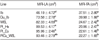
Tables 2 and 3 represent the results and the statistical differences of the DXA and MRI measurements, respectively. At an average age of 154 days, the different breeding groups had – with exception of Ib (P < 0.01) and partially Pi_Ha (P ⩾ 0.08) – a similar average BW calculated by DXA as TTM.
LSM and standard errors of estimation (±) of body composition traits in different pig breeding groups estimated by DXA

LSM = least squares means; DXA = dual-energy X-ray absorptiometry; DXA-%FM = DXA-fat percentage; DXA-FM = DXA-fat mass; DXA-%LM = DXA-lean percentage; DXA-LM = DXA-lean mass; DXA-BMC = DXA-bone mineral content; DXA-BMD = DXA-bone mineral density; DXA-BMP = DXA-bone mineral percentage; DXA-TTM = DXA-total tissue mass; Ib = Cerdo Ibérico; Du = Duroc; WSL = White Sow Lines (German Landrace + German Large White); Pi = Pietrain; Ha = Hampshire.
LSM with different superscripts in one column differ significantly (P < 0.05).
#P = 0.068; *P = 0.059; §P = 0.057; ∼P = 0.079; °P = 0.073; ∧P = 0.077.
LSM and standard errors of estimation (±) of loin eye muscle and back fat areas in different pig lines calculated from magnetic resonance images
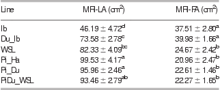
LSM = least squares means; MRI = magnetic resonance imaging; MRI-LA = MRI-loin eye muscle area; MRI-FA = MRI-fat area (above the loin eye muscle area); Ib = Cerdo Ibérico; Du = Duroc; WSL = White Sow Lines (German Landrace+German Large White); Pi = Pietrain; Ha = Hampshire.
LSM with different superscripts in one column differ significantly (P < 0.05).
The highest mean DXA-%FM was estimated for Ib and Du_Ib. WSL, PiDu_WSL, Pi_Du and Pi_Ha had lower DXA values of fat tissue percentage (P < 0.01), although Pi_Ha represented the leanest genetic line in terms of DXA fat mass. According to these results, Ib and Du_Ib had the lowest DXA-%LM compared with Pi_Ha representing the highest DXA-%LM. All other lines were in between.
Bone mineral showed the highest density in the Du_Ib line (P < 0.01). The results for the area calculations of the MRI images are shown in Table 3. Ib and Du_Ib had the smallest MRI-LA combined with the largest MRI-FA. In contrast, Pi_Ha, Pi_Du, PiDu_WSL had the largest MRI-LA covered by the smallest FA. WSL had an intermediate MRI-LA and MRI-FA, respectively.
Discussion
During this study, a combination of DXA and MRI was used to determine differences among different pig lines with regard to their body composition. Screening the literature, tissue composition analyses in animals are often performed by imaging techniques such as CT or MRI alone (Monziols et al., Reference Monziols, Collewet, Bonneau, Mariette, Davenel and Kouba2006; Kongsro et al., Reference Kongsro, Røe, Aastveit, Kvaal and Egelandsdal2008), and in order to predict carcass composition in vivo usually whole-body scans are performed (Kolstad and Vangen, Reference Kolstad and Vangen1996). The intention to evaluate the combination of an imaging and a quantitative analysis technique, however, results from the fact that all imaging techniques – even CT – require complex and time-consuming image analyses for each single image (Kolstad and Vangen, Reference Kolstad and Vangen1996; Brenøe and Kolstad, Reference Brenøe and Kolstad2000; Dønnem et al., Reference Dønnem, Eknæs and Randby2011). Furthermore, the accuracy of the prediction of body composition, however, depends, in addition to technical details, on the number of evaluated cross-sectional images, the slice thickness and the distance between single slices (Allen and Leymaster, Reference Allen and Leymaster1985). DXA, in contrast, provides immediate information regarding the soft lean tissue, fat tissue and bone mineral of the whole body by avoiding subjective differences in data evaluation (Albanese et al., Reference Albanese, Diessel and Genant2003; Suster et al., Reference Suster, Leury, Ostrowska, Butler, Kerton, Wark and Dunshea2003). Therefore, it was decided to evaluate what kind of beneficial effects are provided by combining an imaging technique to examine defined body regions and a quantitative analysis technique for the whole body instead of using only one of these techniques.
Evaluating the practicability of the combination of the chosen techniques, the measurement results must be analyzed with caution. The pigs were selected from different breeding groups, representing male, female and neutered purebreds and crossbreds of fat and lean lines. As the overall aim of the study was to evaluate the output of the combination of DXA and MRI in estimating carcass values in live pigs, the sample of animals was chosen in order to maximize the variability of different body composition. To provide meaningful results, it was decided to form six groups according to the different genetic origin and the intended use of the pigs: Ib, Du_Ib, WSL, Pi_Ha, Pi_Du and PiDu_WSL. Ib and Du_Ib as very fat lines; WSL representing typical dam lines; Pi_Ha representing a lean sire line; PiDu_WSL representing terminal crosses; and Pi_Du, as it was decided not to pool Pi_Du with Pi_Ha, as Duroc stands out by its high intramuscular fat content. The arrangement of the pigs according to their intended use resulted partly in groups with a rather small number of animals (especially Ib and Pi_Ha). Therefore, the results regarding the comparison of the groups’ different body compositions should be considered as preliminary results.
The most uncommon breeding groups for countries outside Spain were Cerdo Ibérico (Clemente et al., Reference Clemente, Membrillo, Azor, Dorado, Rodero and Molina2006; Rueda Sabater and Diéguez Carbayo, Reference Rueda Sabater and Diéguez Carbayo2007; Daza et al., Reference Daza, Olivares, Rey, Ruiz and López-Bote2008) and Du_Ib. Compared with Landrace, Iberian pigs show a distinct metabolite and hormone profile (Fernández-Fígares et al., Reference Fernández-Fígares, Lachica, Nieto, Rivera-Ferre and Aguilera2007), lower average daily gain (Nieto et al., Reference Nieto, Miranda, García and Aguilera2002), higher back fat thickness (48.1 mm v. 20.7 mm), intramuscular fat content (3.91% v. 0.66% in longissimus muscle) and pH24 (Serra et al., Reference Serra, Gil, Pérez-Enciso, Oliver, Vázquez, Gispert, Díaz, Moreno, Latorre and Noguera1998). Du_Ib also represents a very fat line as crossbreeding Ib with Duroc increases the prolificacy and feed efficiency of Iberian pigs without reducing the quality of their dry-cured meat products (Carrapiso et al., Reference Carrapiso, Bonilla and García2003). In addition, a Spanish law allows the use of pure Iberian pigs, as well as 50% Iberian × Duroc crosses for producing dry-cured meat products labeled Iberian (B.O.E., 2001). Therefore, crossbreds of those breeds are widely spread in Spain.
The combination of DXA and MRI helped to explain differences in body composition and carcass quality traits in the live pigs between Ib and Du_Ib lines and the conventional breeds as well. Du_Ib showed a higher BW (74.59 kg), calculated as DXA-TTM, than did Ib (47.1 kg, P < 0.01). Nevertheless, the relative proportions of fat and lean tissue of these genotypes did not differ. Similarly, the comparison of the MRI-FA of both lines could not establish a difference between the genotypes (P = 0.97). On the other hand, supported by MRI, an essential difference regarding carcass quality could be verified: Du_Ib had an MRI-LA of 73.58 cm2 compared with Ib representing an MRI-LA of 46.19 cm2 (P < 0.01). This means a remarkable improvement of the carcass quality, regarding the valuable loin cut. The close relationship among the weights of distinct muscles calculated from MRI images and from carcass dissection was shown by Baulain (Reference Baulain1997) and Mitchell et al. (Reference Mitchell, Scholz, Wange and Song2001b). Baulain (Reference Baulain1997) reported a correlation between muscle volume derived from MRI and muscle weight by dissection of 0.95, whereas Mitchell et al. (Reference Mitchell, Scholz, Wange and Song2001b) calculated a coefficient of determination of R 2 = 0.93 between the MRI-derived volume of the longissimus muscle and its weight after carcass dissection. Crossbreeding of slowly growing Cerdo Ibérico with Duroc results in a similar BW (DXA-TTM) to WSL (72.72 kg, P > 0.05), Pi_Du (74.25 kg, P > 0.05), PiDu_WSL (71.56 kg, P > 0.05) and slightly higher than Pi_Ha (64.00 kg, P = 0.08), the slowest growing line among the conventional breeding groups. Generally, crossbreeding of Ib with Du leads to an increased mass of valuable cuts (loin) without loss in percentage of body fat (Tejeda et al., Reference Tejeda, Gandemer, Antequera, Viau and GarcIa2002).
DXA (Mitchell et al., Reference Mitchell, Conway and Potts1996; Mitchell et al., Reference Mitchell, Scholz and Pursel2001a; Lösel et al., Reference Lösel, Kremer, Albrecht and Scholz2010) and MRI (Mitchell et al., Reference Mitchell, Scholz, Wange and Song2001b; Tholen et al., Reference Tholen, Baulain, Henning and Schellander2003) are accurate methods to measure differences in body composition in vivo and carcass traits among different pig lines. Scholz (Reference Scholz2002) confirmed these results by calculating high coefficients of determination (R 2 > 0.89) for in vivo MRI and DXA measurement results in relation with the chemical lipid content (%) of right carcass halves (including head and intestines) in pigs. Iberian pigs (Ib and Du_Ib) were fatter (P < 0.01) than the rest of breeding groups. No differences could be found among the conventional breeding groups for DXA-%LM. In contrast, MRI-LA in WSL was distinctly smaller than in Pi_Ha and Pi_Du (P < 0.05). Remarkably, Du_Ib representing a fat line had a lower DXA-%LM than WSL (P < 0.01), but did not differ (P = 0.45) from WSL in the MRI area of the valuable cut loin (MRI-LA). In addition, the results corresponded with the conclusions drawn from the above-mentioned findings by Serra et al. (Reference Serra, Gil, Pérez-Enciso, Oliver, Vázquez, Gispert, Díaz, Moreno, Latorre and Noguera1998); Nieto et al. (Reference Nieto, Miranda, García and Aguilera2002) and Fernández-Fígares et al. (Reference Fernández-Fígares, Lachica, Nieto, Rivera-Ferre and Aguilera2007) comparing carcass traits of intensively housed Iberian pigs and Landrace pigs. The combined examination by DXA and MRI showed a higher back fat thickness (estimated as MRI-FA) and fat percentage of the body of the Iberian pigs compared with WSL without slaughtering and carcass dissection.
As a further result of this study, DXA-derived BMC and BMD could be compared among the different breeding groups. Measurement of BMC and BMD provides information about the mineralization of bones. Fractures caused by bone softening can be a problem in second-litter gilts. Exhausting lactations of big litters can lead to osteoporosis, which occurs if the sow's demand of calcium cannot be met by the diet. Calcium is removed from the bones to supply the suckling piglets (Jackson and Cockcroft, Reference Jackson and Cockcroft2007). Leg weakness is generally becoming a concern in the pig industry (Guo et al., Reference Guo, Ai, Ren, Wang, Wen, Mao, Lan, Ma, Brenig, Rothschild, Haley and Huang2009). Therefore, it could be helpful to take BMD into consideration for swine selection as suggested by Laenoi et al. (Reference Laenoi, Uddin, Cinar, Große-Brinkhaus, Tesfaye, Jonas, Scholz, Tholen, Looft, Wimmers, Phatsara, Juengst, Sauerwein, Mielenz and Schellander2011). They found quantitative trait loci for BMD and BMC in a conventional Duroc × Pietrain F2 population on chromosomes 2 and 3 (P < 0.05 on chromosome-wide level). Among the conventional lines in the present study, the dam breeding group (WSL) had the highest whole-body BMD, whereas the lowest values were found in the sire lines Pi_Ha (P = 0.12) and especially terminal lines PiDu_WSL (P < 0.01). In addition, it could be shown that crossbreeding Ib with Du results in an increase of BMD, when compared with Ib (P < 0.01).
Conclusions
DXA and MRI, and especially their combination, are very suitable methods to reveal differences in body composition and carcass traits among different pig lines in vivo. The current study reliably identified differences among the native, adipogenic line Ib, their cross with Duroc and different conventional, leaner breeding groups regarding important body composition and carcass traits. Although the results are partly based on a small number of animals and therefore should be treated as preliminary results, the combination of quantitative DXA analysis and MRI demonstrated that expectedly Ib is a very fat, slowly growing and less economic line with regard to their lean yield under conventional circumstances. Du_Ib showed no difference compared with Ib with regard to the quantitative distribution of fat and lean compartments but a significantly improved proportion of the loin and a higher growth rate comparable with the conventional lines in our study. In addition, DXA-BMC and DXA-BMD showed a remarkable increase in Du_Ib compared with Ib. Our results support the crossbreeding of Iberian with Duroc.
Pi_Ha was the leanest and slowest growing line of the conventional breeding groups. Finally, WSL stood out by the highest BMD among the conventional breeding groups. The low BMD in the lean sire lines (Pi_Ha) is an important detail, which should be pointed out as it could be of major concern in the according dams. The data on hand, however, require further investigation because of the small number of animals.
A critical review of these observations leads to the conclusion that whole-body composition measurements performed alone by a quantitative technique such as DXA may not be sufficient to reliably distinguish among different breeding groups in terms of their carcass composition. Whole-body DXA provided no information about the distribution of lean and fat tissue and the exact proportion of valuable lean cuts in vivo. The combination of DXA representing a non-destructive quantitative analysis method and nuclear MRI as a non-destructive ‘dissection’ technique in vivo is a very promising and highly complementary method. It could replace progeny testing combined with carcass dissection of potential breeding animals, which is very expensive, time consuming and ultimately fatal.