1. Introduction
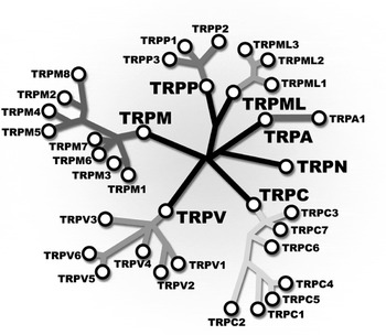
The transient receptor potential (TRP) channel story began in the late 1960s with the report of Cosens & Manning (Reference Cosens and Manning1969) of a spontaneously occurring Drosophila mutant with an abnormal response to light stimuli. In the mutant fly, the photoreceptor potential shows a transient response during prolonged illumination. Twenty years later, this finding lead to the discovery of the first TRP ion channel from Drosophila (Montell & Rubin, Reference Montell and Rubin1989). Montell & Rubin (Reference Montell and Rubin1989) reported that trp encoded a large membrane protein containing 1275 amino acids displaying eight putative transmembrane (TM) spanning segments. The amino acid sequence did not present any clear homology with any other known ion channel-forming protein. It was realized later that a new gene dubbed trp-like (trpl) showed structural homologies with Drosophila trp and also with the superfamily of voltage-gated channel genes, raising the possibility that trpl encoded subunits of a calcium or of a nonspecific cation channel (Phillips et al. Reference Phillips, Bull and Kelly1992). At the same time, Hardie & Minke (Reference Hardie and Minke1992) demonstrated that in trp flies the permeability ratio P Ca/P Na is reduced about 10-fold, strongly suggesting that the trp gene coded for a highly Ca2+-permeable channel. Despite the claims that TRP and TRPl were Drosophila photoreceptor-specific proteins (e.g. Montell & Rubin, Reference Montell and Rubin1989), Petersen et al. (Reference Petersen, Berridge, Borgese and Bennett1995) identified partial sequences of TRP homologues from Xenopus and mice using cDNA libraries, thereby showing that TRP-related proteins were also expressed outside of the invertebrates. Moreover, in the same year, the full sequence of the first human homologue (TRPC1) was reported (Wes et al. Reference Wes, Chevesich, Jeromin, Rosenbergt, Stetten and Montell1995; Zhu et al. Reference Zhu, Chu, Peyton and Birnbaumer1995). trpc1, sharing a 37% identity with the Drosophila trp gene, is present in most human tissues. Today, 18 years after the first TRP channel was obtained, we can list more than 85 channels within this family (invertebrate and vertebrate combined; Montell, Reference Montell2005b) organized by their sequence homology into seven subfamilies (Fig. 1): (1) The TRPC (classical) subfamily encompasses channels presenting a large number of different activation modes. Some are store-operated and activated by stretch, whereas others are Gq/11 receptor-operated channels activated by the production of diacylglicerol or redox processes. TRPC proteins also control growth cone guidance in both mammalian and amphibian model systems. (2) The TRPM (melastatin-related) subfamily contains channels designated as chanzymes since their C-terminal contains catalytic domains able to show protein kinase activities (TRPM6 and TRPM7). As discussed below, some members of this subfamily play an important role in detecting temperature changes. (3) The TRPV (vanilloid) subfamily has members activated by temperature. They are also involved in sensing osmolarity changes and mechanical stimuli. (4) TRPA (ankyrin) contains only one member, TRPA1, which is activated by noxious cold and a myriad of different chemical compounds including allyl isothiocyanate (AITC), the pungent principle of wasabi and other mustard oils. TRPA1 appears to have a central role in the pain response to volatile irritants like the one found in tear gas. (5) TRPN (NOMP-C homologues) has a single member that can be found in worms, flies, and zebrafish. TRPN proteins have a role in mechanotransduction. (6) The TRPP (polycystin) subfamily is formed by the polycystic kidney disease (PKD) proteins or polycystins. Heteromeric complexes TRPP2–TRPP1 form Ca2+-permeable nonselective cation channels. (7) The TRPML (mucolipin) subfamily consists of channels that appear to be only present in intracellular vesicles and two of its members (TRPML1 and 2) have been implicated in hearing. Mutations in the TRPP1 or TRPP2 are responsible for such human diseases as autosomal-dominant PKD, and mutations in TRPML1 are associated with neurodegenerative lysosomal storage disorder, mucolipidosis type 4. (For general reviews on TRP channels, see Clapham, Reference Clapham2003; Montell, Reference Montell2005a; Ramsey et al. Reference Ramsey, Delling and Clapham2006a; Venkatachalam & Montell, Reference Venkatachalam and Montell2007; for a review on Drosophila TRPs, see Montell, Reference Montell2005b; for a review on updated nomenclature of TRP channel families, see Clapham et al. Reference Clapham, Montell, Schultz and Julius2005.)
Phylogeny of TRP channels. The phylogenetic tree shows the relationship between different TRP subfamilies. Sequence identity can be as low as 20%. TRPC2 is a pseudogene in human but not in rats. TRPN channels have not been identified in mammals.

We now know that TRP channels appeared relatively early in evolution and we can trace their expression from invertebrates to vertebrates, including humans (Harteneck et al. Reference Harteneck, Plant and Schultz2000; Venkatachalam & Montell, Reference Venkatachalam and Montell2007). As TRP channels are directly involved in sensory perception such as vision, taste, olfaction, hearing, touch, thermal perception, and nociception, a recurrent theme is the idea that multicellular organisms use TRP channel-dependent pathways to sense their surroundings and, at more cellular level, TRP channels allow cells to respond to changes in their local environment. The critical role they play as sensors in cellular physiology make them incredibly attractive puzzles (Clapham, Reference Clapham2003; Voets et al. Reference Voets, Talavera, Owsianik and Nilius2005).
Despite the large body of literature on TRP channels that has accumulated during the past 30 years (more than 4000 articles since 1969; Liedtke & Heller, Reference Liedtke, Heller, Liedke and Heller2007), very little is known about the mechanisms that control the gating process in these amazing molecular machines, and much work remains to be done in the fields of biophysics and protein structure (Gaudet, Reference Gaudet, Liedtke and Heller2007).
2. TRP channel assembly
2.1 General TRP channel architecture
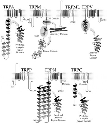
TM segment prediction algorithms indicate that TRP channels are related to the superfamily of voltage-gated cation channels. The putative organization of these channels consists in 6TM with the carboxy- and amino-terminal facing the intracellular side of the membrane (Fig. 2) (Harteneck et al. Reference Harteneck, Plant and Schultz2000; Ramsey et al. Reference Ramsey, Moran, Chong and Clapham2006b). Family relatives are voltage-activated shaker-like channels, cyclic nucleotide gated channels, and hyperpolarization-activated cyclic nucleotide-gated (HCN) channels (Harteneck et al. Reference Harteneck, Plant and Schultz2000). Although originally predicted to be 8 TM (Montell & Rubin, Reference Montell and Rubin1989), shortly thereafter TRP channel researchers reached a consensus about their 6TM organization and similarities with voltage-dependent K+ and Ca2+ channels (Phillips et al. Reference Phillips, Bull and Kelly1992; Wes et al. Reference Wes, Chevesich, Jeromin, Rosenbergt, Stetten and Montell1995; Harteneck et al. Reference Harteneck, Plant and Schultz2000; Ramsey et al. Reference Ramsey, Moran, Chong and Clapham2006b). According to the proposed 6TM architecture, the pore region would be a hydrophobic region somewhere between fifth and sixth segment (for a review, see Owsianik et al. Reference Owsianik, Talavera, Voets and Nilius2006). Studies based on biochemical and optical based methods strongly suggest that TRP channels are formed by four subunits (Jahnel et al. Reference Jahnel, Dreger, Gillen, Bender, Kurreck and Hucho2001; Kedei et al. Reference Kedei, Szabo, Lile, Treanor, Olah, Iadarola and Blumberg2001; Amiri et al. Reference Amiri, Schultz and Schaefer2003), and that active channels could assemble as homo- or heterotetramers (Hoenderop et al. Reference Hoenderop, Voets, Hoefs, Weidema, Prenen, Nilius and Bindels2003; Helwig et al. Reference Helwig, Albrecht, Hartenek, Schultz and Schaefer2005; Schaefer, Reference Schaefer2005).
The seven TRP subfamilies and the gross architecture of their subunits. TRP channels are tetramers consisting of subunits containing six transmembrane (TM) segments (S1–S6). By analogy to K+ channels the central pore is assumed to be formed by the hairpin located in between TM S5 and S6. A variable number of ankyrin domains are present in the N-terminal of most TRP channels with the exception of TRPP and TRPM subfamilies. The crystal structure of this domain has been elucidated in the case of TRPV1, 2, and 6. TRPM6 and 7 are defined as chanzymes owing to the presence of a C-terminal a typical protein kinase domain. TRP and coiled coil domains are present in the C terminus domain (for more details, see text).

TRP channels are diverse in their design and their features change from family to family. Figure 2 shows a schematic view of this diversity, and in the following sections we will give an overview of the structure–function relationships present in this remarkable superfamily of ion channels.
2.2 Viewing TRP channels through the electron microscope
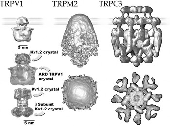
Advances in our knowledge of the crystal structure of TRP channels has been hampered by the lack of adequate expression systems needed to obtain large amounts of protein, and attempts to overexpress these proteins in bacteria have so far been unsuccessful. However, by using mammalian cell (HEK293) transfection or yeast (Saccharomyces cerevisiae), sufficient amounts of protein have been collected to make it possible to determine the structures of three TRP channels by electron microscopy (albeit at low resolution): TRC3 (Mio et al. Reference Mio, Ogura, Hara, Mori and Sato2005, Reference Mio, Ogura, Kiyonaka, Hiroaki, Tanimura, Fujiyoshi, Mori and Sato2007), TRPM2 (Maruyama et al. Reference Maruyama, Ogura, Mio, Kiyonaka, Kato, Mori and Sato2007), and TRPV1 (Moiseenkova & Wensel, Reference Moiseenkova and Wensel2009) (Fig. 3). More recently, Moiseenkova & Wensel (Reference Moiseenkova and Wensel2009) gave a critical appraisal of the different techniques used to provide the first steps in the elucidation of TRP channel structure and speculate on the future of the field. Of particular interest is that the structures of TRPC3 and TRPM2 proved to be very different from that of TRPV1. The TRPC3 channel structure was determined using a single particle reconstruction from images coming from a cryoelectron microscopy (Fig. 3). The bell-shape structure obtained in this manner has an enormous volume mostly conformed by the cytoplasmic domain. This large cytoplasmic domain is made of a sparse external shell that appears as girdles connected through low-density panels. This type of structure explains how the relatively low molecular weight channel-forming protein can present such a large volume. Using electron microscopy in negative stain, a large volume bullet-shape structure was also observed for TRPM2. By contrast, the structure of TRPV1 is in many ways reminiscent of that of the Kv1.2 (Long et al. Reference Long, Campbell and MacKinnon2005; cf. second and third structures in the first column of Fig. 3), which includes a large ‘hanging gondola’ most probably made up of the N and C terminus of the channel (Fig. 3). The TRPV1 volume is several times smaller than that of TRPC3 or TRPM2. It has been argued that the large expansion of the TRP channel cytoplasmic surface allows for the association with several macromolecules. Because TRPV1 has also evolved to be a specialized sensor of various signals, and is likely to be a macromolecular complex, one can only agree with Moiseenkova & Wensel (Reference Moiseenkova and Wensel2009) that, ‘it is not obvious why the TRPC3 and TRPV1 channel structures look so different.’
Comparison of structures of TRP channels derived from electron microscopy studies. Cryoelectron microscopy structures: TRPV1, 19 Å (Moiseenkova et al. Reference Moiseenkova, Stanciu, Iseresheva, Tobe and Wensel2008); TRPC3, 15·3 Å (Mio et al. Reference Mio, Ogura, Kiyonaka, Hiroaki, Tanimura, Fujiyoshi, Mori and Sato2007). A comparison between the TRPV1 and the Kv1.2/β subunit structure (PDB code 2A79) is shown. The crystal structure of Kv1.2 and TRPV1 ankyrin domains were superimposed on the TRPV1 structure obtained using electron microscopy and T domain of Kv1.2 and β subunit were superimposed on the Kv1.2 structure obtained by cryomicroscopy. Negative stain structures, resolution: TRPM2, 37 Å (Maruyama et al. Reference Maruyama, Ogura, Mio, Kiyonaka, Kato, Mori and Sato2007).

2.3 TRP channel multimerization properties
Homo- or heteromultimerization was first tested when Drosophila TRP and TRPL channels were coexpressed in Xenopus oocytes (Gillo et al. Reference Gillo, Chorna, Cohen, Cook, Manistersky, Chorev, Arnon, Pollock, Selinger and Minke1996). When coexpressed TRP and TRPL form active heteromers with a characteristic conductance but low constitutive activity; they also can be coimmunoprecipitated from a preparation of Drosophila eye (Gillo et al. Reference Gillo, Chorna, Cohen, Cook, Manistersky, Chorev, Arnon, Pollock, Selinger and Minke1996; Xu et al. Reference Xu, Li, Guggino and Montell1997). Coexpression of TRP and TRPL leads to a store-operated, outwardly rectifying current distinct from that caused by either TRP or TRPL alone (Minke et al. Reference Minke1996). TRP-gamma, another subunit involved in the photoreceptor response in Drosophila, also interacts with TRPL forming a phospholipase C-sensitive channel (Xu et al. Reference Xu, Chien, Butler, Salkoff and Montell2000).
Regarding mammalian TRP channels, the first report on heteromultimerization came from coimmunoprecipitation experiments on TRPC1 and TRPC3 complexes (Xu et al. Reference Xu, Li, Guggino and Montell1997). Functional evidence of TRPC heteromultimerization came later from TRPC1 and TRPC3 coexpression experiments, which resulted in a diacylglycerol-sensitive Ca2+-regulated cation currents different from the currents produced by the original subunits (Lintschinger et al. Reference Lintschinger, Balzer-Geldsetzer, Baskaran, Graier, Romanin, Zhu and Groschner2000). Clapham and coworkers demonstrated that TRPC1 is able to coassemble with TRPC4 and TRPC5 (Strübing et al. 2001). Interestingly, coexpression of TRPC1 and TRPC4 or TRPC5 gives origin to novel outwardly rectifying (TRPC4 and C5 are doubly rectifying channels), nonselective cation conductances, and the recombinant TRPC1/TRPC5 channel was activated by Gq-coupled receptors and its activity was independent of calcium store depletion. The characteristics of these heteromeric channels resemble those of native Gq-coupled receptor-activated currents present in the hippocampus and the frontal cortex, giving support to the hypothesis that TRPC heteromers form receptor-modulated currents in the mammalian brain. An attempt to determine the combinatorial rules within the TRPC subfamily was made by Hofmann et al. (Reference Hofmann, Schaefer, Schultz and Gudermann2002) using a variety of different experimental approaches. They concluded that TRPC2 is utterly unable to form heteromers with the other TRPC proteins, whereas the other TRPC subunits can form heterodimers, but only with phylogenetically close relatives: TRPC1/TRPC4/TRPC5 representing one group and TRPC3/TRPC6/TRPC7 representing the other. The rules of TRPC channel assembly put forward by Hofmann et al. (2002) appeared to apply in vivo since Strübing et al. (2003) were able to detect the presence of the complex formed by TRPC1/TRPC4/TRPC5 in the prenatal brain. Although the investigations of Hofmann et al. (2002) ruled out heteromerization between distant relatives in the TRPC subfamily, the situation regarding heteromerization is far from clear. The evidence provided by several groups indicates that distant TRPC members can form heteromeric channels. Thus, TRPC3 can assemble with TRPC1 (Lintschinger et al. Reference Lintschinger, Balzer-Geldsetzer, Baskaran, Graier, Romanin, Zhu and Groschner2000; Liu et al. Reference Liu, Bandyopadhyay, Singh, Groschner and Ambudkar2005), and more recently Poteser et al. (Reference Poteser, Graziani, Rosker, Eder, Derler, Kahr, Zhu, Romanin and Groscner2006) reported the existence of a TRPC3/TRPC4 complex able to form redox sensitive channels in endothelial cells.
Regarding the molecular determinants involved in tetramerization, Lapage et al. (Reference Lapage, Lussier, Barajas-Martinez, Bousquet, Blanchard, Francoeur, Dumaine and Boulay2006) used chimeras between TRPC4 and TRPC6 to demonstrate that there are two domains responsible for the association between TRPC4 and TRPC6 (members of distinct subgroups of TRPCs). TRPC4 coimmunoprecipitated with TRPC6 subunits containing the N-terminal ankyrin and coil-coiled domain (residues 1–304) of TRPC4 (domain 1) (Fig. 2). TRPC6/TRPC4 chimeras containing part of the S4–S5 linker and C-terminal tail (residues 516–974) of TRPC4 (domain 2) are also able to interact and form tetramers with TRPC4 subunits.
It is also known that TRPVs can form hetero-oligomers. How and where TRPV channels form heteromultimers are important questions since the coassembly of different TRPV subunits with widely different gating properties may have important repercussions for the coding and regulation of thermosensation. The early work done using TRPV3 channels and based on coimunoprecipitation and electrophysiology suggested that TRPV3 and TRPV1 form heteromultimers (Smith et al. Reference Smith, Gunthorpe, Kelsell, Hayes, Reilly, Facer, Wright, Jerman, Walhin, Ooi, Egerton, Charles, Smart, Randall, Anand and Davis2002). More convincing data regarding the interaction between different TRPV subunits were obtained by Hoenderop et al. (Reference Hoenderop, Voets, Hoefs, Weidema, Prenen, Nilius and Bindels2003) using a variety of different techniques. In a study that included chemical cross-linking, sucrose gradient centrifugation, coimmunoprecipitation, and electrophysiological analyses of concatemeric channels, Hoenderop et al. (Reference Hoenderop, Voets, Hoefs, Weidema, Prenen, Nilius and Bindels2003) confirmed that TRPV channels assemble as tetramers and demonstrated that TRPV5/6 form heteromultimers with novel ion transport properties. These results were further confirmed by a systematic study on TRPVs interaction carried out by Helwig et al. (Reference Helwig, Albrecht, Hartenek, Schultz and Schaefer2005). Using fluorescence energy transfer (FRET) and a biochemical approach, they showed that TRPV5/6 are able to form heteromultimers. However, their data indicated that besides TRPV1/2 and TRPV5/6, all the other members of the TRPV subfamily show a preference to form homo-oligomers (Helwig et al. Reference Helwig, Albrecht, Hartenek, Schultz and Schaefer2005). Moreover, in vivo experiments carried out in rat cerebral cortex cells provided evidence that TRPV1 and TRPV2 interactions do take place in native tissue (Liapi & Wood, Reference Liapi and Wood2005). Cheng et al. (Reference Cheng, Yang, Takanishi and Zheng2007) studied TRPV1–4 interactions using spectra-FRET, an approach similar to that used by Helwig et al. (Reference Helwig, Albrecht, Hartenek, Schultz and Schaefer2005) but is more sensitive. The spectra-FRET approach allows the detection of strong fluorescent signals when the signals are specifically collected from the plasma membrane, thereby efficiently decreasing the intracellular signals. The results confirmed previous reports of heteromeric interactions of TRPV1/2. However, in contrast to the results obtained by Helwig et al. (Reference Helwig, Albrecht, Hartenek, Schultz and Schaefer2005), they also found FRET values corresponding to heteromization when they included TRPV3 and TRPV4. Heteromerization of TRPV1–4 was further confirmed by combining their energy transfer results with single channel recordings. Heteromeric channels show particular intermediate conductance levels and different gating kinetic properties.
There is just one example of a TRPM family channel heteromultimer, which is the case of TRPM6 and TRPM7 channels with the unique characteristic of harboring serine/threonine kinase domains in the distal part of their C termini. TRPM6 appears to be crucial for maintaining the normal Mg2+ homeostasis since mutations in the trpm6 gene causes autosomal recessive hypomagnesemia with secondary hypocalcemia. The study of Chubanov et al. (Reference Chubanov, Waldegger, Mederos, Schnitzler, Vitzthum, Sassen, Seyberth, Konrad and Gudermann2004) strongly suggested that TRPM6 does not conduct currents by itself (cf. Voets et al. Reference Voets, Janssens, Droogmans and Nilius2004). Instead, an association between TRPM7 and TRPM6 was required to incorporate channel complexes in the plasma membrane, and disruption of the complex by the naturally occurring TRPM6 mutation S141L may be the cause of hypomagnesaemia in humans. The association of TRPM6 and TRPM7 channels was also studied by Schmitz et al. (Reference Schmitz, Dorovkov, Zhao, Davenport, Ryazanov and Perraud2005), who found that there is a cross talk between their activities and the need of TRPM7 for TRPM6 functional membrane expression. A more recent study (Li et al. Reference Li, Jiang and Yue2006), however, clearly demonstrates that TRPM6, TRPM7, and heteromultimers of these two channels of the melastatin subfamily form well-defined ion channels with different ionic permeabilities, and with distinctive single channel conductance. These different findings have not been explained but they could be reconciled by the observation of Topala et al. (Reference Topala, Groenestege, Thébault, Van den Verg, Nilius, Hoenderop and Bindels2007) of low TRPM7-like endogenous currents in HEK293 cells.
The molecular nature of the interactions that lead to TRP channel heteromultimerization is unclear (for a review, see Schaefer, Reference Schaefer2005), and whether the formation of heteromeric TRP channel is physiologically relevant remains to be clarified.
2.3.1 The mysterious TRP domain
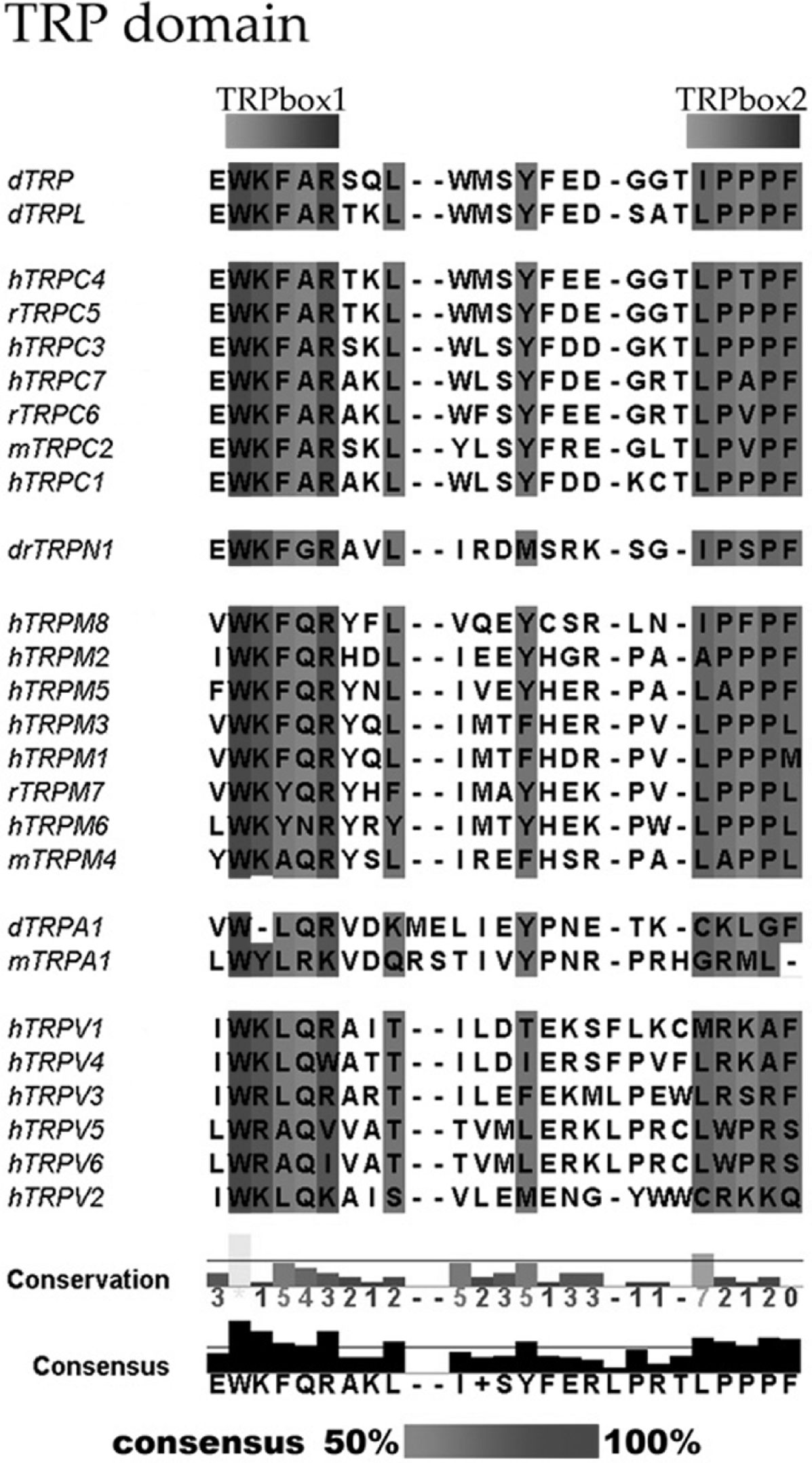
Some TRP subfamilies display a conserved sequence on their proximal C-terminal region that has been used as a signature sequence for TRP channels (Fig. 4). This ~25-amino acid intracellular region, just after the S6 region, is called the TRP domain (Montell, Reference Montell2001). This domain contains the TRP box, a highly conserved region first reported by Zhu et al. (Reference Zhu, Chu, Peyton and Birnbaumer1995) as the ‘EWKFAR motif’ and was found in 11 of the 13 TRP channel sequences known at that time (all of them members or relatives of the mammalian TRPC family). The TRP box is defined by the consensus sequence WKFQR (Fig. 4), where W is the only conserved residue among the TRP families. Positive charges on positions 2 and 5 of that sequence are shared by almost all TRPs that present a TRP domain. It is possible to distinguish a second conserved sequence on the carboxy-terminal end of the TRP domain in the channels belonging to the TRPC and TRPM subfamilies. This second box is proline-rich, sharing the consensus sequence LPPPF (leucine on position one is highly conserved). The main role of the TRP domain remains elusive, but it has been suggested that it may participate in channel assembly (García-Sanz, Reference García-Sanz, Fernández-Carvajal, Morenilla-Palao, Planells-Cases, Fajardo-Sánchez, Fernández-Ballester and Ferrer-Montiel2004) and PIP2 binding (Rohacs et al. Reference Rohacs, Lopes, Michailidis and Logothetis2005; Rohacs, Reference Rohacs2006; Brauchi et al. Reference Brauchi, Orta, Mascayano, Salazar, Raddatz, Urbina, Rosenmann, Gonzalez-Nilo and Latorre2007). Recently, using chimeras between TRPV channels, it has been proposed that the TRP domain may operate as both a tetramerization domain and a transduction domain somehow coupled to channel gating (García-Sanz et al. Reference García-Sanz, Valente, Gomis, Fernández-Carvajal, Fernández-Ballester, Viana, Belmonte and Ferrer-Montiel2007). García-Sanz et al. (Reference García-Sanz, Valente, Gomis, Fernández-Carvajal, Fernández-Ballester, Viana, Belmonte and Ferrer-Montiel2007) showed that the incorporation of certain amino acids that alter the interactions inside the TRP domain may have an important effect on the activity of the channel by affecting the activation energy of channel gating. Interestingly, they propose that the TRP domain works as a ‘negative allosteric modulator’ holding the channel in the closed state. We note here that a small portion of the C-terminal of the Ca2+-activated K+ channel (hSlo) behaves very similarly to the TRP domain of TRPV channels. The interaction of that domain (tail) with the other portion of the hSlo channel inhibits channel opening, and this inhibition is relieved by Ca2+ binding to the tail via an allosteric mechanism (Schreiber et al. Reference Schreiber, Yuan and Salkoff1999).
A signature sequence for TRP channels: the TRP domain. This domain contains – in some cases – a highly conserved region the ‘EWKFAR motif’ (see the TRPC family). The TRP box is defined by the consensus sequence WKFQR, where W is the only conserved residue among TRP families. In channels belonging to the TRPC and TRPM subfamilies, there is a second conserved sequence on the C terminus end of the TRP domain; this second box has the consensus sequence LPPPF.

2.4 Molecular determinants of channel assembly
Structurally, TRPs appear to share a common TM scaffold with four subunits around a central aqueous pore. Recent work shows that TRP channels from different families do not share the same assembly mechanism or tetramerization region (García-Sanz et al. Reference García-Sanz, Fernández-Carvajal, Morenilla-Palao, Planells-Cases, Fajardo-Sánchez, Fernández-Ballester and Ferrer-Montiel2004; Hoenderop et al. Reference Hoenderop, Voets, Hoefs, Weidema, Prenen, Nilius and Bindels2003; Erler et al. Reference Erler, Al-Ansary, Wissenbach, Wagner, Flockerzi and Niemeyer2006; Lapage et al. Reference Lapage, Lussier, Barajas-Martinez, Bousquet, Blanchard, Francoeur, Dumaine and Boulay2006; Tsuruda et al. Reference Tsuruda, Julius and Minor2006). N and C termini and protein moieties located between TM segments 1–6 appear to be involved in TRPV1 channel tetramerization and the N terminus of TRPV4 makes a significant contribution to the assembly of TRPV4 (García-Sanz, 2004; Hoenderop et al. Reference Hoenderop, Voets, Hoefs, Weidema, Prenen, Nilius and Bindels2003). Ankyrin-repeat domains (ARDs) located in the N terminus of TRPV5 and TRPV6 are involved in channel tetramerization (Erler et al. Reference Erler, Hirnet, Wissenbach, Flockerzi and Niemeyer2004; Chang et al. Reference Chang, Gyftogianni, Van De Graaf, Hoefs, Weidema, Bindels and Hoenderop2004). However, in these cases, channel assembly appears not to be determined by direct interaction of ARDs as previously proposed but rather by interaction of the ARDs with other structures still to be determined (Phelps et al. Reference Phelps, Huang, Lishko, Wang and Gaudet2008). Coiled coil domains located on the distal portion of TRPM8 C-terminal (Erler et al. Reference Erler, Al-Ansary, Wissenbach, Wagner, Flockerzi and Niemeyer2006; Tsuruda et al. Reference Tsuruda, Julius and Minor2006), TRPM2 (Mei et al. Reference Mei, Xia, Beech and Jiang2006), and TRPM7 (Fujiwara & Minor, Reference Fujiwara and Minor2008) appear to be important in the assembly process of these channels (but see Phelps & Gaudet, Reference Phelps and Gaudet2007 and below). On the other hand, TRPCs apparently use a combination of N-terminal interactions through ankyrin–coiled coil domains together with C-terminal interactions to form active tetramers (Lapage et al. Reference Lapage, Lussier, Barajas-Martinez, Bousquet, Blanchard, Francoeur, Dumaine and Boulay2006).
3. N-terminal domain
3.1 Ankyrin domains
The 33-amino acid ankyrin repeat (AR) is among the most abundant repeat motifs and is present in different types of proteins (Mosavi et al. Reference Mosavi, Cammett, Desrosiers and Peng2004). Probably because its high specificity, AR are involved in a varied set of cellular functions including regulation of cell cycle, transcription, cell–cell signaling, development, and transport. ARs act as building bricks that pile up side by side, forming a modular and highly efficient and specific protein-binding surface. AR structure is well defined: each repeat folds into two antiparallel helices followed by a beta-hairpin (Mosavi et al. Reference Mosavi, Cammett, Desrosiers and Peng2004; Sedgwick & Smerdon, Reference Sedgwick and Smerdon1999; see below).
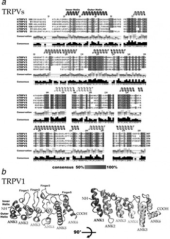
TRP channels from TRPA, TRPN, TRPV, and TRPC subfamilies present ankyrin domain repeats on their amino-terminal region (Fig. 2; Montell, Reference Montell2005a; Gaudet, Reference Gaudet2008). ARDs are highly conserved in TRPV channels and the six members of this subfamily contain a six-repeat ARD. The complete structure resembles the shape of a cupped hand were the helices form a palm and the hairpins (or sometimes loops) represent the fingers (Fig. 5a). The ARDs of TRPVs are important for the integrity of the channel, as their deletion impairs assembly and trafficking to the cell membrane (Jung et al. Reference Jung, Lee, Hwang, Cho, Shin, Kang, Kim and Oh2002; Chang et al. Reference Chang, Gyftogianni, Van De Graaf, Hoefs, Weidema, Bindels and Hoenderop2004; Erler et al. Reference Erler, Hirnet, Wissenbach, Flockerzi and Niemeyer2004). TRPV1 N-terminal has been shown to be involved in its calmodulin regulation and peptides from the N-terminal bind to calmodulin in a Ca2+-dependent manner (Numazaki et al. Reference Numazaki, Tominaga, Takeuchi, Murayama, Toyooka and Tominaga2003; Rosenbaum et al. Reference Rosenbaum, Gordon-Shaag, Munari and Gordon2004). Interestingly, protein kinase C (PKC) potentiation of the thermosensitive TRPV1 activity is linked to interactions with SNARE complex-related proteins through its N-terminal domain. Morenilla-Palao et al. (Reference Morenilla-Palao, Planells-Cases, García-Sanz and Ferrer-Montiel2004) found that TRPV1 interacts specifically (in vitro and in vivo) with two vesicular proteins, Snapin and Synaptotagmin IX, and that this interaction allows the recruitment of receptors located in vesicular compartments to the plasma membrane by an orchestrated exocytosis process. TRPV2 N-terminal was found to interact with the recombinase gene activator protein, which associates with the channel during biosynthesis and early trafficking (Barnhill et al. Reference Barnhill, Stokes, Koblan-Huberson, Shimoda, Muraguchi, Adra and Turner2004; Stokes et al. Reference Stokes, Wakano, Del Carmen, Koblan-Huberson and Turner2005). The N-terminal region in TRPV2 also interacts with the adapter protein, Acyl CoA binding domain protein, (ACBD)3, allowing the recruitment of protein kinase A (PKA) into the channel micro environment (Stokes et al. Reference Stokes, Wakano, Del Carmen, Koblan-Huberson and Turner2005).
Ankyrin repeat domains in the TRPV subfamily. (a) Alignment of the ARD of mammalian TRPV channels. Residues conforming the inner and outer helices of the ankyrin domains are indicated as helices above each repeat. (b) Crystal structure of the ARD domain of TRPV1. Mofified from Lishko et al. (Reference Lishko, Procko, Jin, Phelps and Gaudet2007) and Phelps et al. (Reference Phelps, Huang, Lishko, Wang and Gaudet2008).

Two separate groups determined the crystal structure for TRPV2-ARD (McCleverty et al. Reference McCleverty, Koesema, Patapoutian, Lesley and Kreusch2006; Jin et al. Reference Jin, Touhey and Gaudet2006), and Lishko et al. (Reference Lishko, Procko, Jin, Phelps and Gaudet2007) solved the crystal structure of the TRPV1-ARD (residues 101–364) (Fig. 5b). Although previous predictions place four ARD's on the N-terminal sequence of TRPVs, the crystal structure clearly shows the presence of six-repeat ARD motifs that appear to be conserved throughout the TRPV subfamily (Fig. 5a). ARDs on TRPV channels do not follow a standard pattern. Repeats 1–3 have long and flexible fingers, whereas repeats 5 and 6 have long outer helices. Moreover, a large counterclockwise turn was observed in the stacking of repeats 4 and 5. These modifications affect the overall ARD structure/symmetry and may alter its functional behavior. TRPV1-ARD and TRPV2-ARD do not form hetero-oligomers but apparently ARDs play a role in promoting tetrameric assembly of TRPV5 and TRPV6 (Chang et al. Reference Chang, Gyftogianni, Van De Graaf, Hoefs, Weidema, Bindels and Hoenderop2004; Erler et al. Reference Erler, Hirnet, Wissenbach, Flockerzi and Niemeyer2004). It would be of interest to explore the nature of the interaction in other TRPV channels and especially in TRPA1 as ARDs are being used for interactions with agonists (see below). Interestingly, the TRPV2-ARD structure predicted that phosphorylation sites in TRPV1’s N terminus (S116, Y200) should be on the surface of interaction of ARD with other proteins. Phosphorylation events could, therefore, modulate the binding of regulatory factors to the ARD (Jin et al. Reference Jin, Touhey and Gaudet2006).
TRPV1 presents at least two mechanisms that lead to the modulation of its responsiveness to noxius stimuli: desensitization and tachyphylaxis. Although both are forms of desensitization, desensitization proper is a short-term phenomenon and in the case of TRPV1, it is produced upon acute channel activation by capsaicin or protons. By contrast, tachyphylapsis is a loss of channel sensitivity after multiple separate challenges with an agonist. The crystal structure of TRPV1-ARD solved by Lishko et al. (Reference Lishko, Procko, Jin, Phelps and Gaudet2007) provided several hints about how the tachyphylapsis phenomenon comes about. In contrast to TRPV2-ARD, the TRPV1-ARD structure revealed a binding site able to accommodate ATP, and biochemical studies demonstrated that calmodulin binds to the same site. The site is noncanonical, being formed by a concave surface of repeats 1–3, and containing several hydrophobic and positively charged residues. In particular, R115, K155, and K160 interact with the ATP triphosphate moiety. Mutations K155A and K160A completely eliminate nucleotide interaction with the TRPV1-ARD, sensitization by ATP, and a remarkable reduction in tachyphylapsis. These apparently contradictory results were explained by Lishko et al. (Reference Lishko, Procko, Jin, Phelps and Gaudet2007) on the basis of a model that assumes that in the sensitized state TRPV1-ARD is bound to ATP and that the Ca2+ influx through the open channel causes release of ATP and binding of the Ca–calmodulin complex to the TRPV1-ARD, which induces desensitization.
Solved at 1.7 Å resolution, the crystal structure of the ARD of TRPV6 showed many similarities with that of TRPV1 and TRPV2 (Phelps et al. Reference Phelps, Huang, Lishko, Wang and Gaudet2008), and revealed structural characteristics unique to TRPV ARDs. For example, unusually long finger loops and amino acid residues conserved in TRPV proteins diverge from the AR sequence consensus, causing a twist between the fourth and 5th repeat. There are, however, several important differences between these ARD domains that should be noted since they may have important functional consequences. In particular, unlike the TRPV1-ARD, the TRPV6-ARD is unable to bind ATP or calmodulin (CaM) to any appreciable extent – suggesting that CaM modulates TRPV channels using more than one mechanism. Moreover, there is ample evidence that CaM is able to interact with a site in the C terminus of TRPV6. CaM facilitates channel inactivation, a process that can be overcome by a phosphorylation of the CaM binding site mediated by PKC (Niemeyer et al. Reference Niemeyer, Bergs, Wissenbach, Flockerzi and Trost2001).
3.2 TRP channel activation through covalent modification of N-terminal cysteines
TRPA1 is activated by a number of pungent natural compounds used in cooking including wasabi and mustard, which owe their pungency to isothyocyanate compounds (see Fig. 13e) (Bandell et al. Reference Bandell, Story, Hwang, Viswanath, Eid, Petrus, Earley and Patapoutian2004; Jordt et al. Reference Jordt, Bautista, Chuang, McKemy, Zygmunt, Hogestatt, Meng and Julius2004). This list of compounds has recently been extended to include a series of environmental irritants such as air pollutants and chemotherapeutic agents (Bautista et al. Reference Bautista, Jordt, Nikai, Tsuruda, Read, Poblete, Yamoah, Basbaum and Julius2006; Hinman et al. Reference Hinman, Chuang, Bautista and Julius2006; Macpherson et al. Reference Macpherson, Hwang, Miyamoto, Dubin, Patapoutian and Story2006, Reference Macpherson, Dubin, Evans, Marr, Schultz, Cravatt and Patapoutian2007; McNamara et al. Reference McNamara, Mandel-Brehm, Bautista, Siemens, Deranian, Zhao, Hayward, Chong, Julius, Moran and Fanger2007; Trevisani et al. Reference Trevisani, Siemens, Materazzi, Bautista, Nassini, Campi, Imamachi, Andre, Patacchini, Cottrell, Gatti, Basbaum, Bunnett, Julius and Geppetti2007). A fascinating new discovery is that cysteine residues in TRP channels can be covalently modified by a great variety of chemically unrelated agonists, whose only similarity is that they are electrophyles, as part of their mechanism of activation (Hinman et al. Reference Hinman, Chuang, Bautista and Julius2006; Macpherson et al. Reference Macpherson, Dubin, Evans, Marr, Schultz, Cravatt and Patapoutian2007; Bang & Hwang, Reference Bang and Hwang2009). Thus, unlike other natural agonists such as menthol or capsaicin that appear to act in a lock-and-key fashion, TRPA1 agonists act by covalently modifying their target cysteines and binding ‘irreversibly’ to the channel. Mutagenesis experiments showed that modification of only three cysteines of the 31 present in the cytoplasmic aspect of mouse TRPA1 led to agonist activation. All three cysteines are located in the N-terminal region of mouse TRPA1 channels (C415, C422, and C622; Macpherson et al. Reference Macpherson, Dubin, Evans, Marr, Schultz, Cravatt and Patapoutian2007). Two of these cysteines are inside one of the 18 ARD present in TRPA1 (C415, C422), and C622 is in the linker that joins together the last ARD with TM domain 1. Three cysteines were also found to be responsible for TRPA1 agonist activation by Hinman et al. (Reference Hinman, Chuang, Bautista and Julius2006) and they mapped to C619, C639, and C663, all of them located in the ARD-TM1 linker of the human TRPA1 channel. Thus, the results obtained by Macpherson et al. (Reference Macpherson, Dubin, Evans, Marr, Schultz, Cravatt and Patapoutian2007) and Hinman et al. (Reference Hinman, Chuang, Bautista and Julius2006) share only one cysteine residue in common, C619; this cysteine residue in the human TRPA1 corresponds to C622 in mouse TRPA1. Recalling that isothiocyanates also modify primary amines, Hinman et al. (2007) found that a residual response of the triple cysteine mutant to AITC was due to a covalent modification of a lysine side chain since the mutant K708R produces TRPA1 channels that are completely insensitive to AITC.
How the free energy of ligand binding is converted into mechanical work (pore opening) is still a mystery, but the results of Chen et al. (Reference Chen, Zhang, Kort, Huth, Sun, Miesbauer, Cassar, Neelands, Scott, Moreland, Reilly, Hajduk, Kym, Hutchins and Faltynek2008) may have identified some of the pieces of the puzzle. Using a high-throughput screen, Chen et al. (Reference Chen, Zhang, Kort, Huth, Sun, Miesbauer, Cassar, Neelands, Scott, Moreland, Reilly, Hajduk, Kym, Hutchins and Faltynek2008) found electrophilic thioaminal-containing compounds that activated the rat TRPA1 but inhibited the human TRPA1. Notably, the opposite functional responses to these compounds are due to differences in some key amino acid residues contained in the S6 TM segments. Alanine 946 and methionine 949 are found to be critical for channel activation in rTRPA1 whereas the equivalent residues in hTRPA1 (serine 943 and isoleucine 946) are responsible for channel block. Moreover, point mutations of residue A946 can lead to activation (rA946G), inhibition (rA946T), or no response (rA946D). These data strongly suggest that the pore region of TRPA1 is involved in the coupling between the covalent binding of electrophilic compounds and channel gating.
In summary, all the data presented above strongly suggest that TRPA1 works as a chemical nociceptor in the nervous system, able to detect chemicals that mediate tissue damage and raise the voice of alarm to the whole organism by allowing the propagation of the pain response.
TRPA1 is not the only TRP channel to be activated by covalent modification of cysteines mediated by pungent compounds and, actually, it was known that allicin, one of the constituents of garlic, was able to activate both TRPA1 and TRPV1 (MacPherson et al. Reference Macpherson, Geierstanger, Viswanath, Bandell, Eid, Hwang and Patapoutian2005; cf. Bautista et al. Reference Bautista, Jordt, Nikai, Tsuruda, Read, Poblete, Yamoah, Basbaum and Julius2006). This observation was recently confirmed by Salazar et al. (Reference Salazar, Llorente, Jara-Oseguera, Garcia-Villegas, Munari, Gordon, Islas and Rosenbaum2008), who further showed that the allicin behaves as a TRPV1 agonist through the covalent modification of a single cysteine localized in the N-terminal of the channel at position C157. Keeping Section 3.1 in mind, it is important to note that this residue is located near the amino acids that form part of the ATP binding site, a region that can undergo substantial structural conformational coupled to channel gating.
3.3 TRPM homology regions (MHRs)
Unlike the AR expressing channels, TRPMs share four high homology regions in their N-terminal cytoplasmic domain (Clapham, Reference Clapham2003). Although MHRs are highly homologous within TRPM channels, they do not show similarities outside of the TRPM family. As stated above, TRPM6 MHR mutation on Serine 141 (S141L) severely alters channel behavior, preventing proper channel assembly and causing hereditary hypomagnesemia (Walder et al. Reference Walder, Landau, Meyer, Shalev, Tsolia, Borochowitz, Boettger, Beck, Englehardt, Carmi and Sheffield2002; Schlingmann et al. Reference Schlingmann, Weber, Peters, Niemann Nejsum, Vitzthum, Klingel, Kratz, Haddad, Ristoff, Dinour, Syrrou, Nielsen, Sassen, Waldegger, Seyberth and Konrad2002; Chubanov et al. Reference Chubanov, Waldegger, Mederos, Schnitzler, Vitzthum, Sassen, Seyberth, Konrad and Gudermann2004). Given the low similarity to known protein domains, it is difficult to speculate about the structural organization of MHR domains.
4. TM regions
Primary sequence analyses suggested that the 6TM domain architecture is the most probable for TRP channels (Montell, Reference Montell2005a; Ramsey et al. Reference Ramsey, Delling and Clapham2006a). Although there is evidence supporting this hypothesis, it is all circumstantial and we cannot discard at present the existence of some exceptions since predictions for TRP channel TM domains often return more than 6TMs (Ramsey et al. Reference Ramsey, Delling and Clapham2006a). An appropriate example to illustrate this point is the voltage- and calcium-activated potassium channel (BK). This potassium channel belongs to the S4 superfamily that includes the six TM domains of voltage-dependent K+ (Kv) channels but has an extra TM called S0 joining the standard 6TM segments, which allows the N-terminal to face the extracellular medium (for a review, see Latorre & Brauchi, Reference Latorre and Brauchi2006). To our knowledge, the Vannier et al. (Reference Vannier, Zhu, Brown and Birnbaumer1998) report is the only detailed topological study performed on a TRP (TRPC3) channel. In the case of TRPC3, the h1 segment, a seventh N-terminal hydrophobic domain predicted for members of the TRPC, TRPM, TRPV families, is intracellular and does not constitute a real TM segment.
The general scaffold for a 6TM domain channel considers that the TM region would be divided in two main modules: the voltage sensor (S1–S4) and the pore region (S5–S6) (Gaudet, 2007; Latorre et al. Reference Latorre, Vargas, Orta, Brauchi, Liedtke and Heller2007b). For the case of TRP channels, there is growing evidence supporting an S5–S6 conductive pore module. One line of evidence supporting this topology is that the pore sequence is reasonably conserved among members of the same TRP subfamily. Another argument is that mutations introduced on the S6 segment may produce dominant negative forms of TRP channels (Kuzhikandathil et al. Reference Kuzhikandathil, Wang, Szabo, Morozova, Blumberg and Oxford2001; Krapivinsky et al. Reference Krapivinsky, Mochida, Krapivinsky, Cibulsky and Clapham2006). The strongest evidence in support of the S5–S6 conductive pore module, however, is that mutations of the residues within this consensus region affect permeation properties of the channels (see ‘Pore’ section and Owsianik et al. Reference Owsianik, Talavera, Voets and Nilius2006 for a review on permeation of TRPs).
Although the analogy with Shaker type of ion channels suggests that the region between the helices S1 and S4 may be responsible for voltage sensing, there is a lack of convincing data showing that the voltage dependence is related to this particular module. Even if the voltage sensor is located in the S1–S4 domain, there is no reason to believe that the coupling between the sensor displacement and the pore opening is similar to that of the Shaker channel.
TRP channels are known to be responsive to several natural as well as synthetic compounds (Voets et al. Reference Voets, Talavera, Owsianik and Nilius2005) and, as pointed out by Dhaka et al. (Reference Dhaka, Viswanath and Patapoutian2006), most of the compounds with oral sensory qualities different from olfaction and taste can be attributed to the opening of TRP channels. This behavior is understood, in general, as part of their role as molecular sensors for external stimuli. Interestingly, several binding sites have been found within the TM region (Bandell et al. Reference Bandell, Dubin, Petrus, Orth, Mathur, Hwang and Papapoutian2006; Jordt & Julius, Reference Jordt and Julius2002). The mechanism allowing the transformation of the chemical energy of binding into the mechanical energy necessary to open the pore is still unknown, but an allosteric hypothesis which relates this process with the channels' voltage dependence has been suggested (Nilius et al. Reference Nilius, Karel, Owsianik, Prenen, Droogmans and Voets2005; Voets et al. Reference Voets, Owsianick, Jansens, Talavera and Nilius2007; Latorre et al. Reference Latorre, Vargas, Orta, Brauchi, Liedtke and Heller2007b).
4.1 Searching for the voltage sensor
The 6TM domain voltage-gated ion channels play a pivotal role in muscle contraction, neuronal excitability, and secretion. The voltage sensor domain (VSD) is the key element of voltage-gated ion channels for sensing TM potential, and has been studied for more than 50 years (Hodgkin & Huxley, Reference Hodgkin and Huxley1952) at the levels of both, biophysics and protein structure (e.g. Bezanilla, Reference Bezanilla2000; Long et al. Reference Long, Tao, Campbell and MacKinnon2007).
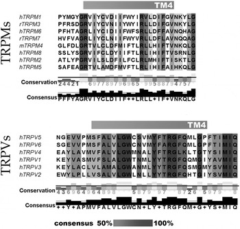
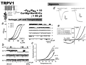
The main feature of VSD is the special array of positive charges observed in S4 and, in particularlin voltage-dependent K+ (Kv) channels, in the S2 TM segment (Noda et al. Reference Noda, Shimizu, Tanabe, Takai, Kayano, Ikeda, Takahashi, Nakayama, Kanaoka and Minamino1984; Papazian et al. Reference Papazian, Shao, Seoh, Mock, Huang and Wainstock1995; Seoh et al. 1996; Jiang et al. Reference Jiang, Lee, Chen, Ruta, Cadene, Chait and MacKinnon2003). These charges allow the voltage sensor to be displaced inside the membrane electric field in response to changes in membrane potential (Hodgkin & Huxley, Reference Hodgkin and Huxley1952; Hille, Reference Hille2001). The VSD is present in the membrane not only in voltage-dependent channels, but also in voltage-dependent enzymes (Murata et al. Reference Murata, Iwazaki, Sasaki, Inaba and Okamura2005) and incorporated into voltage-dependent proton channels (Sasaki et al. 2006; Ramsey et al. Reference Ramsey, Moran, Chong and Clapham2006b). Because some TRP channels are voltage-activated (Nilius et al. Reference Nilius, Karel, Owsianik, Prenen, Droogmans and Voets2005; Latorre et al. Reference Latorre, Vargas, Orta, Brauchi, Liedtke and Heller2007b), it is tempting to suggest the existence of a similar module in voltage-dependent TRP channels. In contrast to Kv channels, however, the voltage dependence of thermoTRPs is predicted to be very weak (Brauchi et al. Reference Brauchi, Orio and Latorre2004; Voets et al. Reference Voets, Janssens, Droogmans and Nilius2004, Reference Voets, Owsianick, Jansens, Talavera and Nilius2007; Nilius et al. Reference Nilius, Karel, Owsianik, Prenen, Droogmans and Voets2005; Latorre et al. Reference Latorre, Vargas, Orta, Brauchi, Liedtke and Heller2007b). One possible explanation for the weak voltage dependence shown by thermoTRPs (apparent number of gating charges, z=0.6–0.7 for TRPV1 and TRPM8, compared with z=13 in Shaker K+ channels) may be the scarcity of positive charges in the S4 domain: the S4 of the TRPV subfamily contains a single arginine residue and there are two arginines in the S4 of the TRPM subfamily (Fig. 6).
Predicted TM segment 4 (S4) in human TRPM and TRPV subfamily. Alignments were performed by consensus using T-COFFEE v. (Notredame et al. Reference Notredame, Higgins and Heringa2000) and CLUSTALW. The sequences used in the alignments were extracted from NCBI GenBank.

The first hints that the positively charged residues in the S4 domain may be involved in voltage sensing were obtained by exchanging the arginines and lysines by neutral amino acids in the S4 domain of Shaker K+ channels (Aggarwal & MacKinnon, 1996; Seoh & Bezanilla, 1996). The same mutagenesis strategy was followed by Voets et al. (Reference Voets, Owsianick, Jansens, Talavera and Nilius2007), who neutralized all the charged residues, including histidine 845, contained in the S4 domain and in the S4–S5 linker of the TRPM8 channel. Using the limiting slope method (Almers, Reference Almers1978; Sigg & Bezanilla, Reference Sigg and Bezanilla1997), they found that the total apparent number of gating charges (zδ) per TRP8 channel is, on average, 0.85 e− and that neutralization of residues R842 or K866 was able to decrease the number of gating charges to about 0.6 e−. Notably, the zδ obtained with the limiting slope method is very similar to that obtained by fitting the Po–V curve to a Boltzmann function and there is a decrease in zδ obtained with the neutralization of a lysine K866 located in the S4–S5 linker. This first observation tends to support the use of a two-state model and, of course, for the sake of economy, this result validates its use. However, this simplest model does not explain the single channel electrical activity that comes in bursts separated by long quiescent periods. The most simple kinetic model able to account for this type of channel gating needs to assume the existence of more than one closed and at least two open states (Latorre et al. Reference Latorre, Brauchi, Orta, Zaelzer and Vargas2007a). If this is the case, the determination of the zδ using the limiting slope may not be the best experimental approach since this method breaks down for kinetic models containing more than one open state connected through voltage-dependent transitions (Sigg & Bezanilla, Reference Sigg and Bezanilla1997). The second result is surprising given that, assuming a Kv channel structure valid as a template for TRPM8, the S4–S5 linker would be excluded from the electric field. There is, however, no reason to dismiss the possibility that the structure of the TRPM8 is different from that of Kv channels and that the decrease in zδ when K856 is neutralized can be accounted for if the S4 is long enough to leave K856 inside the electric field (Latorre et al. Reference Latorre, Vargas, Orta, Brauchi, Liedtke and Heller2007b). Direct measurements of VSD movements in the form of gating currents and/or fluorescence quenching are at present needed to prove what parts of the S1–S4 module are involved in voltage sensing.
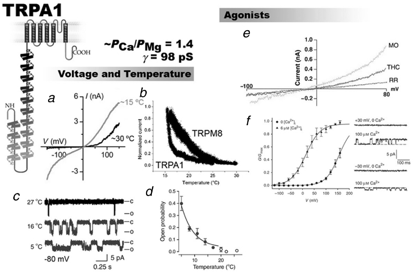
A closer inspection of the predicted S4 segment of TRPV subfamily channels reveals only one basic residue and, unexpectedly, TRPV1, TRPV3, and TRPV4 are outward rectifiers (Nilius et al. Reference Nilius, Karel, Owsianik, Prenen, Droogmans and Voets2005), and TRPV6 behaves as weak inward rectifier in the absence of Mg2+ (Voets et al. Reference Voets, Janssens, Prenen, Droogmans and Nilius2003). More surprising is the fact that TRPA1 is also voltage-dependent, albeit with a lower zδ (~0.4; Karashima et al. Reference Karashima, Talavera, Everaerts, Janssens, Kwan, Vennekens, Nilius and Voets2009), despite the fact that it does not have positively charged residues in its S4 segment. These observations strongly suggest that the actual location of the voltage sensor in TRP channels is still a mystery.
4.2 Agonist binding sites inside TM region
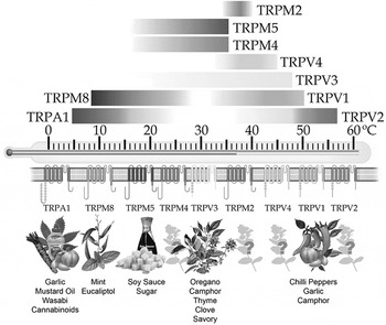
Mammalian TRP channels are also the target of several very diverse series of pungent and other chemical compounds (Caterina et al. Reference Caterina, Schumacher, Tominaga, Rosen, Levine and Julius1997; Patapoutian et al. Reference Patapoutian, Peier, Story and Viswanath2003; Hinman et al. Reference Hinman, Chuang, Bautista and Julius2006; Bandell et al. Reference Bandell, MacPherson and Patapoutian2007; Macpherson et al. Reference Macpherson, Dubin, Evans, Marr, Schultz, Cravatt and Patapoutian2007). Temperature-sensitive TRP channels (thermoTRP) channels are the best example of TRP channels able to act as sensors of a myriad of chemical stimuli. The list of compounds includes capsaicin (active component of hot chili peppers), menthol, icilin, camphor, allicin (one of the active components of garlic), mustard oil, and others, and is increasing every day (Fig. 9) (Voets et al. Reference Voets, Talavera, Owsianik and Nilius2005; Dhaka et al. Reference Dhaka, Viswanath and Patapoutian2006).
A comparison between mammalian and avian (heat-sensitive but capsaicin-insensitive) TRPV1 channels resulted in a map of the structural determinants for capsaicin interaction. The results revealed that residues Y511 and S512, located at the transition between the second intracellular loop and the third TM domain (Jordt & Julius, Reference Jordt and Julius2002), are required for capsaicin binding. Rabbits are also insensitive to capsaicin and the rabbit TRPV1 is about 100-fold less sensitive to vanilloid activation than the human or the rat clone. Comparison between the primary sequences of the rabbit, rat, and human TRPV1 channels confirmed the results of Jordt & Julius (Reference Jordt and Julius2002) that the capsaicin and resiniferatoxin (RTX, an ultra potent vanilloid from the plant Euphorbia resinifera) binding sites lie between TM regions 3 and 4 (Gavva et al. Reference Gavva, Klionsky, Qu, Shi, Tamir, Edenson, Zhang, Viswanadhan, Toth, Pearce, Vanderah, Porreca, Blumberg, Lile, Sun, Wild, Louis and Treanor2004). Gavva et al. (Reference Gavva, Klionsky, Qu, Shi, Tamir, Edenson, Zhang, Viswanadhan, Toth, Pearce, Vanderah, Porreca, Blumberg, Lile, Sun, Wild, Louis and Treanor2004) argue that since the residue Y511 is conserved across species, it cannot explain the difference in vanilloid sensitivity between rat and rabbit TRPV1, and found that a single nonconserved residue is able to restore capasaicin binding (I550T) located in S4. On the other hand, the double mutation I550T/L547M restores the high binding affinity of RTX.
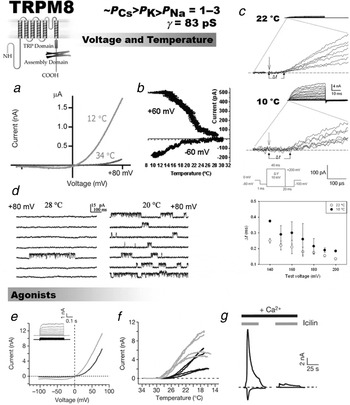
The same species comparison approach revealed the amino acid residues essential for the binding of icilin a super-cooling agent to the cold receptor TRPM8 (Chuang et al. Reference Chuang, Neuhausser and Julius2004). The determinants of icilin sensitivity include residues located in the intracellular loop connecting TM domains 2 and 3. Note that the residues important for icilin sensitivity reside in a position that also correspond to the capsaicin binding site in TRPV1, suggesting that the protein structures involved in agonist interaction appear to be conserved across TRP channel subfamilies. Interestingly, icilin is unable to activate TRPM8 in the absence of external Ca2+ (Fig. 11). Therefore, for full channel activation icilin requires the presence of another stimulus, for example, a decrease in temperature, and in this regard TRPM8 behaves a coincidence detector (Chuang et al. Reference Chuang, Neuhausser and Julius2004). Using a high-throughput assay, which includes and compares thousands of random mutations, Bandell et al. (Reference Bandell, Dubin, Petrus, Orth, Mathur, Hwang and Papapoutian2006) identified two distinct sites in TRPM8 required for sensitivity of the TRPM8 agonist menthol. The main binding site was proposed to be located in the S2 segment (Y745), while the C-terminal site (TRP domain, residues Y1005, L1009) would be required for geometrical specificity and presumably for coupling to the gate opening. Nevertheless, Brauchi et al. (Reference Brauchi, Orta, Mascayano, Salazar, Raddatz, Urbina, Rosenmann, Gonzalez-Nilo and Latorre2007) found that a chimera made of a TRPM8 channel with the C-terminal of TRPV1 has essentially the same menthol sensitivity as the wild-type, suggesting that the C-terminal is not an absolute requirement for menthol binding. This conclusion is supported by the fact that binding studies using radiolabeled menthol demonstrated that the mutation L1009R in the TRPM8 C-terminal resulted in normal radiolabeled menthol binding (Voets et al. Reference Voets, Owsianick, Jansens, Talavera and Nilius2007). Voets et al. (Reference Voets, Owsianick, Jansens, Talavera and Nilius2007) confirmed Y745 as an important residue for menthol binding, but also located a different set of residues on the S4 and inside the S4–S5 intracellular loop that affect the TRPM8 menthol sensitivity and the affinity to radiolabeled menthol. In particular, the mutation of residue R842A, located in S4, decreased both TRPM8 menthol sensitivity (as determined from activation curves) and binding using radiolabeled menthol.
4.3 Gates and dynamic properties of the selectivity filter
If the TRP channel pore follows an architectural design similar to that of the Kv channels, we expect to find a large internal vestibule able to accommodate large organic cations. If this were the case, quaternary ammonium (QA) ions can be used to investigate the gross architecture of this channel region and to determine the location of the structure (gate) in charge of hindering the passage of ions when the channels are closed (Armstrong, Reference Armstrong and Eisenman1975). Jara-Oseguera et al. (Reference Jara-Oseguera, Llorente, Rosenbaum and Islas2008) followed this strategy in the case of the TRPV1 channel and found that QA ions of different sizes, from tetraethylammonium (TEA) to tetrapentylammonium, were able to block the TRPV1 channel when applied internally. These results are consistent with a diameter for the inner vestibule of about 12 Å in its widest section. They further found that closed channels cannot be blocked and that the inner pore can accommodate a TEA molecule when the channel is in its closed configuration – TEA can be trapped inside the channel when it closes. Therefore, as is the case with Kv channels, the evidence suggests the existence of an internally located gate. Clearly, this type of experiments revealed a gate able to hinder the path to large cations of the size of TEA, but does not tell us if this gate also avoids permeation to small cations such as K+, Na+, or Ca2+.
Salazar et al. (Reference Salazar, Jara-Oseguera, Hernandez-Garcia, Llorente, Arias-Olguin, Soriano-Garcia, Islas and Rosenbaum2009) have added a new twist to the TRPV1 gating story by discovering that TRPV1 closing implies the formation of two constrictions in the internal vestibule channel. The most intracellular constriction was found to be at the level of residue L681 of the S6 segment of the rTRPV1 being the one in charge of preventing the passage of large molecules and implying a decrease in the internal vestibule diameter from 12 to about 6 Å. About in the middle of the S6 segment (Y671), they found a second narrowing of the internal vestibule able to restrain the passage to small permeating ions. Experimentally, they used a cysteine-less TRPV1 mutant and the cysteine scanning mutagenesis technique to test the state-dependent accessibility of thiol reagents such as 2-(trimethylammonium) ethyl methenethiosulfonate (MTSET) and 2-aminoethyl methanethiosulfonate (MTSEA). These compounds were used as calipers to determine the approximate diameter of the first constriction these charged molecules encounter when coming from the intracellular side of the channel; with a diameter of 3.6 Å, MTSEA is much smaller that MTSET (~6 Å). State-dependent accessibility was found for MTSET when using the L681C mutant, but not for MTSEA, setting the maximum diameter of the most intracellular constriction at ~6 Å. On the other hand, state-dependent accessibility for the small ion Ag+ can be only detected beyond S6 residue Y671, strongly suggesting that the gating access for small permeant cations is located in this region of the S6 segment. Notably, the S6 segment of TRPV1 lacks the region proline-X-proline found in Kv (Webster et al. Reference Webster, Del Camino, Dekker and Yellen2004) or the glycine residues found in some Ca2+-activated K+ channels (Jiang et al. Reference Jiang, Lee, Chen, Ruta, Cadene, Chait and MacKinnon2002) that allow the S6 helix to attain the bend configuration found in the open channel structures. The PVP region in the S6 of Kv channels aligns well with a conserved tyrosine residue in the S6 of TRPV1 and other channels of the TRPV family. Whether this residue can play the role of a gating hinge remains to be explored further.
Since the early seventies it has been clear that K+ and Na+ channels should contain in some region of their conduction machinery a structure known as a selectivity filter, which is able to endow the channel with the ability to discriminate the nature of the ion trying to go through once the gate opens (for a nice historical account, see Armstrong, Reference Armstrong and Eisenman1975). A fantastic picture of this domain was given by Zhou et al. (Reference Zhou, Morais-Cabral, Kaufman and MacKinnon2001). The determination at 2.0 Å resolution, allowed us to see even ‘a K+ ion disrobing’ (as excitedly stated by Armstrong, Reference Armstrong and Eisenman1975) in the selectivity filter of the KcsA channel. In K+ channel from different families, the selectivity filter sequence constitutes a distinctive signature since the GYGD motif is present in every potassium selective pore. By contrast, TRP channels do not show a clear consensus on their putative selectivity filter sequence. Moreover, most of them do not discriminate between monovalent cations, and for that reason they are categorized as nonselective cation channels (exceptional cases are TRPV5 and TRV6, which are calcium selective). Dogma dictates, on the other hand, that permeability properties of the pore are one structural–functional fingerprint for a given channel and should remain constant under different physiological or experimental conditions. Apparently, as we discuss below, a completely different picture appears to be true for several TRP channels. (The interested reader should consult Owsianik et al. Reference Owsianik, Talavera, Voets and Nilius2006, for a detailed revision regarding TRP channels permeation properties.)
We would like to focus this section on TRP channels pore dilation process. We consider this a remarkable new finding that will force researchers to revise their hypothesis, built considering static pore architectures. Additionally, we note that this finding provides another regulatory element to ion channel function and an element that should be considered in future models for activation.
The work on ATP-gated ion channels of the P2X family suggested that prolonged agonist exposure increased the pore size, leading to enhanced permeability of the channel. This phenomenon, known as pore dilation, has been largely relegated to P2X receptors until now. However, a general mechanism underlying pore dilation on P2X channels remains controversial. Several models have been proposed, including direct changes in the selectivity filter, formation of new channel multimers, and recruitment of a nonselective pore (Fujiwara & Kubo, Reference Fujiwara and Kubo2004; Locovei et al. Reference Locovei, Scemes, Qiu, Spray and Dahl2007; Egan et al. Reference Egan, Samways and Li2006).
During the past few years, various researchers have provided evidence for the existence of a large pore in the case of TRPV1 channels, allowing permeation of large organic cations including spermine, NMDG+, Yo-Pro2+, quaternary amonium ions, gentamycin, and QX-314. These results suggest either a big static pore or changes on their pore diameter during activation (Ahern et al. Reference Ahern, Wang and Miyares2006; Meyers et al. Reference Meyers, MacDonald, Duggan, Lenzi, Standaert, Corwin and Corey2003; Jara-Oseguera et al. Reference Jara-Oseguera, Llorente, Rosenbaum and Islas2008; Binshtok et al. Reference Binshtok, Bean and Woolf2007; Myrdal & Steyger Reference Myrdal and Steyger2005; Hellwig et al. Reference Hellwig, Plant, Janson, Schafer, Schultz and Schaefer2004). Moreover, the detailed study of Chung et al. (Reference Chung, Guler and Caterina2005) showed that the permeability properties of TRPV3 channels change upon stimulation with 2-aminoethyl diphenylborinate (2-APB), diphenylboronic anhydride, and temperature. Several properties such as rectification, permeability, Ca2+ blocking, gating kinetics, and temperature dependence are affected after persistent stimulation.
TRPV5, one of the calcium selective TRP channels, presents a pore dilation process that is dependent of internal and external pH. Using a well conducted set of experiments, Yeh et al. (Reference Yeh, Kim, Jabbar and Huang2005) demonstrated that the pore of TRPV5 channels changes its diameter (giving an estimate of 6–12 Å expansion), that the pore helix twist upon stimulation, and that a residue located on the C-terminal region is responsible for intracellular pH-induced pore dilation. In the same way, TRPV1 undergoes time- and agonist-dependent pore dilation after stimulation, allowing permeation of large organic cations such as Yo-Pro2+ and NMDG+. The channel also presents differential ionic selectivity changes when stimulated by different agonists. One explanation for the dynamic changes in P Ca/P Na may be that it results from altered rates of Ca2+ interactions with the Ca2+ binding sites that are induced by activation-dependent conformational changes, rather than being a consequence of changes in TRPV1 pore size. As in TRPV5, a C-terminal modification in TRPV1 (PKC-dependent phosphorilation) affects selectivity (Chung et al. Reference Chung, Guler and Caterina2008). In the case of TRPV1, different activation modalities affect Ca2+ permeability; for example, Ca2+-driven currents evoked by protons are smaller than those evoked by capsaicin. Site-directed mutagenesis experiments showed that charges D646, E648, and E651, all located on the outer mouth of the pore, are involved in such a process (Samways et al. Reference Samways, Khakh and Egan2008). Pore dilation phenomena appears not to be a property of only TRPV channels since it has recently been shown that TRPA1 activation results in dynamic changes of pore permeability to NMDG+ and Yo-pro2+ uptake. Moreover, TRPA1 agonist such AITC induced Yo-Pro2+ uptake in a concentration-dependent fashion (Chen et al. Reference Chen, Kim, Bianchi, Cavanaugh, Faltynek, Kym and Reilly2009). These changes in TRP channel permeability may have effects on channel function through calcium-dependent desensitization and also on other neighboring proteins that are modulated by intracellular calcium. Interestingly, in cells expressing TRPM8 channels, neither menthol nor icilin induced permeability changes (Chen et al. Reference Chen, Kim, Bianchi, Cavanaugh, Faltynek, Kym and Reilly2009). The fact that pore dilation occurs in TRPA1, but not in TRPM8, may provide an opportunity to understand which are the molecular determinants of pore dilation.
5. C-Terminal domain
5.1 TRP domain, intracellular linkers, PIP2, and allosteric control
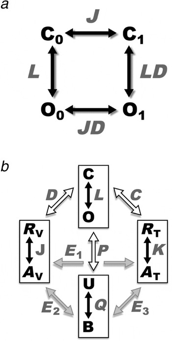
As discussed above, a two-state model (Nilius et al. Reference Nilius, Vriens, Prenen, Droogmans and Voets2004; Voets et al. Reference Voets, Owsianick, Jansens, Talavera and Nilius2007) is the simplest scheme to describe the effects of temperature on TRP channels opening. It is clear, however, that this model represents an oversimplification of the full gating intricacies of voltage-dependent thermoTRPs. In fact, although this two-state scheme describes fairly well the overall equilibrium behavior of voltage-dependent thermoTRPchannels, some results cannot be adequately accounted for by the two-state model. These include results showing that both TRPV1 and TRPM8 can gate independently of voltage (Brauchi et al. Reference Brauchi, Orio and Latorre2004; Matta & Ahern, Reference Matta and Ahern2007) and that the voltage-dependent transitions are poorly temperature-dependent (Latorre et al. Reference Latorre, Vargas, Orta, Brauchi, Liedtke and Heller2007b). These facts are well explained by the use of allosteric models of channel activation (Latorre et al. Reference Latorre, Brauchi, Orta, Zaelzer and Vargas2007a, Reference Latorre, Vargas, Orta, Brauchi, Liedtke and Hellerb; Matta & Ahern, Reference Matta and Ahern2007), and we will describe them in more detail in Section 6. Here, we will try to give a mechanistic vision of the possible nature of the coupling factors that participate in the gating of these channels.
A recent report from García-Sanz et al. (Reference García-Sanz, Valente, Gomis, Fernández-Carvajal, Fernández-Ballester, Viana, Belmonte and Ferrer-Montiel2007) further investigates the role of the TRP domain among TRPV channels. The authors found that chimeras constructed by replacing the TRP domain of TRPV1 with the TRP domain of the other members of this subfamily (TRPV2–TRPV4) affects channel function by altering the sensitivity to the stimuli. TRPV1 channels containing the TRP domain of either TRPV5 or TRPV6 reached the plasma membrane but did not show electrical activity. Notably, the authors suggest that the TRP domain might work as the coupling structure between the stimuli and the activation gate. As discussed earlier in this review, the TRP domain appears to be the, or part of the, tetramerization domain forming coiled coil structures (García-Sanz et al. Reference García-Sanz, Fernández-Carvajal, Morenilla-Palao, Planells-Cases, Fajardo-Sánchez, Fernández-Ballester and Ferrer-Montiel2004). Based on this observation and the results obtained with the TRP domain chimeras, García-Sanz et al. (Reference García-Sanz, Valente, Gomis, Fernández-Carvajal, Fernández-Ballester, Viana, Belmonte and Ferrer-Montiel2007) have proposed a model in which this channel region acts as a negative allosteric modulator. In other words, by forming a coiled coil structure the TRP domain keeps the channel closed. This hypothesis is attractive because if the stimuli (temperature, agonists, etc.) modify the interaction surfaces between subunits, they would probably induce an expansion of the ‘gating ring ’ formed by the C-terminals that would be translated into channel opening. This mechanism would be similar to that proposed for the MthK channel in which the gating ring uses the energy of Ca2+ binding to perform the necessary mechanical work to open the pore (Jiang et al. Reference Jiang, Lee, Chen, Ruta, Cadene, Chait and MacKinnon2002).
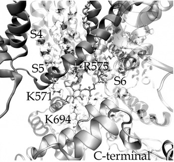
We described TRP channels as 6TM proteins, in which voltage sensing occurs somewhere between segments S1 and S4. That region and perhaps some part of the membrane facing pore region (S5–S6 helices) also participate in agonist binding and, in a still unknown manner, couples the chemical energy of binding to the gating process. Additionally, we know that in both TRPV1 and TRPM8 there is a region inside the C-terminal domain that is responsible for, or at least has the role in, coupling the thermal energy and the PIP2 binding energy to channel opening (Vlachova et al. Reference Vlachova, Teisinger, Susankova, Lyfenko, Ettrich and Vyklicky2003; Rohacs et al. Reference Rohacs, Lopes, Michailidis and Logothetis2005; Brauchi et al. Reference Brauchi, Orta, Salazar and Latorre2006, Reference Brauchi, Orta, Mascayano, Salazar, Raddatz, Urbina, Rosenmann, Gonzalez-Nilo and Latorre2007). Predictions (Clapham, Reference Clapham2003) and subsequent work on PIP2 binding site in TRPM8 (Rohacs et al. Reference Rohacs, Lopes, Michailidis and Logothetis2005) identify the amino acids involved in channel activation by this lipid in the TRP domain. Functional analyses on chimeric channels, molecular dynamics, and docking methods were used to demonstrate that the TRP domain region also serves as a PIP2 binding site in TRPV1 (Brauchi et al. Reference Brauchi, Orta, Mascayano, Salazar, Raddatz, Urbina, Rosenmann, Gonzalez-Nilo and Latorre2007). Molecular modeling shows a close interaction of PIP2 phosphates outside the C-terminal domain specifically with amino acids R575 and R579 located in the S4–S5 linker (of the neighbor subunit), a region of critical importance in the coupling of voltage sensing properties and gate function on potassium channels (Fig. 7; Long et al. Reference Long, Campbell and MacKinnon2005). Interaction of PIP2 with those charges may, in turn, decrease the electrical energy necessary to displace the voltage sensor, thereby promoting the observed leftward shift in the conductance–voltage curve. In this scenario, the C-terminal domain and the VSD are connected trough PIP2, suggesting that phospholipid binding might work as a chemical switch that modulates the activity of the channel. We note here that charged residues located in the S4–S5 intracellular linker of TRPM8 affects voltage sensitivity (although they are supposedly outside the dielectric field!), menthol, and temperature activation (Voets et al. Reference Voets, Owsianick, Jansens, Talavera and Nilius2007). Thus, we speculate that the complex – linker S4–S5/PIP2/TRP domain – may work as an allosteric modulator of TRPV channels, using the phosphate coordination with charged residues as the structure that controls their polymodal activation (Fig. 7). This concept might be extended, at least, to TRPMs and possibly to other TRP channels.
Homology model for the TRPV1 channel reveals a PIP2-binding site. Ribbon diagram of the TRPV1 channel depicting one subunit (S4, S5), a neighboring one (C-terminal), and one bound PIP2 molecule. The model predicts that K571 and R579 in the S4–S5 linker of one subunit as well as K694 in the C-terminal from the neighboring subunit are in close contact with the PIP2 molecule. TRPV1 homology models were built by using as reference structure the crystal structures of Kv1.2 (PDB:2A79) and HCN2 (PDB:1Q43) as templates for TM and C-terminal regions respectively. The construction of topology file of the PIP2 molecule is described by Brauchi et al. (Reference Brauchi, Orta, Mascayano, Salazar, Raddatz, Urbina, Rosenmann, Gonzalez-Nilo and Latorre2007).

5.2 Coiled coil domains and TRP tetramerization
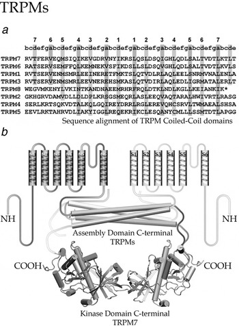
Coiled coil is a protein structure domain in which α-helices wrap around each other to form a particularly stable structure. This structure, first proposed by Crick (Reference Crick1952), can form when the α-helices have most of their hydrophobic amino acids on one side (for the solution structure of a C-terminal tetramerization domain, see Nooren et al. Reference Nooren, Kaptein, Sauer and Boelens1999). In this case, α-helices can twist around each other with their hydrophobic side facing inward forming a supercoil twisted structure. Thus, coiled coil structures function as oligomerization domains for a variety of proteins including structural proteins, transcription factors, motor proteins, and ion channels. The coiled coil structure is highly conserved among different living organisms. Sequences with a propensity to assume coiled coil structures are characterized by a heptad repeat pattern of amino acids (abcdefg)n, where a and d are hydrophobic, and e and g are charged or polar (see Fig. 8a). Coiled coils may interact with each other and with other coiled coil domains (Nooren et al. Reference Nooren, Kaptein, Sauer and Boelens1999). Coiled coil domains have been implicated in subunit interaction and assembly of ion channels, including potassium channels (e.g. Jenke et al. Reference Jenke, Sanchez, Monje, Stuhmer, Weseloh and Pardo2003; Schwake et al. Reference Schwake, Athanasiadu, Beimgraben, Blanz, Beck, Jentsch, Saftig and Friedrich2006). The TRP channels TRPV1 (García-Sanz et al. 2004), TRPM8 (Erler et al. Reference Erler, Al-Ansary, Wissenbach, Wagner, Flockerzi and Niemeyer2006; Tsuruda et al. Reference Tsuruda, Julius and Minor2006), and TRPM2 (Mei et al. Reference Mei, Xia, Beech and Jiang2006) have also been found to assemble in what may be considered a coiled coil-dependent fashion.
The coiled coil structure of the distal C terminus in TRPM channels. (a) Sequence alignments of TRPM coiled coil domains. Heptad repeat pattern of amino acids abcdefg is shown on top of the coiled coil domains and hydrophobic amino acids d and a denoted by vertical rectangles. Note that the coiled coil sequences are ordered according to their length and hydrophobic matching producing the two groups: TRPM7, 6, 1, 3 and TRPM8, 2, 4, 5. (b) The TRPM7 TM domains of two subunits, their respective C-terminals containing the antiparallel coiled coil assembly domain and the dimeric kinase domains are shown. Modified from Fujiwara & Minor (2008).

Coiled coil domains present in the C terminus region immediately after the channel pore were found to be crucial for the proper assembly and function of TRPV1 channels (García-Sanz et al. Reference García-Sanz, Fernández-Carvajal, Morenilla-Palao, Planells-Cases, Fajardo-Sánchez, Fernández-Ballester and Ferrer-Montiel2004). The region between Glu 684 and Arg 721 of the TRP domain of TRPV1 adopts a coiled coil shape that is conserved within the TRPV family. A series of deletion experiments and the use of a dominant negative form of TRPV1 suggest that this region is critical for channel self-association. Coiled coil domains are part of TRPM channels but, in contrast to TRPV1, they are located in the distal part of the C-terminal (Fig. 8a; Launay et al. Reference Launay, Cheng, Srivatsan, Penner, Fleig and Kinet2004; Mei et al. Reference Mei, Xia, Beech and Jiang2006; Erler et al. Reference Erler, Al-Ansary, Wissenbach, Wagner, Flockerzi and Niemeyer2006; Tsuruda et al. Reference Tsuruda, Julius and Minor2006; Fujiwara & Minor, Reference Fujiwara and Minor2008). Truncation of the first 177 amino acid residues of the N-terminal and of the last 160 residues containing the coiled coil domain of the C-terminal domains of the TRPM4 showed that only the latter behaves as a dominant negative, preventing homomultimerization between the wild-type TRPM4 and the ΔC-TRPM4 (Launay et al. Reference Launay, Cheng, Srivatsan, Penner, Fleig and Kinet2004). These results only suggest that the last 160 amino acids of the C-terminal region of TRPM4 are required for multimerization but do not necessarily imply that the coiled coil region of the TRPM4 channel is directly involved in channel assembly. Evidence that this structure is involved in channel function was given by Tsuruda et al. (Reference Tsuruda, Julius and Minor2006), who showed that the coiled coil domain of TRPM8, comprising residues 1064–1104 (last 40 amino acids; Fig. 8a), works as dominant negative when coexpressed with WT channels, and was necessary for channel maturation and trafficking. In addition, when the coiled coil domain is expressed by itself, it is capable of forming tetramers. A dominant-negative can be also produced by attaching the coiled coil region to the S6 TM domain of TRPM8; finally, a single mutation L1089P within a central a position in the coiled coil region results in nonfunctional channels (Erler et al. Reference Erler, Al-Ansary, Wissenbach, Wagner, Flockerzi and Niemeyer2006).
The crystal structure of the coiled coil domain of TRPM7 has recently been elucidated (Fujiwara & Minor, Reference Fujiwara and Minor2008). Despite the fact that TRP channels appear to have a fourfold rotational symmetry around the pore, the homotetrameric crystal structure of TRPM7 coiled coil shows a twofold symmetry. In solution, the TRPM7 coiled coil is a tetramer assembled with an antiparalallel orientation (Fig. 8b): two α-helical strands going in one direction and two strands going in the opposite direction. This twofold symmetry, however, matches the structural symmetry of the kinase domain (Fig. 8b), a dimer held together by an exchange of the N terminus α-helix of each monomer (Yamagushi et al. Reference Yamagushi, Matsushita, Nairn and Kuriyan2001) which follows the C terminus of the TRPM7 coiled coil. Notably, an analysis of the primary structure of the coiled coil domains of other members of this subfamily suggest that they can be divided into two groups: TRPM1, 3, 6, 7, and TRPM2, 4, 5, 8 (see alignments in Fig. 8). The large differences in the coiled coil lengths and the lack of matching between hydrophobic core compositions suggests that the quaternary structure of the coiled coil domain of these two groups will be different. These observations predict that it is unlikely that heterotetramers will form between groups. In other words, coiled coil domains impose restrictions to the type of channel that can be assembled.
It should be noted, however, that deletions of the C-terminal region prevent TRPM8 function but not tetramerization. Indeed, TRPM8 lacking the whole C-terminal region are able to reach the membrane and form tetramers but are nonfunctional (Phelps & Gaudet, Reference Phelps and Gaudet2007). Even in the absence of N- or C-terminal regions, the channel is able to tetramerize and reach the membrane. Combining immunostaining and confocal microscopy, electrophysiology, and biochemistry, Phelps & Gaudet (Reference Phelps and Gaudet2007) also showed that, to be functional, TRPM8 need the coiled coil domain located at the end on the C-terminal as well as a region between the amino acids 40 and 86 of the intracellular N-terminal. It should be noted here that, contrary to what is found in TRPM8, deletion of the TRPM2 coiled coil domain indeed affects assembly and trafficking (Mei et al. Reference Mei, Xia, Beech and Jiang2006).
6. ThermoTRP channels
One of the most fascinating features of TRP channels is the presence in at least three different families of channels exquisitely sensitive to temperature, the thermoTRP channels. Understanding the molecular nature of the temperature-dependent conductance offers a challenge to biophysicists, as attractive as was the unveiling of the molecular nature of the voltage sensor in voltage-dependent channels. In this and the next section, we describe some of the biophysical properties which we speculate account for the temperature sensitivity of these fantastic molecular machines, and we end the review by giving a description of what we have learned from the use of knockout models.
6.1 A brief detour on thermoTRP channels
Nine members of the mammalian TRP ion channels show different temperature thresholds: TRPV1–4, TRPM2, TRPM4, and TRPM5 are heat-activated, whereas TRPM8 and TRPA1 are activated by cold (Fig. 9) (Talavera et al. Reference Talavera, Yasumatsu, Voets, Droogmans, Shigemura, Ninomiya, Margolskee and Nilius2005; Dhaka et al. Reference Dhaka, Viswanath and Patapoutian2006; Huang et al. Reference Huang, Zhang and Mcnaughton2006; Togashi et al. Reference Togashi, Hara, Tominaga, Higashi, Konishi, Mori and Tominaga2006; for a recent review on ThermoTRP channels tissue distribution, agonists, antagonists, and modulators, see Mandadi & Roufugalis, Reference Mandadi and Roufogalis2008). The first member of this class of channels that was cloned was TRPV1 (Caterina et al. Reference Caterina, Schumacher, Tominaga, Rosen, Levine and Julius1997). TRPV1 is expressed mainly in small diameter nociceptive neurons of the trigeminal and dorsal root ganglia (DRG). Capsaicin (the active compound of ‘hot’ chili peppers), protons, depolarizing voltages, and noxious thermal stimuli (~42°C) all activate TRPV1 (Fig. 10) (Caterina et al. Reference Caterina, Schumacher, Tominaga, Rosen, Levine and Julius1997; Voets et al. Reference Voets, Janssens, Droogmans and Nilius2004). The channel is cationic with a relatively high Ca2+ permeability (P Ca/P Na ~10). TRPV1 can be activated by protons at room temperature when the pH is decreased below 6. The decrease in temperature threshold for activation promoted by protons is hypothesized to be involved in inflammatory pain because tissue acidification accompanies ischemia and inflammation (Tominaga et al. Reference Tominaga, Caterina, Malmberg, Rosen, Gilbert, Skinner, Raumann, Basdbaum and Julius1998). TRPV1 is also activated by pungent extracts of onion and garlic, compounds that also activate the TRPA1 channel (Macpherson et al. Reference Macpherson, Geierstanger, Viswanath, Bandell, Eid, Hwang and Patapoutian2005; Salazar et al. Reference Salazar, Llorente, Jara-Oseguera, Garcia-Villegas, Munari, Gordon, Islas and Rosenbaum2008). TRPV2 has a temperature threshold of about 53°C and is expressed in DRG neurons of intermediate size (Caterina et al. Reference Caterina, Rosen, Tominaga, Brake and Julius1999). Its expression, however, is not restricted to the nervous system; it is also found in the spleen and the small intestine. This channel is also permeable to Ca2+ (P Ca/P Na~3) but is capsaicin-insensitive and is not activated by protons. Unlike the other thermoTRPs, TRPV3 and TRPV4 are mainly expressed in keratinocytes (Chung et al. Reference Chung, Lee, Mizuno, Suzuki and Caterina2004). TRV3 is also expressed in the tongue and nose, and TRPV4 in the central nervous system (CNS). Both channels are activated at innocuous temperatures (33–35°C) and are therefore activated at body temperature (Güler et al. 2002; Peier et al. Reference Peier, Moqrich, Hergarden, Reeve, Andersson, Story, Barley, Dragoni, Mclntyre, Bevan and Patapoutian2002a; Smith et al. Reference Smith, Gunthorpe, Kelsell, Hayes, Reilly, Facer, Wright, Jerman, Walhin, Ooi, Egerton, Charles, Smart, Randall, Anand and Davis2002; Watanabe et al. Reference Watanabe, Davis, Smart, Jerman, Smith, Hayes, Vriens, Cairns, Wissenbach, Prenen, Flockerzi, Droogmans, Benham and Nilius2002; Xu et al. Reference Xu, Ramsey, Kotecha, Moran, Chong, Lawson, Ge, Lilly, Silos-Santiago, Xie, DiStefano, Curtis and Clapham2002). Camphor, a natural compound that modulates sensation of warmth in humans, activates TRPV3 in keratinocytes but not sensory neurons (Moqrich et al. Reference Moqrich, Hwang, Earley, Petrus, Murray, Spencer, Andahazy, Story and Patapoutian2005). Components of plants such as carvacrol, eugenol, and thymol strongly activate and sensitize TRPV3 (Xu et al. Reference Xu, Delling, Jun and Clapham2006). TRPM2 is expressed mainly in the brain but is also present in the heart, liver, and pancreas (Perraud et al. Reference Perraud, Fleig, Dunn, Bagley, Launay, Schmitz, Stokes, Zhu, Bessman, Penner, Kinet and Scharenberg2001; Togashi et al. Reference Togashi, Hara, Tominaga, Higashi, Konishi, Mori and Tominaga2006). The channel discriminates poorly between monovalent and divalent cations, and contains a C-terminal domain homologous to the ADP ribose phosphohydrolase (Nudix domain; Perraud et al. Reference Perraud, Fleig, Dunn, Bagley, Launay, Schmitz, Stokes, Zhu, Bessman, Penner, Kinet and Scharenberg2001). TRPM2 is constitutively activated at body temperature (threshold ~35°C at −60 mV) and its activation is strongly potentiated by cyclic ADP ribose. By regulating Ca2+ entry into pancreatic β cells depending on the availability of cyclic ADP ribose, TRPM2 appears to play an important role in regulating insulin secretion. TRPM5 is a cationic channel impermeable to Ca2+ that is selectively expressed in taste receptor cells and has a key role in the perception of bitter, sweet, and umami taste (Pérez et al. Reference Pérez, Huang, Rong, Kozak, Preuss, Zhang, Max and Margolskee2002; Hofmann et al. 2003). TRPM5 is voltage- and temperature-dependent; current increases steeply in the 15–35°C range (Talavera et al. Reference Talavera, Yasumatsu, Voets, Droogmans, Shigemura, Ninomiya, Margolskee and Nilius2005). The temperature dependence of this channel may explain why taste perception in human is modulated by temperature and, in particular, why the perception of sweet is enhanced as temperature is increased. TRPM8, which was cloned independently by two groups and characterized as a cold- and menthol-sensitive receptor (hence its other name, CMR1) (Fig. 11) (McKemy et al. Reference McKemy2002; Peier et al. Reference Peier, Moqrich, Hergarden, Reeve, Andersson, Story, Barley, Dragoni, Mclntyre, Bevan and Patapoutian2002a), is expressed selectively in a small population of primary sensory neurons. It is directly activated by slight cooling (threshold ~22–30°C) and depolarizes sensory neurons (McKemy, Reference McKemy2005; Reid, Reference Reid2005). TRPM8 is also activated by the super-cooling agent icilin and by eucalyptol, albeit with lower efficacy and potency than menthol (McKemy et al. Reference McKemy, Neuhausser and Julius2002). Another compound of interest and of pharmacological importance is 2-aminoethoxydiphenyl borate since it activates TRPV1, TRPV2, and TRPV3, but inhibits the activity of TRPM8 induced by menthol (Hu et al. Reference Hu, Gu, Wang, Colton, Tang, Kinoshita-Kawada, Lee, Wood and Zhu2004). Strong cooling activates (<17°C) TRPA1 channels and has been suggested to be important in the transduction of strong (painful) cooling stimuli (Story et al. Reference Story, Peier, Reeve, Eid, Mosbacher, Hricik, Earley, Hergarden, Andersson, Hwang, McIntyre, Jegla, Bevan and Patapoutian2003; Bandell et al. Reference Bandell, Story, Hwang, Viswanath, Eid, Petrus, Earley and Patapoutian2004); however, the involvement of this channel in the cold sensitivity of sensory neurons has been questioned (Babes et al. Reference Babes, Zorzon and Reid2004; Jordt et al. Reference Jordt, Bautista, Chuang, McKemy, Zygmunt, Hogestatt, Meng and Julius2004; but see Sawada et al. Reference Sawada, Hosokawa, Hori, Matsumura and Kobayashi2007; Karashima et al. Reference Karashima, Talavera, Everaerts, Janssens, Kwan, Vennekens, Nilius and Voets2009; see below). Isothiocyanate compounds, the active component of wasabi and mustard, activates TRPA1 channels, identifying this channel as a molecular target for the pungent action of mustard oils (Bandell et al. Reference Bandell, Story, Hwang, Viswanath, Eid, Petrus, Earley and Patapoutian2004; Jordt et al. Reference Jordt, Bautista, Chuang, McKemy, Zygmunt, Hogestatt, Meng and Julius2004).
Schematic representation of the thermoTRPs that function in temperatures ranging from noxious heat to noxious cold. Proposed membrane topology and functionally important domains are represented. Various plant compounds activate the thermoTRPs: TRPV1 is activated by capsaicin, the pungent ingredient in chilly peppers; TRPM8 is activated by menthol, the cooling compound from mint; TRPA1 is activated by various pungent compounds such as allicin, the active ingredient in garlic, and isothiocyanates, the pungent ingredients found in wasabe.

Polymodal nature of TRPV1. (a) Left panel. Plots showing the normalized conductance in function of voltage at the indicated temperatures. Solid lines correspond to the best fit to Boltzmann functions (Brauchi et al. Reference Brauchi, Orta, Salazar and Latorre2006). Right panel. single-channel currents recorded at different membrane potentials from an oocyte heterologously expressing TRPV1. Notice the progressive reduction in P o with hyperpolarisation (Premkumar et al. Reference Premkumar, Agarwal and Steffen2002). (b) Heat-evoked single channel activity in oocytes expressing TRPV1. The temperature was elevated from 25 to 50°C. Measurable activity was detected at ~42°C. Note the marked increase in burst duration as temperature is increased. Data were obtained from an inside-out patch at a holding potential of −60 mV (Liu et al. Reference Liu, Hui and Qin2003). (c) Protons lower the threshold and increase the magnitude of heat-evoked currents in TRPV1-expressing cells. Currents were normalized to responses at 45°C (pH 7·6) (Tominaga et al. Reference Tominaga, Caterina, Malmberg, Rosen, Gilbert, Skinner, Raumann, Basdbaum and Julius1998). (d) VR1 responds to purified vanilloids and pepper extracts. Activation of TRPV1 by capsaicin and resinferatoxin. Membrane currents were recorded in the whole-cell voltage-clamp configuration. Bars denote duration of agonist application (Caterina et al. Reference Caterina, Schumacher, Tominaga, Rosen, Levine and Julius1997). (e) Concentration dependence of the activation of TRPV1 by camphor (Xu et al. Reference Xu, Blair and Clapham2005).

Polymodal nature of TRPM8. (a) TRPM8 is activated by cold temperature (12°C) and exhibits strong outward rectification (McKemy et al. Reference McKemy, Neuhausser and Julius2002; Chuang et al. Reference Chuang, Neuhausser and Julius2004). (b) Current–temperature recordings at holding potentials of −60 and +60 mV (Brauchi et al. Reference Brauchi, Orio and Latorre2004). Note that currents can be elicited with a threshold of about 18°C at −60 mV, suggesting that temperature sensors can be activated when the voltage sensors are at rest (see panel a). (c) The time course TRPM8 channel activation is well described by an initial lag followed by an exponential current increase. Top: Family of macroscopic TRPM8 ionic currents at two (22 and 10°C) different temperatures elicited in response to the variable depolarizing voltages shown in the pulse protocol. TRPM8 currents were measured using the whole-cell configuration of the patch clamp technique. Below each family of current records selected current records are shown in an enlarged time scale that allows appreciating the initial lag (Δt) of the ionic currents at the two temperatures studied. The single arrow indicates the time at which the test voltage pulse was applied. Below: Delay duration (Δt) is plotted versus test voltage pulse for three different experiments at two different temperatures. The delay has low temperature dependence (Q 10=1·6 at +200 mV). (d) TRPM8 single-channel recordings at two temperatures: 28°C (low P o) and 20°C (high P o). Short- and long-duration closed states can be discerned. Recordings were obtained in cell-attached conditions; patches were performed on HEK293 transfected cells (Latorre et al. Reference Latorre, Vargas, Orta, Brauchi, Liedtke and Heller2007b). (e) Voltage activation curves. Responses evoked by menthol (50 mm, grey) and cold (16 to 8°C, black) show strong outward rectification. Inset, baseline currents (black) and menthol-evoked responses (grey) in nominally Ca2+-free bath solution at room temperature. (f) Temperature–response curves (from 33 to 16°C) were determined for trigeminal neurons in the presence (grey) or absence (black) of 10 mm menthol. Menthol potentiated the size of cold-evoked currents and shifted thermal thresholds from 27·1±0·5 to 29·6±0·3°C. (g) Activation and current desensitization of icilin (2 mm) dependent on calcium (McKemy et al. Reference McKemy, Neuhausser and Julius2002).

6.2 Allosteric models and thermoTRP channel activation
In TRPM8 channels, we found that the data could be explained using an allosteric mechanism for both voltage and temperature dependence (Brauchi et al. Reference Brauchi, Orio and Latorre2004). This type of mechanism assumes the existence of independent temperature and voltage sensors both having resting (R) and activated (A) states coupled to channel opening. For clarity, we discuss first the simple model depicted in Fig. 12a. In this case, in the absence of stimuli the channel is confined to the equilibrium between states C0 and O0, with a small equilibrium constant L that makes channel opening very improbable. The equilibrium between states C0 and C1 represents the activation of voltage sensors and is governed by the voltage-dependent constant J. These two equilibria are coupled by the allosteric factor D, so that when voltage sensors are active, the opening of the channel (equilibrium between states C1 and O1) has a higher equilibrium constant, LD. When the channel is open, the activation of voltage sensors (O0–O1 equilibrium) is JD. Therefore, when the temperature is high, the maximum open probability achievable by depolarization will be determined by the LD value.
An allosteric kinetic model to account for the thermoTRP channel gating. (a) We consider first the simple case in which the concentration of the agonist is zero and the system is at a temperature that is unable to activate the channel, in this case the gating mechanism includes an allosteric interaction between channel opening (C–O), voltage sensor activation (R V–A V). L is the C–O equilibrium constant when the voltage sensor is in the resting state (R V) and J is the R V–A V voltage-dependent equilibrium. D is the allosteric interaction factor such as the C–O equilibrium constant that increases D-fold when the channel opens. (b) A general allosteric gating mechanism includes an allosteric interaction between channel opening (C–O), voltage sensor activation, temperature sensor activation (R T–A T) and agonist activation (U–B). L would be, in this case, the C–O equilibrium constant when voltage sensors and temperature sensors are in the resting state (R V, R T) and the agonist binding sites are unliganded (U); K is the R T–A T temperature-dependent equilibrium constant; and Q is the agonist binding constant defined as Q=[A]/K D, where [A] is the agonist concentration and K D is the dissociation constant. Allosteric interactions of temperature sensors and agonist binding with channel opening are determined by allosteric factors P and C respectively. Allosteric interaction between voltage and temperature sensor, between agonist binding and voltage sensors, and between agonist binding and temperature sensors are determined by allosteric factors E 1, E 2, and E 3 respectively.

It is important to note here that, being a homotetramer, the channel is likely to contain four voltage sensors. However, because there is no evidence about their molecular nature and whether they move independently, we are making a simplifying assumption that the voltage sensors move in concert.
In the more general case shown in Fig. 12b, temperature sensors and voltage sensors are coupled by an allosteric factor E and to channel opening by means of allosteric factors D and C respectively (Latorre et al. Reference Latorre, Brauchi, Orta, Zaelzer and Vargas2007a; Matta & Ahern, Reference Matta and Ahern2007). The model can be generalized to include, for example, the effect of ligand activation governed by an equilibrium constant Q and coupled to channel gating by an allosteric factor P. This type of model has received experimental support and some of the most important results are described below. In TRPV1 and TRPM8, the single channel activity occurs in bursts separated by gaps (Liu et al. Reference Liu, Hui and Qin2003; Brauchi et al. Reference Brauchi, Orio and Latorre2004; Figs 10b and 11 c respectively). In the case of TRPV1, temperature mainly affects burst length (Q 10=32); the gaps (Q 10=7) and single-channel dwell time histograms show the presence of 2–6 closed and 2–5 open components across temperature (Liu et al. Reference Liu, Hui and Qin2003). The individual time constants of the different components show a much weaker temperature dependence with Q 10<3, but their distribution is dramatically modified by temperature. For example, at a low temperature (40°C), short closures are as frequent as long ones, whereas at 50°C the long closures are almost absent from the single channel records, indicating that temperature mainly modifies the equilibrium between the different components rather than their transition rates. More importantly, short openings and long quiescent periods are coupled, indicating that the kinetic scheme is branched and that transitions between open and closed states occur in loops, as is the case of the allosteric models proposed by Brauchi et al. (Reference Brauchi, Orio and Latorre2004) and Matta & Ahern (Reference Matta and Ahern2007) (Fig. 12a and b).
As pointed out by Latorre et al. (Reference Latorre, Brauchi, Orta, Zaelzer and Vargas2007a), having multiple states does not rule out the possibility that both temperature and voltage sensors move simultaneously (i.e., they have infinite cooperativity). There are at least four different sets of data that are in disagreement with this hypothesis but in concordance with the allosteric model in which voltage and temperature sensors are independent structures coupled to channel gating. First, a model with n identical sensors that are highly temperature- and voltage-dependent demands that the voltage at which P o=1/2, V 1/2 be linear in the whole temperature range. In TRPM8, however, V 1/2 saturates at high and low temperatures (Latorre et al. Reference Latorre, Brauchi, Orta, Zaelzer and Vargas2007a); second, the time course of TRPM8 channel activation is well described by an initial lag followed by an exponential increase in ionic current. This delay is shortened as the prepulses reach more depolarized values (Fig. 11c) (Cole-Moore shift; Cole & Moore, Reference Cole and Moore1960), indicating that the channel transits between several voltage-dependent closed states and that large hyperpolarizing voltages will populate those closed states further removed from the open state. The delay, and hence the voltage–dependent steps, show a very weak temperature dependence (Q 10=1.6) strongly suggesting that the voltage and temperature sensors are different entities; third, the results of Brauchi et al. (Reference Brauchi, Orta, Mascayano, Salazar, Raddatz, Urbina, Rosenmann, Gonzalez-Nilo and Latorre2007) clearly demonstrate that voltage sensors can function normally in TRPM8 channel in which specific mutagenesis made in the C terminus abolish temperature sensitivity; and fourth, Matta & Ahern (Reference Matta and Ahern2007) found that voltage does not fully activate TRPM8 and TRV1, as would be the case for channels intrinsically voltage-dependent and temperature and/or agonist act by modifying the voltage-dependent equilibria (see, e.g. Voets et al. Reference Voets, Janssens, Droogmans and Nilius2004). Furthermore, TRPV1 channel in the presence of resiniferatoxin (Winter et al. Reference Winter, Dray, Woods, Yeats and Bevan1990; Caterina et al. Reference Caterina, Schumacher, Tominaga, Rosen, Levine and Julius1997), an extremely potent channel agonist, becomes essentially voltage-independent, a clear demonstration that this thermoTRP channel can gate independently of voltage (Matta & Ahern, Reference Matta and Ahern2007).
From the discussion above it is clear that voltage, temperature, and agonists act separately to modulate channel activity and that remains to be demonstrated quantitatively, which is the degree of allosteric coupling between different sensors. On the other hand, the molecular nature of the temperature sensor is still a mystery and the unveiling of the structures involved in temperature detection will be crucial for determining how the thermal energy is converted into mechanical (pore opening) energy.
6.3 In Search of the temperature sensor
Evidence suggests that the C-terminal plays a role either in containing or strongly modulating the temperature sensor (Brauchi et al. Reference Brauchi, Orta, Salazar and Latorre2006, Reference Brauchi, Orta, Mascayano, Salazar, Raddatz, Urbina, Rosenmann, Gonzalez-Nilo and Latorre2007). In particular, the C-terminal domain of TRPM8 and TRPV1 is modular and confers the channel phenotype regarding temperature sensitivity, channel gating kinetics, and PIP2 sensitivity. A chimera in which the proximal part of the C terminus of TRPV1 replaces the equivalent section of TRPM8 is heat- and PIP2-activated. In addition, PIP2 activation, but not temperature sensitivity, disappears when positively charged amino acid residues contained in the exchanged region are neutralized. These findings and those described above strongly suggest that temperature, voltage, and agonists interact allosterically, and only the proximal region of the C terminus is essential for conferring channel phenotype regarding temperature sensitivity. These results also agree with the TRPV1 C terminus truncation experiments of Vlachova et al. (Reference Vlachova, Teisinger, Susankova, Lyfenko, Ettrich and Vyklicky2003) and Liu et al. (Reference Liu, Ma and Qin2004). In particular, Liu et al. (Reference Liu, Ma and Qin2004) reported that a mutant with a deletion encompassing the last 88 residues of the distal TRPV1 C terminus is fully functional regarding its temperature sensitivity.
The recent results of Grandl et al. (Reference Grandl, Hu, Bandell, Bursulaya, Schimdt, Petrus and Patapoutian2008) show that in the case of TRPV3, mutations in the pore region and the S6 TM domain can render the channel utterly insensitive to temperature. Using the same strategy used to identify residues that are important for menthol binding in the case of TRPM8 (Bandell et al. Reference Bandell, Dubin, Petrus, Orth, Mathur, Hwang and Papapoutian2006), Grandl et al. (Reference Grandl, Hu, Bandell, Bursulaya, Schimdt, Petrus and Patapoutian2008) screened 14 000 random mutant clones and identified three point mutations – I644 and N647 in pore loop and Y661 in S6 – which render TRPV3 temperature-insensitive. Remarkably, these mutations did not modify channel activation by 2-APB. These observations can be most economically accounted for by assuming that thermoTRP channels behave as polymodal allosterically gated receptors. These are exciting findings, but it is clear that we are far from having unequivocally identified the temperature sensor. As is the case of the C terminus of TRPV1 and TRPM8, the pore loop and S6 in TRPV3 can be essential for the gating transition from an upstream temperature-sensing domain. It is also possible that, as pointed out by Grandl et al. (Reference Grandl, Hu, Bandell, Bursulaya, Schimdt, Petrus and Patapoutian2008), the temperature sensor is different in different thermoTRP channels.
The possibility that membrane lipids act as the temperature-sensing molecular machinery cannot at present be dismissed. The data available until now suggest, however, that the membrane lipid composition affects the temperature threshold but not its temperature sensitivity. Compelled by the question of whether the heat sensitivity of TRPV1 is intrinsic to the lipid bilayer, Liu et al. (Reference Liu, Hui and Qin2003) incubated the cells in a high cholesterol solution or altered the amount of cholesterol in the membrane of TRPV1-expressing HEK cells β cyclodestrin (MeβCD). It is well known that cholesterol can affect the physical properties of lipid bilayers by changing the fluidity of the lipid bilayer and can also modulate the function of membrane proteins (e.g. Ikonen, Reference Ikonen2008). In the case of incubating the HEK cells in high cholesterol, Liu et al. (Reference Liu, Hui and Qin2003) found a rightward shift in the TRPV1 channel temperature activation curve toward higher temperatures with no effect on temperature sensitivity. Cholesterol depletion induced by MeβCD, on the other hand, had no effect on the channel heat response.
Morenilla-Palao et al. (Reference Morenilla-Palao, Pertusa, Meseguer, Cabedo and Viana2009) reported that the TRPM8 channel associates with cholesterol-rich membrane domains known as lipid rafts both in sensory neurons and ion heterologous expression systems. This association is favored by glycosylation of an asparagine residue located in the pore-forming loop of the channel. Lipid raft disruption with MeβCD shifted the threshold for current activation by about 3°C toward warmer temperatures, with no change in Q 10. Thus, in two very different lipid environments, the TRPM8 channel has essentially the same temperature sensitivity albeit different temperature activation thresholds, suggesting that membrane fluidity and/or lipid–protein interaction modify the closed–open temperature-independent equilibrium.
6.4 What have we learned from knockout models?
TRPV1. Caterina et al. (Reference Caterina, Leffler, Malmberg, Martin, Trafton, Petersen-Zeitz, Koltzenburg, Basbaum and Julius2000) reported that neurons lacking the TRPV1 channel are severely impaired in their responses to stimuli such as capsaicin, protons, or temperature. Trpv1-null mice are also unable to react to injections of capsaicin or resiniferatoxin into the plantar skin of the hind paw and do not avoid solutions containing capsaicin; moreover, subcutaneous injections of capsaicin do not evoke the typical decrease in body temperature that occurs in the trpv1 +/+ mice (Jancsó-Gábor et al. Reference Jancsó-Gábor, Szolcsányl and Jancsó1970; Caterina et al. Reference Caterina, Leffler, Malmberg, Martin, Trafton, Petersen-Zeitz, Koltzenburg, Basbaum and Julius2000). Actually, this is the first observation suggesting that TRPV1, in addition to its role in detecting the environmental temperature, was also involved in the regulation of body temperature (see below). Simultaneously, Davis et al. (2000) found essentially the same results as those reported by Julius’ group in neurons of trpv1 −/− mice. It is important to note here that, whereas it is clear that trpv1-null mice display deficits in thermally evoked pain, they are not insensitive to noxious heat. Data from Caterina et al. (Reference Caterina, Leffler, Malmberg, Martin, Trafton, Petersen-Zeitz, Koltzenburg, Basbaum and Julius2000) clearly suggest that TRPV1 alone cannot account for all the perception of noxious heat. It is possible, however, that some the residual nocifensive response to noxious heat stimuli in trpv1-null mice is mediated by the capsaicin-insensitive TRPV2 channel. This hypothesis needs to be tested using TRPV2-deficient mice; unfortunately, such mutant mice have not yet been reported in the literature. Thus, TRPV1 appears to be essential for sensitization to thermal stimulus (e.g. Tominaga, Reference Tominaga, Caterina, Malmberg, Rosen, Gilbert, Skinner, Raumann, Basdbaum and Julius1998) but not for the normal sensation of noxious heat.
The finding that trpv1-null mice have a normal body temperature was taken as evidence that this channel is not involved in determining body temperature. However, Steiner et al. (2007) found that TRPV1 has a suppressing effect on body temperature (T b) (for a review, see Montell & Caterina, 2007). Steiner et al. (2007) synthesized and tested a potent (IC50<1 nm) TRPV1 antagonist dubbed AMGO347. Remarkably, this antagonist inhibits TRPV1 regardless of whether channel activation is induced by capsaicin, low pH, or temperature. AMGO347 induces hyperthermia in rat and mice, an effect that, most importantly, is completely abolished when trpv1-null mice are used. The antagonist causes hyperthermia through tail-skin vasoconstriction and thermogenesis without affecting behavioral thermoregulation. The main conclusion of this study is that tonic activation of TRPV1 in the abdominal viscera by unknown factors is responsible for the suppressive effect on T b.
TRPV1 serves as a temperature detector in the CNS in addition to the body surface and gut. TRPV1 channels appear to be involved in the release of vasopressin (VP; antidiuretic hormone) from the neurohypophysis through a temperature-activated depolarization of hypothalamic neurons (Sharif-Naeini et al. 2008). In fact, the VP-releasing hypothalamic neurons express a TRPV1 splice variant that lacks part of the N-terminal (Sharif-Naeini et al. 2008). The VP release serves as a negative feedback to the evaporative heat lost induced by hyperthermia. The thermal activation threshold for TRPV1 in VP neurons was found to be below that found in recombinant systems (35 versus ~43°C) and it is the TRPV1-induced depolarization evoked by hyperthermia that causes VP release. It is clear that the trpv1 gene is involved in the temperature-dependent VP release since trpv1 −/− neurons have an impaired thermosensitivity (Q10=8.5 versus Q 10=19 in wild-type neurons) and an impaired heat-evoked VP release in vivo.
TRPV3. TRPV3 is activated by warm temperatures (>33°C) and it shows an increased response at higher (noxious) temperatures (reviewed by Dhaka et al. Reference Dhaka, Viswanath and Patapoutian2006). Unlike most thermoTRP channels, this ion channel is expressed in keratinocytes (Peier et al. Reference Peier, Reeve, Andersson, Moqrich, Barley, Hergarden, Story, Colley, Hogenesch, Mclntyre, Bevan and Patapoutian2002b). trpv3-null mice present strong deficits in their response to innocuous and noxious heat as demonstrated by behavioral tests (Moqrich et al. Reference Moqrich, Hwang, Earley, Petrus, Murray, Spencer, Andahazy, Story and Patapoutian2005), and the activation of the TRPV3 channel by the agonist camphor in utterly lost in trpv3 −/− keratinocytes. Thus, when animals are made to chose between one plate at room temperature and another heated to 35°C, wild-type mice spend on average 92% of the time on the side heated at 35°C, whereas the trpv3 −/− spent only 64% of the time on the warm side. This difference is highly significant; yet the mutant mice still show a small bias toward the warm side, suggesting that other thermoTRP channels (TRPV4?) may be involved in innocuous thermosensation. On the other hand, mice lacking TRPV3 also show an impaired response to noxious heat as determined by the onset of nociceptive behavior to a tail immersion test or in their avoidance of a hot plate. In the first case, significant differences were observed at temperatures >48°C and in the second differences between wild-type and mutant animals can be only observed at 55°C. Together, these observations strongly suggest that TRPV3 in skin cells are essential for mammalian thermosensation.
TRPV4. TRPV4 is expressed in the circumventricular organs of the mammalian CNS, a site important for osmotic sensing (Liedtke et al. 2000). Using a trpv4-null model, Liedtke & Friedman (Reference Liedtke and Friedman2003) found that the trpv4 −/− mice drank less water and became more hyperosmolar than the wild-type controls. Response to both hyper- and hyposmolar stimuli is impaired in trpv4 −/− mice. Another trpv4 −/− mouse model (Mizuno et al. Reference Mizuno, Matsumoto, Imai and Susuki2003), however, shows a state akin to diabetes insipidus, making clear the need for more detailed water balance studies to establish where the differences between the two models reside. In transfected cells, TRPV4 is only activated by a hypotononic challenge (Guler et al. 2002; Watanabe et al. Reference Watanabe, Davis, Smart, Jerman, Smith, Hayes, Vriens, Cairns, Wissenbach, Prenen, Flockerzi, Droogmans, Benham and Nilius2002). TRPV4 appears, therefore, to be necessary for the normal response to changes in osmotic pressure, and probably plays the role of an osmotic sensor in the CNS (Strotmann et al. 2000, 2003). trpv4 −/− mice were reported to display no change in escape latency from acute painful stimuli on the hot plate or upon radiant paw heating compared to wild-type (Liedtke & Friedman, Reference Liedtke and Friedman2003; Susuki et al. 2003). In contrast to these results, Todaka et al. (Reference Todaka, Taniguchi, Satoh, Mizuno and Suzuki2004) found that TRPV4 knockout mice displayed longer latencies to escape compared to the wild-type from plates heated 35–45°C when hyperalgesia was induced, indicating that this channel determines the sensitivity rather than the threshold for painful heat. Moreover, the data of Lee et al. (Reference Lee, Iida, Mizuno, Suzuki and Caterina2005) show that on a thermal gradient, TRPV4 knockout mice prefer the warmer temperatures and, in a two temperature choice test, trpv4 −/− mice show a strong bias for the higher temperature whereas the wild-type mice fail to discriminate between 30 and 34°C. Lee et al. (Reference Lee, Iida, Mizuno, Suzuki and Caterina2005) also found that knockout mice exhibited longer latencies during acute tail heating and that trpv4 −/− and wild-type mice exhibit similar modifications in behavior on a thermal gradient after paw inflammation. Dhaka et al. (Reference Dhaka, Viswanath and Patapoutian2006) have argued that the inconsistencies between the two set of results may be attributable to differences in the sensitivity to hot plate (paws; Todaka et al. Reference Todaka, Taniguchi, Satoh, Mizuno and Suzuki2004) versus tail flick (Lee et al. Reference Lee, Iida, Mizuno, Suzuki and Caterina2005) assays and/or how these assays are scored. It is also probable that TRPV4 behaves as a mechanosensitive channel in hair cells in the cochlea involved in hearing. trpv4 −/− mice have a delayed onset hearing loss and an increased susceptibility to acoustic injury (Tabuchi et al. Reference Tabuchi, Sususki, Mizuno and Hara2005). TRPV4 also has a role in peripheral nociceptor sensitization in vivo since C-type fibers in TRPV4-deficient mice show a pronounced impairment in their response to an inflammatory cocktail composed of prostaglandin E2 and serotonin. Compared to wild-type mice, C-type fibers of the TRPV4-null mice present a large decrease in spontaneous activity and mechanical threshold, indicating that TRPV4, as well as TRPV1, is involved in nociceptor sensitization (Cheng et al. Reference Cheng, Yang, Takanishi and Zheng2007).
TRPM8. The ability to sense a decrease in temperature is fundamental for the survival of warm-blooded animals. However, the ionic mechanisms of nonnoxious cold transduction have remained elusive until recently and, in particular, the contribution of TRPM8 to cold detection in vivo was unclear (see, e.g., Viana et al. Reference Viana2002; Madrid et al. Reference Madrid, Donovan-Rodríguez, Meseguer, Acosta, Carlos and Viana2006). This situation has recently changed radically with the appearance of three reports in which the ability of TRPM8-deficient mice to detect cold temperatures was studied (Bautista et al. Reference Bautista, Movahed, Hinman, Axelsson, Sterner, Hogestatt, Julius, Jordt and Zygmunt2005; Colburn et al. Reference Colburn, Lubin, Stone, Wang, Lawrence, D'andrea, Brandt, Liu, Flores and Qin2007; Dhaka et al. Reference Dhaka, Murray, Mathur, Earley, Petrus and Patapoutian2007). These studies show that mice lacking TRPM8 show severe behavioral deficits in response to cold. The TRPM8-deficient mice show a reduction in their ability to discriminate between cold and warm temperatures, lack behavioral response to cold-induced icilin application, and exhibit a significant reduction in the nocifensive reaction to acetone-evoked evaporating cooling of the hind paw. TRPM8 appears to contribute to the detection of noxious cold, defined as temperatures below 15°C since even at these low temperatures the mutant mice explored the cold surface of a plate for longer times than did wild-type mice (Bautista et al. Reference Bautista, Movahed, Hinman, Axelsson, Sterner, Hogestatt, Julius, Jordt and Zygmunt2005). However, TRPM8 null mice are still able to react with a normal nocifensive reaction to temperatures below 0°C, suggesting the presence of a different receptor able to sense more extreme noxious cold temperatures (Dhaka et al. Reference Dhaka, Murray, Mathur, Earley, Petrus and Patapoutian2007). It can be also that noxious cold is elicited by inhibition of warm-sensing fibers. In this regard, an interesting observation is reported by Roza et al. (Reference Roza, Belmonte and Viana2006), who found that cold-insensitive sensory fibers developed sensitivity to cold when exposed to the K+ channel blocker 4-amino pyridine, an effect that was not modified by menthol. At the cellular level, in the TRPM8 null mice there is a selective loss of TRPM8 channels in dorsal root and trigeminal ganglia, cornea, and spinal cord. On the other hand, sensory nerve fibers from TRPM8-deficient mice show a diminished firing rate and the sensory neurons of these rodents are totally insensitive to menthol and icilin, and have a much diminished response to cold.
TRPA1. TRPA1 channels are expressed in the sensory epithelium of the utricle, a membranous structure located in the vestibule of the inner ear. In addition, antibodies directed against the TRPA1 C-terminal react with mouse vestibular and cochlear hair cells (reviewed by Corey, 2006). Because of its site of expression, the appearance of TRPA1 mRNA in hair cell epithelia simultaneously with the onset of mechanosensitivity, and its biophysical and pharmacological properties prompted Corey et al. (2004) and Nagata et al. (2005) to propose that this channel plays a central role in hearing by mediating hair cell transduction (but see below). TRPA1 is also expressed in DRG neurons, which behave as polymodal nociceptors since they also express TRPV1 (Story et al. Reference Story, Peier, Reeve, Eid, Mosbacher, Hricik, Earley, Hergarden, Andersson, Hwang, McIntyre, Jegla, Bevan and Patapoutian2003; Bandell et al. Reference Bandell, Story, Hwang, Viswanath, Eid, Petrus, Earley and Patapoutian2004; Jordt et al. Reference Jordt, Bautista, Chuang, McKemy, Zygmunt, Hogestatt, Meng and Julius2004).
6.4.1 The puzzle of the cold sensitivity of TRPA1 channels
Conflictive results on the exact role of this channel started when this channel was identified in sensory neurons that were also activated by capsaicin. Jordt et al. (Reference Jordt, Bautista, Chuang, McKemy, Zygmunt, Hogestatt, Meng and Julius2004), in contrast to the findings of Story et al. (Reference Story, Peier, Reeve, Eid, Mosbacher, Hricik, Earley, Hergarden, Andersson, Hwang, McIntyre, Jegla, Bevan and Patapoutian2003), were unable to obtain reliable responses of TRPA1 expressing neurons to noxious cold. The Patapoutian and Julius groups (and everybody else) agreed, however, that TRPA1 is the molecular target of pungent natural compounds as are, for example, the isothiocyanates contained in mustard oil and wasabi (Fig. 13 and see above). In agreement with the results of Jordt et al., Nagata et al. (2005) also reported a lack of sensitivity to noxious cold of the TRPA1 channel. Furthermore, two groups have reported that TRPA1 is activated directly by internal Ca2+ and that the apparent cold sensitivity occurs indirectly since there is a cytosolic Ca2+ increase during cooling in heterologous systems (Doemer et al. 2007; Zurborg et al. 2007; Fig. 13f). Whether TRPA1 is actually gated by noxious cold still leaves a big question mark, but gains support with the results of Sawada et al. (Reference Sawada, Hosokawa, Hori, Matsumura and Kobayashi2007), which show that TRPA1 can be activated by deep cooling in inside-out patches in Ca2+-free solutions (Fig. 13c and d). The results should, nevertheless, be contrasted with those of Zurborg et al. (2007), who were unable to activate TRPA1 in the absence of internal Ca2+. Moreover, they found that in the presence of Ca2+, cold inhibited the TRPA1 activation induced by the divalent cation. We note here that the experiments carried out by Zurborg et al. (2007) were performed in the absence of Ca2+ but used whole-cell patch clamping, whereas Sawada et al. (Reference Sawada, Hosokawa, Hori, Matsumura and Kobayashi2007) conducted their single-channel current recordings in inside-out patches. Sawada et al. (Reference Sawada, Hosokawa, Hori, Matsumura and Kobayashi2007) argued that the differences between their observations and those of Jordt et al. (Reference Jordt, Bautista, Chuang, McKemy, Zygmunt, Hogestatt, Meng and Julius2004) and Nagata et al. (2005) could be attributable to the length of the cooling stimuli (activation of TRPA1 by cooling is slow) or the transfection procedures (cell expressing tetracycline-inducible mTRPA1 versus transient transfected cells). More recently, Karashima et al. (Reference Karashima, Talavera, Everaerts, Janssens, Kwan, Vennekens, Nilius and Voets2009) have confirmed and extended Sawada et al.’s (Reference Sawada, Hosokawa, Hori, Matsumura and Kobayashi2007) findings. In particular, they showed that in Ca2+-free solutions, increasing temperature has the effect of shifting the conductance–voltage relation leftward, a behavior reminiscent to that of TRPM8 (Fig. 13a).
Polymodal nature of TRPA1. (a) Current–voltage relations obtained at ~30°C (black trace) ~15°C (grey trace) using Ca2+-free intracellular and extracellular solutions (Karashima et al. Reference Karashima, Talavera, Everaerts, Janssens, Kwan, Vennekens, Nilius and Voets2009). (b) Comparison of the temperature activation curves for TRPA1 and TRPM8 (Story et al. Reference Story, Peier, Reeve, Eid, Mosbacher, Hricik, Earley, Hergarden, Andersson, Hwang, McIntyre, Jegla, Bevan and Patapoutian2003). (c) Single-channel recordings show that TRPA1 is activated by deep cooling. Inside-out patch recordings (−80 mV) in membrane excised from TRPA1-expressing HEK293 cells. (c, closed state; o, open state). Downward deflections show inward current. (d) Open probabilities (P o) of the deep cooling-induced currents for 5–30 s, data as shown in (c) are plotted against temperature (Sawada et al. Reference Sawada, Hosokawa, Hori, Matsumura and Kobayashi2007). (e) THC and mustard oil activate a subset of sensory neurons. Electrophysiological response of a representative neuron to 20 mm THC or 20 mm mustard oil (MO) in the absence (MO) or presence (RR) of 5 mm ruthenium red. Voltage ramps from −100 to +80 mV (200 ms) were applied every 2 s. THC-evoked currents display slight outward rectification. Mustard oil activates identical currents of larger amplitude in the same cell. Ruthenium red (10 mm) blocks THC- and mustard oil-evoked currents (Jordt et al. Reference Jordt, Bautista, Chuang, McKemy, Zygmunt, Hogestatt, Meng and Julius2004). (f) Left panel. TRPA1 is activated by intracellular Ca2+. Voltage activation curves are shown in the absence and presence of Ca2+. Right panel. Single-channel recordings from inside-out patches at the indicated potentials and internal Ca2+ concentrations (Zurborg et al. Reference Zurborg, Yurgionas, Jira, Caspani and Heppenstall2007).
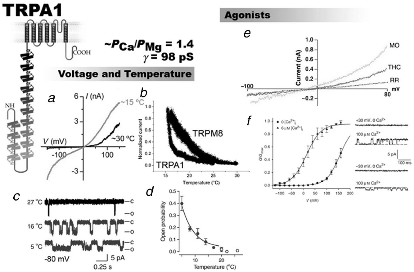
Unfortunately, the answer to this puzzle did not come from trpa1 knockout mice because the behavioral responses of trpa1 −/− knockout mice to noxious cold temperatures are also conflicting (e.g. Kwan & Corey, Reference Kwan and Corey2009). Kwan et al. (Reference Kwan, Allchorne, Volltrath, Christensen, Zhang, Woolf and Corey2006) reported that trpa1 −/− knockout mice are deficient in their behavioral responses to 0°C and mustard oil, but that their hearing is normal. Although these results clearly support the role of TRPA1 as the noxious cold detector, they are difficult to reconcile with the proposal that this channel is the transduction channel in hair cells (Corey et al. 2004). In agreement with Kwan et al. (Reference Kwan, Allchorne, Volltrath, Christensen, Zhang, Woolf and Corey2006), Karashima et al. (Reference Karashima, Talavera, Everaerts, Janssens, Kwan, Vennekens, Nilius and Voets2009) showed that there is a subset of cold-sensitive trigeminal ganglion neurons that are not present in the trpa1 knockout mice, and cold plate and tail-flick behavioral experiments revealed TRPA1-dependent, cold-induced, nociceptive behavior in mice.
However, Bautista et al. (Reference Bautista, Jordt, Nikai, Tsuruda, Read, Poblete, Yamoah, Basbaum and Julius2006) found that the cold- or menthol-evoked responses in trigeminal neurons were identical in ganglia of wild-type and trpa1 −/− mice. Behaviorally, TRPA1-deficient mice showed the same responses as wild-type mice to evaporative cooling and paw withdrawal from a surface cooled from 20 to −10°C. The knockout TRPA1 model reported by Bautista et al. (Reference Bautista, Jordt, Nikai, Tsuruda, Read, Poblete, Yamoah, Basbaum and Julius2006), as is the case of the trpa1 −/− mice reported by Kwan et al. (Reference Kwan, Allchorne, Volltrath, Christensen, Zhang, Woolf and Corey2006), have normal auditory responses. The use of trpa1 −/− mice is also important in demonstrating that the target of the active compounds of garlic and mustard oil is the TRPA1 channel, but given the results of Salazar et al. (Reference Salazar, Llorente, Jara-Oseguera, Garcia-Villegas, Munari, Gordon, Islas and Rosenbaum2008) that showed activation of TRPV1 by pungent extracts of onion and garlic, TRPA1 does not appear to be the sole target.
We conclude that the most recent evidence tends to support direct activation of TRPA1 by cold. In particular, the experiments of Sawada et al. (Reference Sawada, Hosokawa, Hori, Matsumura and Kobayashi2007) and those of Karashima et al. (Reference Karashima, Talavera, Everaerts, Janssens, Kwan, Vennekens, Nilius and Voets2009) demonstrating that TRP1 can be activated by noxious cold in cell-free patches are very convincing.
7. Conclusion
Despite the considerable advances in the TRP channel field that have occurred during the past few years, we are still missing most of the pieces that will give us the solution to the puzzle of how these molecular machines are able to integrate all the stimuli that modify their electrical behavior. It is clear, as is the case of voltage-dependent K+ channels, that TRP channel-forming proteins are modular in nature and that the existence of these modules determines, for example, their modulation by agonists and their ion and selectivity properties. This modularity may help provide a structural basis for the notion, which is well supported by mutagenesis and electrophysiological evidence, that TRP channels are allosterically gated polymodal receptors. Mutagenesis experiments have also substantially helped in the localization of the binding sites for several agonists, but how the free energy of ligand binding is converted into mechanical energy (pore opening) remains a mystery. Some data, however, suggest that in the case TRPA1, the pore region is involved in coupling the covalent binding of electrophilic compounds to channel gating. The pore domain has attracted much attention lately because agonists are able to modify channel gating and evoke ionic selectivity changes. In other words, the pore is not a static structure but its properties are dynamically modulated by agonists during channel activation. The crystal structures of the ARD domains of TRPV channels and the coiled coil domain present in the C terminus of TRPM7 have provided important hints about channel assembly and the mechanisms that lead to the modulation of TRPV1 responses to noxious stimuli. The first steps toward the elucidation of the TRP channel structure have been provided using the electron microscopy and the conclusion of these observations is that the different TRP channel subfamilies may not share the same structural design.
Acknowledgements
We thank Dr. John Ewer for helpful comments on the manuscript and Ms. Luisa Soto for excellent technical assistance. The authors gratefully acknowledge grant support from FONDECYT (#11070190 for S.B. and #1070049 for R.L.).