The diagnosis of Kawasaki disease in an infant less than 6 months of age is difficult as they rarely fulfil the standard diagnostic criteria. This group of children with incomplete Kawasaki disease is at high risk of developing coronary artery abnormalities if not treated early.Reference You, Hye and Sung1 Therefore, it is crucial to have a high index of suspicions and to make an early diagnosis as this systemic vasculitis potentially can cause coronary artery aneurysms in up to 25% if left untreated, and rarely systemic aneurysms that can be seen in 2.2% of the Kawasaki disease cases.Reference Newburger, Takahashi and Gerber2 Here, we report a case of an infant with incomplete Kawasaki disease with multiple coronary and systemic arteries aneurysms complicated with internal iliac arteries thrombosis.
Case report
A previously well and thriving 2-month-old girl was referred to our Paediatric Cardiology Unit on day 21 of illness following a history of persistent fever with appearance of a rash on day 4 of fever, lymph nodes swelling, and indurated Bacillus Calmette–Guérin scar. However, there were no conjunctivitis, oral, or extremity changes. She was initially treated at the referring hospital for infective lymphadenitis but noted to have a persistent fever for more than 2 weeks despite multiple courses of antibiotics. Initial echocardiography on day 15 of illness showed normal coronary arteries. Doppler ultrasound abdomen was done because of the persistent fever showed fusiform dilatation of bilateral common iliac, internal iliac, and common femoral arteries with partial thrombus within the right internal iliac artery. She was then referred to us for suspected atypical Kawasaki disease with systemic arteries aneurysms complicated with internal iliac artery thrombosis.
Upon our assessment, the child was afebrile for the past 48 hours. She was irritable, there were red lips, indurated Bacillus Calmette–Guérin scar; however, there was no desquamation noted. Her temperature 37.2 °C, blood pressure 75/48 mmHg, respiratory rate 40 breaths per min, and heart rate was 140 beats per minute with palpable all distal pulses. Her other examination findings were unremarkable.
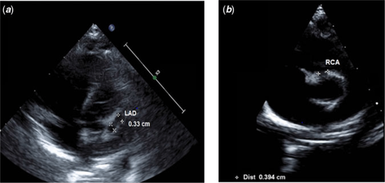
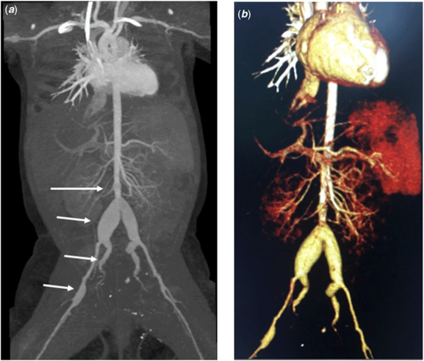
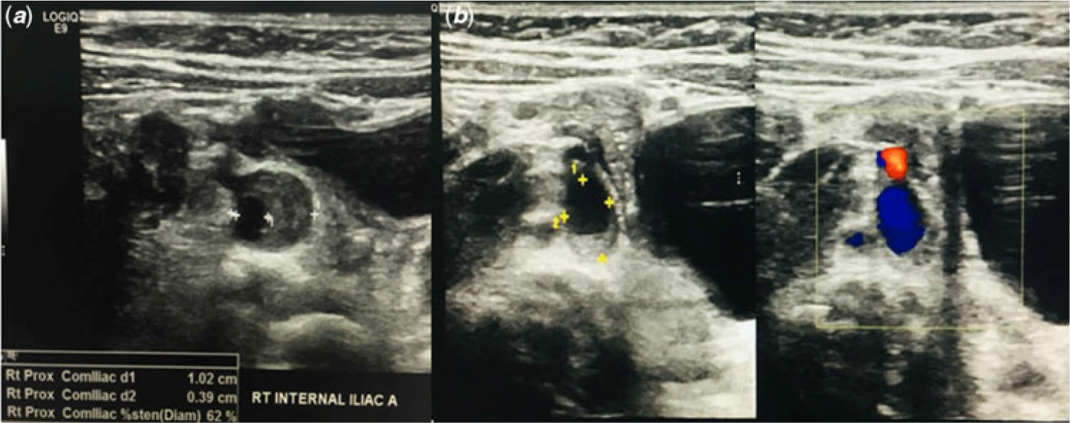
The investigations that were done were as follows: haemoglobin 13.7 g/dL, platelet 399 × 109/L, white blood cells 6.3 × 10⁹/L, C-reactive protein 108.75 mg/L, erythrocyte sedimentation rate 90 mm/hour, albumin 22 g/L, urine culture and sensitivity: sterile pyuria. Repeated echocardiography showed left anterior descending artery (LAD) aneurysm 3.3 mm (z-score: 7.49) (Fig 1) and dilated right coronary artery (RCA) 3.9 mm (z-score: 8.47). These findings were confirmed by the CT angiography which also revealed saccular aneurysm at the level of inferior mesenteric artery (0.8 × 0.6 cm), fusiform dilatation of bilateral common iliac arteries measuring 1.5 × 1.3 cm (right) and 1.0 × 0.9 cm (left), bilateral internal iliac arteries (0.6 cm on both sides), and right common femoral artery aneurysms (0.57 cm) with suspicious filling defect seen at both internal iliac arteries suggestive of thrombus (Fig 2). Her Doppler ultrasound demonstrated that the thrombus causes partial occlusion (62%) of both internal iliac arteries (Fig 3a).
(a and b) Echocardiography images showing the left anterior descending artery (LAD) aneurysm and dilated right coronary artery (RCA).

(a and b) CT angiography showing saccular anerysm at the level of inferior mesentric artery (0.8 × 0.6 cm) and fusiform dilatation of bilateral common iliac arteries measuring 1.5 × 1.3 cm (right) and 1.0 × 0.9 cm (left), bilateral internal iliac arteries (0.6 cm on both sides), and right common femoral artery aneurysms (0.57 cm).

(a) Vascular ultrasound demonstrating thrombus at right internal artery causing partial occlusion (62%). (b) Vascular ultrasound showing small residual thrombus on the right internal iliac artery measuring 0.3 cm in thickness (previously 1 cm).

She was given a high dose of intravenous immunoglobulin (2 gm/kg over 10 hours) and was started on aspirin 25 mg daily (3 mg/kg/day). To prevent further thrombus formation, she was started on intravenous heparin infusion (20 unit/kg/hour) with aim activated partial thromboplastin time twice than the baseline. A comprehensive discussion with vascular surgeons for the management of the internal iliac artery thrombosis decided for conservative management as surgical intervention may carry higher risks of morbidity and mortality given the age of the patient. It was decided that alteplase (tissue plasminogen activator) would be given only if any evidence of limb ischaemia due to risk of bleeding.
Following intravenous immunoglobulin infusion, the child was less irritable and repeated inflammatory markers were normalised. The intravenous heparin infusion was then substituted to subcutaneous enoxaparin 8 mg twice daily (1.5 mg/kg/day). She was allowed discharged well after 16 days of admission with aspirin and subcutaneous enoxaparin.
Following discharged, she remained well with no complication from the antithrombotic therapy. Her femoral and distal pulses were well felt. The coronary artery aneurysm was resolved upon clinic review at 2 months. The RCA: 1.70 mm (z-score: 0.90) and LAD: 1.70 mm (z-score: 1.10). Her vascular ultrasound showed unchanged dilatation over the common iliac arteries measuring 1.4 cm (right) and 0.8 cm (left) in diameter and both internal iliac arteries with small residual thrombus on the right internal iliac artery measuring 0.3 cm in thickness (previously 1 cm) and resolved left iliac artery thrombus (Fig 3b).
Discussion
Kawasaki disease has been reported as the leading cause of acquired heart disease in developed countries.Reference Newburger, Takahashi and Gerber2 It is an acute, self-limiting vasculitis predominantly occur under 5 years of age with the highest incidence in East Asian children. The diagnosis of Kawasaki disease in a younger infant is challenging due to fewer clinical criteria and no specific diagnostic test. Most often, the diagnosis was delayed resulting in late treatment with intravenous immunoglobulin as seen in our case. Ideally, intravenous immunoglobulin should be given in the first 10 days of illness to reduce the risk of severe cardiovascular complications effectively. A study by Yoon et al. reported that younger infants less than 6 months had a higher rate of incomplete Kawasaki disease and cardiac complication with 23 versus 2.9% and 30 versus 12%, respectively, compared to the older children group.Reference You, Hye and Sung1 The most devastating cardiac complication is coronary artery aneurysm that may lead to myocardial infarction and sudden death. Other reported complications include myopericarditis, pericardial effusion, arrythmia, and valvular regurgitation.
Systemic medium- or large-sized artery involvement has been rarely reported, but their presence suggestive of severe form of Kawasaki disease’s vasculitis may increase the likelihood of severe cardiac sequelae.Reference Mario, Michael, Benjamin and Shyam3 Other differentials of systemic arteries aneurysms in children are infective vasculitis, Ehlers-Danlos Syndrome, Takayasu’s arteritis, fibromuscular dysplasias, and congenital-idiopathic aneurysms.Reference Rajabrata, Arnold and Robert4 The risk factors for severe course of illness as seen in this case include younger age, a longer duration of fever, the elevated C-reactive protein, erythrocyte sedimentation rate, hypoalbuminaemia, and sterile pyuria.Reference Ram Krishna, Sundaram and Dhanalakshmi5 In younger patients with incomplete Kawasaki disease, the late diagnosis leading to delay in intravenous immunoglobulin administration is also contributing to the establisment of coronary and systemic aneuryms.Reference Brian, Anne and Jane6 A genetic component to the disease pathogenesis could be analysed as there has been increasing evidence that an individual’s immune response to the inciting event or pathogen may exert a significant influence on the severity of presentation in Kawasaki disease based on their genetic predisposition.Reference Sadeep, Howard and Brahim7
The iliac arteries thrombosis improved with heparin infusion and prevention of further thrombus formation is crucial to avoid vascular occlusion. The choice of anti-antithrombotic therapy for this patient took into account the patient’s age and multiple aneurysms. Aspirin with low-molecular weight heparin was chosen as administering warfarin will be tedious in a small infant.Reference Brian, Anne and Jane6 A patient with involvements of the systemic arteries should undergo long-term serial assessments of both their coronary and peripheral arteries.Reference Rajabrata, Arnold and Robert4, Reference Brian, Anne and Jane6 The decision to intervene surgically in this patient should be considered in preventing any aneurysm-related complications, for instance, in the presence of stenosis or dissections.Reference William, Matthew and Jeffrey8 The outcomes of systemic arteries aneurysms depend on their diameter in the acute phase and they resemble those of coronary artery lesions whereby some may regress (4%) and others persist (0.4%).Reference Kei, Toshiaki, Yuki, Nobuyuki and Shiro9–Reference Hoshino, Etsuko and Osamu11
In conclusion, the potential for coronary and systemic arteries complication following severe vasculitis in incomplete Kawasaki disease highlights the importance of careful examination and a high index of suspicions for early diagnosis and treatment. Incomplete Kawasaki disease should be suspected in any young infant with persistent high-grade fever even if the main clinical features are not adequately presented to avoid unnecessary delay in diagnosis that may lead to serious cardiovascular complications and mortality.
Acknowledgements
The authors would like to acknowledge the staff of Paediatric Cardiology Unit of Serdang Hospital for managing this patient.
Financial Support
This research received no specific grant from any funding agency, commercial, or not-for-profit sectors.
Conflicts of Interest
None.