Introduction
During the Ordovician, the region of Baltoscandia was occupied by the epicontinental Baltic palaeobasin (Männil, Reference Männil1966; Jaanusson, Reference Jaanusson1973). Its deposits are exposed or can be accessed by boreholes in Northeastern Europe, including the northwest of Russia (Männil, Reference Männil1966; Paškevičius, Reference Paškevičius1997). Within the paleobasin, different lithofacies are arranged in facies zones (according to Männil, Reference Männil1966) or confacies belts (according to Jaanusson, Reference Jaanusson and Bassett1976), which are characterized by comparatively stable complexes of litho- and biofacies. Only the two outer confacies belts contain Dapingian bryozoans (i.e., the North Estonian belt, including the north Estonia and Leningrad Oblast’ of north-western Russia) (Bassler, Reference Bassler1911; Modzalevskaya, Reference Modzalevskaya1953; Männil, Reference Männil1959; Dzik, Reference Dzik1981; Goryunova, Reference Goryunova [Gorjunova]1985, Reference Goryunova [Gorjunova]1992, Reference Goryunova [Gorjunova]1996; Goryunova and Lavrentjeva, Reference Goryunova [Gorjunova] and Lavrentjeva1987, Reference Goryunova [Gorjunova] and Lavrentjeva1993; Taylor and Rozhnov, Reference Taylor and Rozhnov1996; Pushkin and Popov, Reference Pushkin and Popov2001; Gorjunova, Reference Gorjunova2009; Koromyslova, Reference Koromyslova2011), and the Lithuanian belt, where numerous bryozoans showing strong secondary dolomitization preventing their taxonomic determination have been found in boreholes in northern Belarus (Pushkin, Reference Pushkin, Bogdanova and Khozatsky1987). Outside Baltoscandia, records of Dapingian bryozoans are rather scarce. They are known from the Kanosh Formation of western Utah, United States, and the Tourmakeady Limestone of Ireland (Taylor and Curry, Reference Taylor and Curry1985; Ernst et al., Reference Ernst, Taylor and Wilson2007; Buttler et al., Reference Buttler, Wyse Jackson, Ernst and McKinney2013; Ernst, Reference Ernst2018); the taxonomic composition of bryozoans at these localities differs from that in northwestern Russia and Estonia.
The Dapingian host sediments in both belts of the Baltic paleobasin are represented mostly by a solid glauconitic limestone, from which fossils are very difficult to extract. As a result, the Dapingian bryozoans here have been poorly studied and their taxonomic composition remains unclear. Fortunately, clayey-calcareous mudmounds, called ‘Hecker-type mudmounds’ (Dronov and Fedorov, Reference Dronov and Fedorov1994), with compacted clayey mud in the cores are occasionally found within the solid limestone; the mud contains numerous small fossils showing good preservation, including small bryozoans. New data on bryozoans from one of the Dapingian Hecker-type mudmounds in northwestern Russia are presented here.
The aims of this paper are: (1) to describe new bryozoan taxa from the Dapingian of northwestern Russia using a combination of scanning electron microscopy (SEM), X-ray microcomputed tomography (micro-CT), and traditional light microscopy of thin sections; (2) to discuss the distribution of early cystoporate bryozoans; and (3) to discuss the paleoecology of the studied bryozoans.
Geological setting
Ordovician deposits are widespread in the vicinity of St. Petersburg. Ordovician limestones and dolomites form the west-east trending elongated Ordovician Plateau elevated above the surrounding terrain (Fig. 1.1). These carbonate rocks dip at a very low angle (2–3 m/km) to the S or SE (Selivanova and Kofman, Reference Selivanova and Kofman1971) and underlie sandy and clayey Devonian deposits. A Cenozoic erosional escarpment, known as the Baltic-Ladoga Klint (Lamanskiy, Reference Lamanskiy1905), restricts the Ordovician strata in the north. The plain north of the Baltic-Ladoga Klint consists mostly of clayey rocks of Ediacaran and Terreneuvian age. The edge of the Klint consists of glauconitic limestone, which forms the base of the carbonate section of the Ordovician Plateau. Outcrops of the glauconitic limestone are confined to river valleys crossing the Baltic-Ladoga Klint and to limestone quarries.
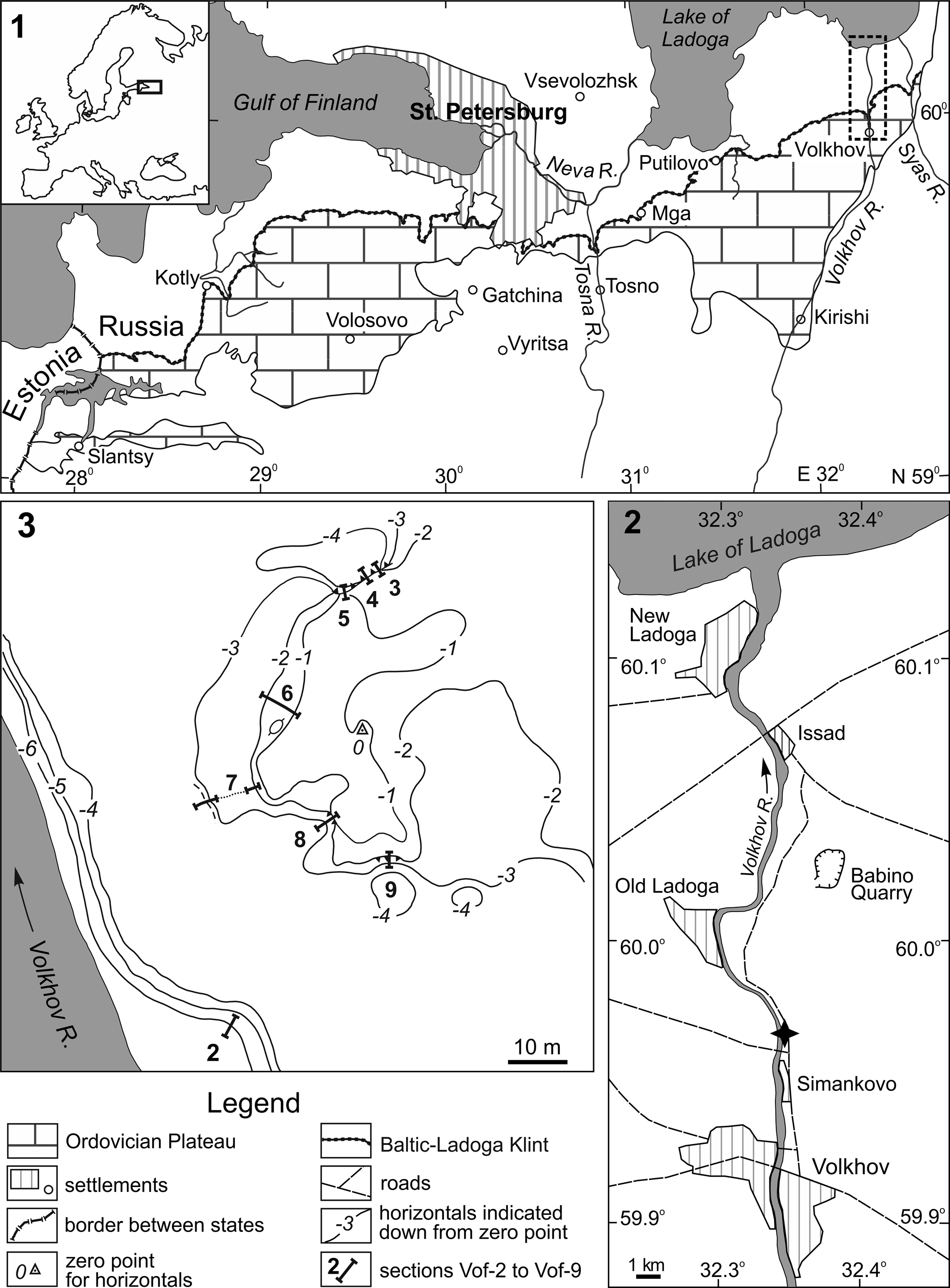
Location and surface topography of the studied Hecker-type mudmound. (1) Schematic geological map of the St. Petersburg area modified after Sokolova (Reference Sokolova and Selivanova1971); insert (2) is shown with a dashed rectangle; (2) schematic map of the lower course of the Volkhov River showing locations of the mudmound and the Babino limestone quarry; (3) topographical plan of the mudmound residual, showing stratigraphic sections. Note: contour lines are starting down from maximal elevation of the hill; contour interval 1 meter.

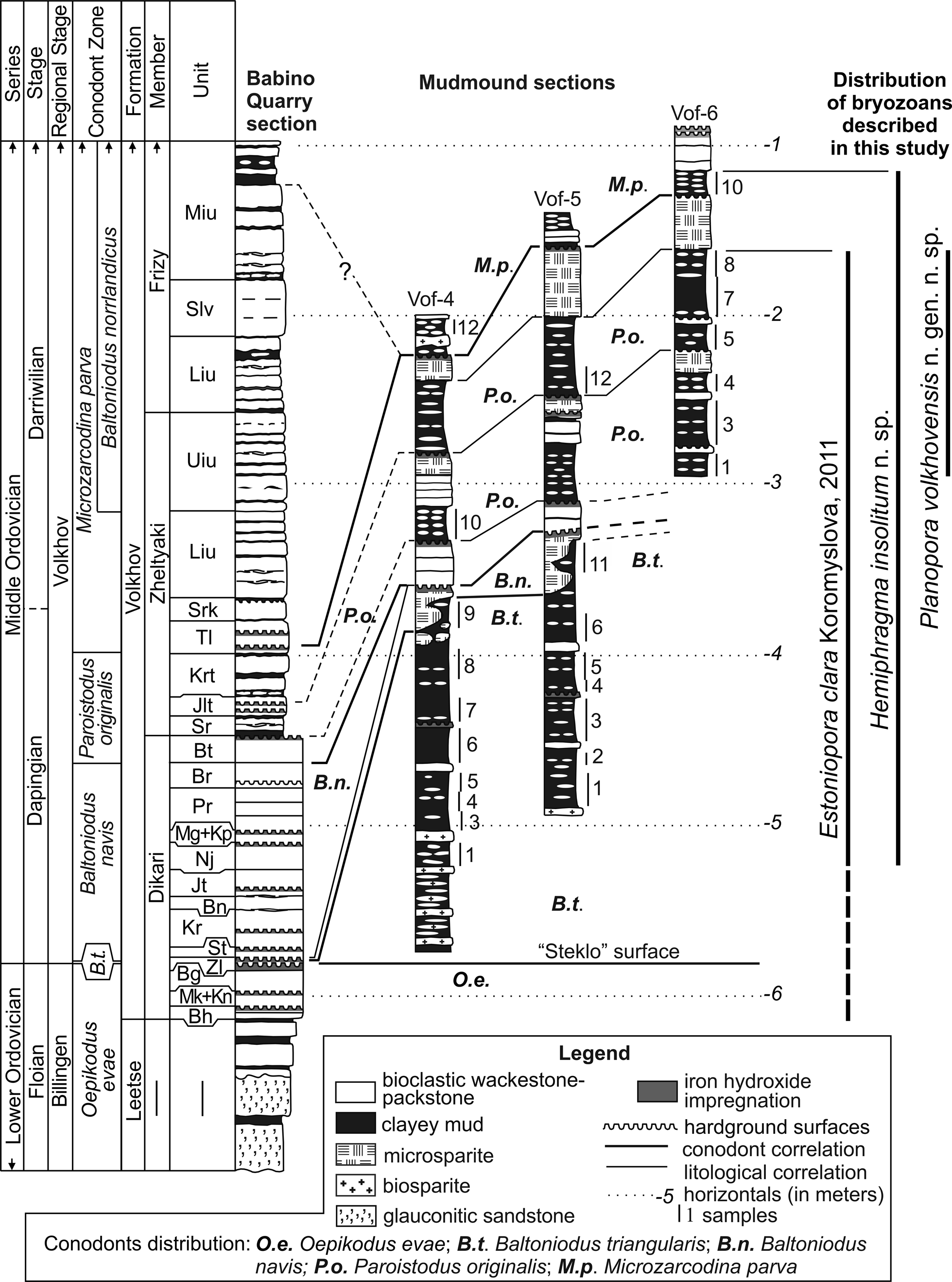
The glauconitic limestone belongs to the Volkhov Formation (uppermost Floian–lowermost Darriwilian). It is underlain by the Leetse Formation (Floian) consisting of glauconitic sandstone, clay, and limestone (Fedorov et al., Reference Fedorov, Koromyslova and Martha2017) and overlain by limestone of the Sillaoru Formation (lower Darriwilian), which is rich in ferruginous ooids, or the clayey limestone of the Lynna Formation; the two latter formations are stratigraphically equivalent (Lindskog et al., Reference Lindskog, Eriksson, Rasmussen, Dronov and Rasmussen2020). Historically, the Volkhov Formation is divided into three members with informal names: Dikari, Zheltyaki, and Frizy, which, in turn, are subdivided into thin units (Fig. 2), recognized and named by quarry workers back in the 19th century. The members and units are easily traceable in outcrops of the Baltic-Ladoga Klint east of St. Petersburg, varying moderately in thickness (Dronov and Fedorov, Reference Dronov and Fedorov1995; Dronov et al., Reference Dronov, Savitsky, Fedorov and Tsyganova1996). The Volkhov Formation belongs to two regional stages: the Billingen and the Volkhov (see Fedorov et al., Reference Fedorov, Koromyslova and Martha2017 for details). The boundary between these stages and between the Lower and Middle Ordovician is recognized within the Zelenyy Unit, belonging to the Dikari Member of the Volkhov Formation (Fig. 2), by the first appearance of the conodont Baltoniodus triangularis (Lindström, Reference Lindström1955) (Tolmacheva and Fedorov, Reference Tolmacheva and Fedorov2001). The conodont B. triangularis, by which the base of the Middle Ordovician Series is defined (Ogg et al., Reference Ogg, Ogg and Gradstein2016), appears directly above the clearly visible smooth hardground surface impregnated by glauconite and containing Gastrochaenolites oelandicus Ekdale and Bromley, Reference Ekdale and Bromley2001 borings (Vinn and Wilson, Reference Vinn and Wilson2010). This hardground surface, known as the ‘steklo’ (Russian: glass) surface, can be traced throughout the entire Eastern Baltic (Lamanskiy, Reference Lamanskiy1905).
Stratigraphic correlation of the sections of the studied Hecker-type mudmound with the section of the Babino limestone quarry, where the standard Floian–lowermost Darriwilian sequence is exposed showing sampling sites and distribution of identified bryozoans. Indexes of the limestone units are given according to Dronov and Mikuláš (Reference Dronov and Mikuláš2010). Abbreviations: units of the Dikari Member: Bh, Barkhat; Mk, Melkotsvet; Kn, Krasnen'kiy; Bg, Beloglaz; Zl, Zelenyy; St, Staritsky; Kr, Krasnyy; Bn, Butina; Jt, Zheltyy; Nj, Nadzheltyy; Mg, Myagon'kiy; Kp, Konoplyastyy; Pr, Pereplet; Br, Bratvennik; Bt, Butok; units of the Zheltyaki Member: Sr, Serina; Jlt, Zhelten'kiy; Krt, Krasnota; Tl, Tolsten'kiy; Srk, Seren'kiy; Liu, Lower unit of intercalation; Uiu, Upper unit of intercalation; units of the Frizy Member: Liu, Lower unit of intercalation; Slv, Sliven'; Miu, Middle unit of intercalation.

The unique Hecker-type mudmounds described from the Leetse and Volkhov formations (Dronov and Fedorov, Reference Dronov and Fedorov1994; Dronov and Ivantsov, Reference Dronov and Ivantsov1994; Fedorov and Dronov, Reference Fedorov and Dronov1998; Fedorov et al., Reference Fedorov, Dronov and Zavarzin1998; Fedorov, Reference Fedorov1999, Reference Fedorov2000, Reference Fedorov2003) are the oldest known Phanerozoic reefs in Europe and the most high-latitude buildups of the first half of the Ordovician (Webby, Reference Webby, Kiessling, Flügel and Golonka2002). Their inner cores consist mainly of partly hardened fossiliferous clayey mud that slake and disperse when wettened; every core is covered with outer microsparite crust. Thin bioclastic limestone beds and lenses, as well as horizons of microsparite nodules, are traceable within the clayey mud (Fedorov, Reference Fedorov2003). All other mudmounds known from elsewhere in the world consist mainly of autochthonous and allochthonous fine-grained carbonates (e.g., Rodrígues-Martinez, Reference Rodríguez-Martínez, Reitner and Thiel2011); clayey mud does not play any role in their composition.
Large Hecker-type mudmounds appear at the base of the Dapingian and replace simple, small mudmounds formed in the upper Floian. The large mudmounds are complex structures composed of 3–4 vertically amalgamated mounds, each consisting of a separate core crowned by a microsparite crust. The thickest and widest is the lower mound (Fedorov, Reference Fedorov2003). The stratigraphic levels of individual mudmounds that comprise the large complex mudmounds have been determined relative to the local section of the Volkhov Formation both by direct correlation and biostratigraphically (Egerquist, Reference Egerquist2003; Fedorov, Reference Fedorov2003; Tolmacheva et al., Reference Tolmacheva, Fedorov and Egerquist2003).
One of the large Hecker-type mudmounds, located near the village of Simankovo (Fig. 1.2), was partially opened during open-pit limestone mining along the Volkhov River right bank at the beginning of the 20th century (Lamanskiy, Reference Lamanskiy1905). The preserved part of the mudmound is a low hill with steep northern and southern slopes and a gentle western slope facing the river (Fig. 1.3). In the east, the mudmound gently dips under the intact enclosing limestone. Long before our research began, the slopes of the hill were thoroughly covered with artificial tailings as well as weathering products and soil.
Materials and methods
We chipped away a 13.5 m long exposure on the northern slope of a large Hecker-type mudmound, located near the village of Simankovo, and excavated several research trenches at its western and southern slopes (Fig. 1.2). Eight sections were described and sampled, including one section in which the ‘steklo’ surface was exposed at the base of the mudmound (Fig. 1.3, section no. 2). A detailed plan of the hill was drawn up using a mining compass, measuring bar, and measuring tape for lithological correlation of the sections. The topmost point of the hill was chosen as zero for counting the descending horizontals (Fig. 1.3).
Numerous 1–1.5 kg samples of clayey mud were taken from the mudmound cores and from overlying enclosing sediments. The samples were air-dried and then soaked in water-filled 3–5 l containers. Once the mud had been dispersed, they were rinsed with tap water by repeatedly filling the containers and draining the clay suspension until the clay particles were completely removed. After drying of the solid residues, thin bioclasts were removed using a 0.1 mm sieve. Skeletal remains sized over 0.1 mm were sorted out under a stereomicroscope. Zonal conodonts from most of the samples were kindly identified by T. Yu. Tolmacheva (A. P. Karpinsky Russian Geological Research Institute); this ensured the assignment of the samples to the regional conodont biozones. Four conodont biozones were recognized within the entire mudmound: Baltoniodus triangularis, Baltoniodus navis, Paroistodus originalis, and Microzarcodina parva. In the deposits overlying the mudmound slopes, conodonts were very scarce; only representatives of the Microzarcodina parva Biozone were recognized. Trilobites of the Asaphus expansus trilobite Biozone were found in clayey limestones of the Lynna Formation, overlapping the top of the mudmound.
Three sections were selected for the study of bryozoans (Fig. 2). Due to intensive quarrying in the past, not a single outcrop of the enclosing glauconitic limestone remained on the banks of the Volkhov River. Therefore, we correlated these three mudmound sections with the section of glauconitic limestone at the Babino quarry located 7 km to the north-east (Figs. 1, 2).
Twenty-eight samples from the sections yielded over 1,000 bryozoan colonies and fragments, including seven species of esthonioporate bryozoans, Revalotrypa gibbosa (Bassler, Reference Bassler1911), R. papillaris (Modzalevskaya, Reference Modzalevskaya1953), R. krestensis Koromyslova in Gorjunova and Koromyslova, Reference Gorjunova and Koromyslova2008, Esthoniopora lessnikowae (Modzalevskaya, Reference Modzalevskaya1953), E. clara Koromyslova, Reference Koromyslova2011, Dianulites janischevskyi Modzalevskaya, Reference Modzalevskaya1953, and Orbipora cf. O. acanthophora Bassler, Reference Bassler1911; three species of trepostome bryozoans, Hemiphragma insolitum n. sp., Monotrypa sp., and Dittopora sp.; and a cystoporate Planopora volkhovensis n. gen. n. sp. Among these, the species R. gibbosa, E. clara, and H. insolitum n. sp. are represented by more than 100 colonies each and predominate in the studied material, while other bryozoan species are less numerous.
Three bryozoan taxa not previously known from the Dapingian of northwestern Russia were discovered: a cystoporate Planopora volkhovensis n. gen. n. sp., a trepostome Hemiphragma insolitum n. sp., and an esthonioporate Esthoniopora clara Koromyslova, Reference Koromyslova2011 (Fig. 2).
We used scanning electron microscopy (SEM) and X-ray microtomography (micro-CT) to visualize the structure of these bryozoans. SEM was performed at the Centre for Microscopy and Microanalysis of the Research Park of St. Petersburg State University, St. Petersburg, using a Hitachi TM 3000, and at the Borissiak Paleontological Institute of the Russian Academy of Sciences (PIN), Moscow, using a Tescan Vega 2. Because the bryozoan colonies found were very small, we studied the internal morphology of some specimens using traditional thin sections and of other specimens using non-destructive micro-CT. Thin sections of bryozoans were studied and imaged using a Leica M165C stereomicroscope at the PIN and a Leica DM1000 compound microscope at the Centre for X-ray Diffraction Studies of the Research Park of St. Petersburg State University. Selected colonies were scanned at the same facility with a Skyscan 1172 (Bruker Co.) micro-CT scanner operated without a filter at 74 kV and 133 mA. The samples were rotated through 180° at 0.25° and 0.3° steps. The exposure was 640–1080 ms/frame, the pixel resolution varied from 1.59 to 1.79 μm. Raw micro-CT data were processed using the programs NReconn and CTVox; 666 to 781 virtual sections were produced, depending on the size of the object. Virtual three-dimensional (3D) models of the colonies were created from two-dimensional data.
Zooecial measurements were taken from micro-CT images, SEM images, or thin sections. The measurements are given in μm as the observed range and (in parentheses) the arithmetic mean ± standard deviation, the number of specimens (N) and the total number of measurements (n).
Abbreviations
Vof-4, Vof-5, and Vof-6 refer to the sections, with the stratigraphic level indicated in brackets—for example (Vof-4 [6]).
Repository and institutional abbreviation
All specimens are preserved at the Borissiak Paleontological Institute of the Russian Academy of Sciences (PIN), Moscow, Russian Federation.
Systematic paleontology
Phylum Bryozoa Ehrenberg, Reference Ehrenberg1831
Class Stenolaemata Borg, Reference Borg1926
Superorder Palaeostomata Ma, Buttler, and Taylor, Reference Ma, Buttler and Taylor2014
Order Cystoporata Astrova, Reference Astrova1964
Suborder Hexagonellina Morozova, Reference Morozova1970
Family ?Hexagonellidae Crockford, Reference Crockford1947
Genus Planopora new genus
Type species
Planopora volkhovensis n. gen. n. sp., Middle Ordovician, Dapingian, Leningrad Oblast’, northwestern Russia.
Diagnosis
Colony erect, bifoliate with flattened branches, initially encrusting some small object. Mesotheca straight or slightly sinuous, absent in zone of early astogeny; median rods lacking. Autozooecia tubular, inclined from mesotheca at ~35–40°. Hemiphragms present, diaphragms absent. Autozooecial apertures each with lunarium at distal end, subcircular to oval, arranged in longitudinal-diagonal rows. Area between autozooecia and their apertures filled by vesicular tissue and traversed by longitudinal ridges. Heterozooecia, monticules, and other maculae, anastomosing tunnel structures, and communication pores absent.
Occurrence
Dapingian (Middle Ordovician); northwestern Russia.
Etymology
From the Latin, planus, flat, in reference to the form of the colony.
Remarks
The order Cystoporata includes three suborders: Ceramoporina, Fistuliporina, and Hexagonellina. Planopora n. gen. cannot belong to the family Rhinoporidae (suborder Ceramoporina), members of which also form bifoliate colonies, because it lacks anastomosing tunnel structures and communication pores, which are unique to Ceramoporidae (Buttler, Reference Buttler1991; Ernst and Key, Reference Ernst and Key2007). Planopora n. gen. is tentatively referred to the family Hexagonellidae (suborder Hexagonellina), because of the presence of tubular autozooecia and vesicular tissue, but the absence of differentiation into the exo- and endozones (Goryunova, Reference Goryunova [Gorjunova]1992, Reference Goryunova [Gorjunova]1996). Only one genus of the family Hexagonellidae has been recorded from the Ordovician, Pakridictya Goryunova, Reference Goryunova [Gorjunova]1992, with a single species, P. maculata Goryunova, Reference Goryunova [Gorjunova]1992. Planopora n. gen. differs from Pakridictya in having autozooecial apertures with lunaria and in lacking minutopores.
Planopora volkhovensis new species
Figures 3, 4
- Reference Koromyslova, Fedorov and Tolokonnikova2020
?Prophyllodictya sp.; Koromyslova et al., fig. 3.
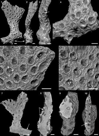
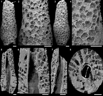
Micro-CT images of Planopora volkhovensis n. gen. n. sp., holotype PIN 5075/2008, mudmound core, Paroistodus originalis conodont Biozone. (1) Erect, flat, bifoliate colony with holdfast (arrowed), front view; (2) same as 1, lateral view; (3) close-up of lateral view, lunarium arrowed; (4) close-up of holdfast, lunarium arrowed; (5) close-up of front view showing apertures of autozooecia, lunaria, and vesicular tissue (arrowed); (6) close-up of opposite front view, lunarian denticle arrowed; (7) longitudinal section parallel to wide side of colony, holdfast arrowed; (8) longitudinal section parallel to narrow side of colony, autozooecial chambers arrowed; (9) transverse section near holdfast; (10) transverse section, mesotheca arrowed. All scale bars represent 250 μm.

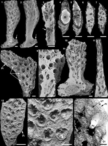
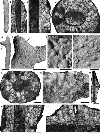
Planopora volkhovensis n. gen. n. sp., mudmound core, Paroistodus originalis conodont Biozone. (1–11) Micro-CT images: (1–7) paratype PIN 5075/2006: (1) front view, holdfast arrowed, (2) longitudinal section, holdfast arrowed, (3) longitudinal section, hemiphragms arrowed, (4–7) successive transverse sections showing formation of mesotheca (arrowed); (8, 9) paratype PIN 5075/2010: (8) front view, longitudinal ridges and marginal plate (Pl) arrowed, (9) longitudinal section near surface, vesicules arrowed; (10, 11) paratype PIN 5075/2009: (10) longitudinal section, holdfast, and, presumably, hemiphragm (H) arrowed, (11) longitudinal section parallel to narrow side of colony. (12–14) SEM images and thin section of paratype PIN 5075/2024: (12) front view, marginal plate (Pl) arrowed, (13) close-up of part of colony showing lunaria, (14) longitudinal section through autozooecia (Az), vesicular tissue (VT), and marginal plate (Pl). Scale bars = 500 μm (12), 250 μm (1–11), 200 μm (13), 100 μm (14).

Holotype
PIN 5075/2008, middle Dapingian, Paroistodus originalis conodont Biozone, Vof-6 [5] of the Hecker-type mudmound core, facies equivalent of the Krasnota Unit (Zheltyaki Member), the environs of the village Simankovo at the right bank of the Volkhov River in Leningrad Oblast’, northwestern Russia.
Paratypes
PIN 5075/2006, PIN 5075/2007, PIN 5075/2009, and PIN 5075/2010 from Vof-6 [5]; PIN 5075/2024, PIN 5075/2025, PIN 5075/2026, PIN 5075/2027, and PIN 5075/2028 from Vof-6 [8]; details as for holotype.
Diagnosis
Planopora n. gen. with lunarium at distal end of each autozooecial aperture; mesotheca without capillaries; hemiphragms rare, diaphragms absent; vesicular tissue traversed by longitudinal ridges.
Occurrence
Middle Dapingian (Middle Ordovician); northwestern Russia.
Description
Colony erect, with flattened bifoliate branches; fragments 2–5 mm long, 1–5 mm wide, and 1 mm thick; originally encrusting some small object, with distinct holdfast (Figs. 3.1–3.4, 3.7, 4.1, 4.2, 4.10). Mesotheca absent during early astogeny and straight or slightly sinuous in zone of astogenetic repetition (Figs. 3.9, 3.10, 4.4–4.7); median rods not observed. No differentiation into endozone and exozone. Autozooecia tubular, arising at 45–60° to mesotheca (Figs. 3.8, 4.3, 4.11). Hemiphragms present, sometimes arranged in alternating order on each wall of autozooecia (Fig. 4.3, 4.10); diaphragms not observed. Autozooecial apertures each with lunarium at distal end, which often not preserved, subcircular to oval, 200–270 μm (236 ± 20 μm; N = 3, n = 23) long with lunarium by 130–260 μm (199 ± 29 μm; N = 3, n = 23) wide, arranged in longitudinal-diagonal rows forming rhombic pattern. Three, rarely four, apertures per 1 mm distance, transversely and longitudinally. Lunaria semicircular, small 40–60 μm (48 ± 8 μm; N = 1, n = 9) long by 80–110 μm (98 ± 9 μm; N = 1, n = 9) wide, with proximo-lateral denticles (Figs. 3.3–3.6, 4.12, 4.13). Autozooecial walls indistinctly laminated (Fig. 4.14). Area between autozooecia and their apertures filled by vesicular tissue and traversed by one to three longitudinal ridges (Figs. 3.5, 3.6, 4.8, 4.9, 4.14). Vesicular tissue often forms the marginal plate, a flattened edge of the colony, up to 100 μm wide (Fig. 4.8, 4.12, 4.14).
Etymology
Named after the type locality, the Volkhov River.
Remarks
Two other cystoporates, Rhinopora? vesiculosa Goryunova, Reference Goryunova [Gorjunova]1992 (family Rhinoporidae Miller, Reference Miller1889, suborder Fistuliporina) and Pakridictya maculata Goryunova, Reference Goryunova [Gorjunova]1992 (family Hexagonellidae Crockford, Reference Crockford1947, suborder Hexagonellina), having bifoliate colonies have been reported from the Darriwilian of Estonia. Planopora volkhovensis n. gen. n. sp. differs from R. vesiculosa in having the autozooecial apertures with lunaria at distal end, instead of the proximal end. The new species differs from P. maculata in having lunaria and lacking median rods (capillaries in Goryunova, Reference Goryunova [Gorjunova]1992) in the mesotheca.
Order Trepostomata Ulrich, Reference Ulrich1882
Suborder Halloporina Astrova, Reference Astrova1978
Family Dittoporidae Vinassa de Regny, Reference Vinassa de Regny1921
Genus Hemiphragma Ulrich, Reference Ulrich1893
Type species
Batostoma irrasum Ulrich, Reference Ulrich1886; Middle Ordovician (Trenton), North America; by original designation.
Remarks
The genus Hemiphragma has an extensive geographical distribution, being known predominantly from the Middle–Upper Ordovician of northwestern Russia, the western Russian Arctic Basin, Taimyr, Estonia, Norway, Italy, USA, and Canada (Bassler, Reference Bassler1911; Fritz, Reference Fritz1957; Astrova, Reference Astrova1965, Reference Astrova1978; Nekhorosheva, Reference Nekhorosheva1965, Reference Nekhorosheva and Bondarev2007; Modzalevskaya, Reference Modzalevskaya1986; Conti, Reference Conti1990; Ernst and Nakrem, Reference Ernst and Nakrem2011).
Hemiphragma insolitum new species
Figures 5–7
Holotype
PIN 5075/2015; Dapingian to lower Darriwilian, Vof-6 [10] of the Hecker-type mudmound core, facial equivalent of the Zheltyaki and Frizy members, the environs of the village Simankovo at the right bank of the Volkhov River in Leningrad Oblast’, northwestern Russia.
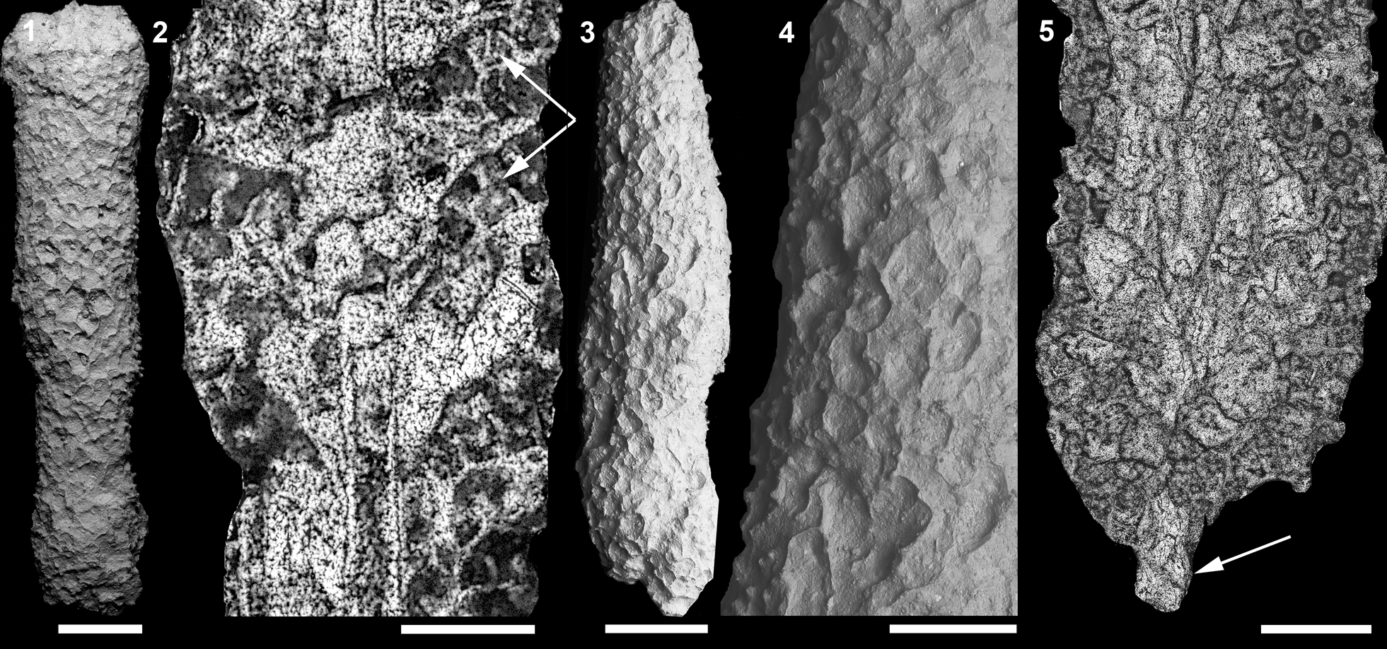
Micro-CT images of Hemiphragma insolitum n. sp. encrusting the apatitic tubule of Sphenothallus sp., holotype PIN 5075/2015, mudmound, Zheltyaki and Frizy members. (1) Rod-shaped colony, Sphenothallus sp. arrowed; (2) zooecia and styles; (3) rod-shaped colony, opposite lateral surface; (4) close-up showing autozooecia, mesozooecia (arrowed), and styles; (5) longitudinal section through central part of colony and Sphenothallus sp. (arrowed); (6) close-up of 5 showing zooecia, hemiphragms, and diaphragms; (7) longitudinal section through central part of colony and Sphenothallus sp., axial tube-like basal lamina arrowed; (8) transverse section through colony and Sphenothallus sp., pair of thickenings arrowed. All scale bars represent 500 μm.

SEM images and thin sections of Hemiphragma insolitum n. sp., mudmound core, Baltoniodus triangularis conodont Biozone. (1–4) Paratype PIN 5075/2016: (1) rod-shaped colony, (2) longitudinal section through colony and Sphenothallus sp. (arrowed), (3) close-up of 2 showing diaphragms and hemiphragms, (4) transverse section through colony and Sphenothallus sp., pair of thickenings arrowed; (5–10) paratype PIN 5075/2017: (5) rod-shaped colony with lateral branch (arrowed), (6) close-up of distal part of colony, lateral branch arrowed, (7, 8) close-up of styles, (9) transverse section through colony and Sphenothallus sp., (10) longitudinal section through zooecia and styles; (11–13) paratype PIN 5075/2019: (11) longitudinal section through colony with lateral branch (arrowed) and Sphenothallus sp., (12) close up of 11 before lateral branch; (13) close-up of 11 through lateral branch. Scale bars = 1 mm (1, 5, 6, 11), 500 μm (2, 12, 13), 200 μm (3, 4, 7, 9), 100 μm (8, 10).

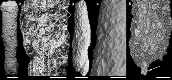
SEM images and thin sections of Hemiphragma insolitum n. sp., mudmound core, Baltoniodus triangularis conodont Biozone. (1, 2) Paratype PIN 5075/2020: (1) rod-shaped colony, SEM, (2) longitudinal section through zooecia and, presumably, Sphenothallus sp., hemiphragms arrowed; (3–5) paratype PIN 5075/2021: (3) rod-shaped colony, SEM, (4) close-up of 3, showing apertures of zooecia and styles, (5) early growth of colony, presumably, Sphenothallus sp. arrowed. Scale bars: 1 mm (1, 3), 500 μm (2, 4, 5).
Paratypes
PIN 5075/2016, PIN 5075/2017, and PIN 5075/2018 from Vof-5 [5]; PIN 5075/2019 from Vof-5 [6]; PIN 5075/2020 and PIN 5075/2021 from Vof-5 [1]; PIN 5075/2022 from Vof-5 [3]; PIN 5075/2023 from Vof-4 [6]; Dapingian, Baltoniodus triangularis conodont Biozone, Hecker-type mudmound core, facies equivalent of the Zelenyy Unit (Dikari Member); location as for holotype.
Diagnosis
Hemiphragma with encrusting colony; autozooecia tubular with rounded or petaloid apertures and moderately numerous diaphragms and hemiphragms; mesozooecia irregular, with polygonal apertures; styles numerous.
Occurrence
Dapingian (Middle Ordovician); northwestern Russia.
Description
Colonies rod-like, formed by growing around some cylindrical object, such as apatitic tubules of possible cnidarian origin (Figs. 5.1, 5.5–5.8, 6.2–6.4, 6.9, 6.11–6.13, 7.2, 7.5); their fragments up to 15 mm long and up to 5 mm in diameter. Some colonies have small lateral branches (Fig. 6.5, 6.6, 6.11, 6.13). No differentiation into endozone and exozone. Autozooecia tubular, budding from axial tube-like basal lamina (Fig. 5.7). Autozooecial walls faintly sinuous. Autozooecial apertures rounded or petaloid (Figs. 5.2, 5.4, 6.6), 230–340 μm (282 ± 35 μm; N = 1, n = 12) in diameter. Three or four apertures per 1 mm distance, transversely and longitudinally. Autozooecial diaphragms and hemiphragms moderately numerous, irregularly distributed (Figs. 5.5–5.8, 6.2, 6.3, 6.12, 6.13, 7.2). Mesozooecia moderately numerous, irregularly located between autozooecia (Fig. 5.4). Mesozooecial apertures polygonal, 110–190 μm (153 ± 23 μm; N = 1, n = 12) in size. Styles numerous, small, surrounding each autozooecial and mesozooecial aperture (Figs. 5.4, 6.6–6.8, 6.10, 7.4).
Etymology
From the Latin, insolitus, unusual, in reference to the unusual colony growth.
Remarks
This species is assigned to the genus Hemiphragma because it has hemiphragms throughout the colony, mesozooecia, and numerous small styles. It differs from the type species of the genus, Hemiphragma irrasum (Ulrich, Reference Ulrich1886), and all other Hemiphragma species in the encrusting colonies, often with the basal lamina of an axial tube-like form as a result of overgrowing a cylindrical object. Only three species of this genus, Hemiphragma priscum Pushkin in Pushkin and Popov, Reference Pushkin and Popov1999, H. rotundatum Bassler, Reference Bassler1911, and H. ottawaense (Foord, Reference Foord1883), are known from the Floian to the lower Darriwilian of Leningrad Oblast’, northwestern Russia (Bassler, Reference Bassler1911; Modzalevskaya, Reference Modzalevskaya1986; Pushkin and Popov, Reference Pushkin and Popov1999). Hemiphragma insolitum n. sp. differs from H. priscum and H. ottawaense, which have ramose colonies, in absent of differentiation into endozone and exozone. Furthermore, H. insolitum n. sp. differs from H. ottawaense in having smaller autozooecial apertures (230–340 μm versus 360–430 μm in H. ottawaense). The new species is distinguished from H. rotundatum, which has subglobular colonies, in having more numerous mesozooecia.
Order Esthonioporata Astrova, Reference Astrova1978
Family Esthonioporidae Vinnassa de Regny, Reference Vinassa de Regny1921
Genus Esthoniopora Bassler, Reference Bassler1911
Type species
Esthoniopora communis Bassler, Reference Bassler1911, Middle–Late Ordovician, Dapingian–Katian, Estonia and Leningrad Oblast, northwestern Russia.
Remarks
The genus Esthoniopora is endemic to the Baltic paleobasin; its representatives are known only from the Ordovician of Leningrad Oblast’ in northwestern Russia and from Estonia. The species E. clara Koromyslova, Reference Koromyslova2011 has previously been described based on several incomplete specimens from the Oepikodus evae conodont Biozone, upper Floian, Lower Ordovician, of Leningrad Oblast’; some of its morphological features have not been observed. During the present study, over 100 colonies of E. clara were found in the Dapingian Hecker-type mudmound, which allowed us to make a more complete description of this species.
Esthoniopora clara Koromyslova, Reference Koromyslova2011
Figure 8
- Reference Goryunova [Gorjunova]1992
Esthoniopora sp.; Goryunova, text-fig. 13a.
- Reference Goryunova [Gorjunova]1996
Esthoniopora clivosa Modzalevskaya, Reference Modzalevskaya1953; Goryunova, pl. 13, fig. 1.
- Reference Goryunova [Gorjunova]1996
Esthoniopora lessnikowae (Modzalevskaya, Reference Modzalevskaya1953); Goryunova, pl. 13, fig. 2.
- Reference Goryunova [Gorjunova]1996
Esthoniopora sp.; Goryunova, pl. 14, fig. 4.
- Reference Koromyslova2011
Esthoniopora clara Koromyslova, p. 923, pl. 5, fig. 3, pl. 6, fig. 1, text-fig. 19e.
- Reference Koromyslova, Fedorov and Tolokonnikova2020
Esthoniopora clara Koromyslova, Reference Koromyslova2011; Koromyslova et al., fig. 2.
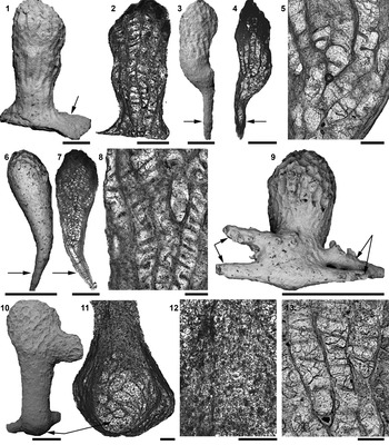
SEM images and thin sections of Esthoniopora clara Koromyslova, Reference Koromyslova2011, mudmound core, Baltoniodus triangularis conodont Biozone. (1, 2) PIN 5075/2011: (1) front view, basal lamina arrowed, (2) autozooecia with diaphragms and hemiphragms; (3–5) PIN 5075/2012: (3) front view, kenozooecium arrowed, (4) kenozooecium (arrowed) and autozooecia, (5) close-up of diaphragms and hemiphragms; (6–8) PIN 5075/2004: (6) front view, kenozooecium arrowed, (7) kenozooecium (arrowed) and autozooecia, (8) close-up of diaphragms and hemiphragms; (9) PIN 5075/2013, front view, holdfasts arrowed; (10–13) PIN 5075/2014: (10) club-shaped colony with lateral branch, holdfast formed by vesicular tissue arrowed, (11) vesicular tissue (arrowed), (12) styles, (13) close-up of diaphragms and hemiphragms. Scale bars = 2 mm (9), 1 mm (3, 4, 6, 7, 10), 500 μm (1, 2), 200 μm (5, 8, 11–13).

Holotype
PIN 3535/2044 from Oepikodus evae conodont Biozone, upper Floian, Lower Ordovician, quarry of Putilovskie lomki, Leningrad Oblast’ of northwestern Russia (Koromyslova, Reference Koromyslova2011, pl. 6, fig. 1).
Emended diagnosis
Esthoniopora with club-shaped colony having basal lamina or holdfast formed by kenozooecia or vesicular tissue; autozooecia tubular, usually growing parallel to each other, apertures rounded, diaphragms and hemiphragms numerous; kenozooecia tubular without diaphragms; vesicular tissue forming no rows; styles numerous in proximal parts of some colonies.
Occurrence
Upper Floian (Lower Ordovician) to Dapingian (Middle Ordovician); northwestern Russia
Description
Colonies club-shaped, 2–10 mm long by 1–5 mm wide, consisting of a small number of autozooecia growing parallel to each other. Some colonies have small lateral branches (Fig. 8.10). Autozooecia budding from basal lamina (Fig. 8.1, 8.2), kenozooecium (Fig. 8.3, 8.4, 8.6, 8.7), or vesicular tissue (Fig. 8.10, 8.11) formed in early astogeny. Kenozooecia and vesicular tissue initially encrusted some small object with distinct holdfast. No differentiation into endozone and exozone. Secondary overgrowths absent. Autozooecia tubular; their apertures located in distal part of colony, rounded, 190–350 μm (253 ± 41 μm; N = 3, n = 18) in diameter. Three or four apertures, transversely and longitudinally, per 1 mm distance. Autozooecial walls faintly sinuous. Autozooecial diaphragms straight, numerous (Fig. 8.2, 8.8, 8.13). Hemiphragms slightly downcurved, numerous; usually arranged alternately at opposite sides of autozooecial walls, sometimes irregularly (Fig. 8.5, 8.8). Vesicular tissue forming no rows, vesicles overlapping one another (Fig. 8.11). Kenozooecia tubular, long; diaphragms absent. Numerous styles observed in proximal parts of some colonies (Fig. 8.12).
Materials
PIN 5075/2011 and PIN 5075/2012 from Vof-5 [1]; PIN 5075/2004 from Vof-5 [2]; PIN 5075/2013 from Vof-4 [7]; PIN 5075/2014 from Vof-3 [1]; Dapingian, Baltoniodus triangularis conodont Biozone, Hecker-type mudmound core, facial equivalent of the Zelenyy Unit (Dikari Member); PIN 5075/2005 from Vof-6 [7]; Dapingian, Paroistodus originalis conodont Biozone, the Hecker-type mudmound core, facial equivalent of the Krasnota Unit (Zheltyaki Member); environs of the village Simankovo at the right bank of the Volkhov River in Leningrad Oblast’, northwestern Russia.
Remarks
This species differs from E. communis Bassler, Reference Bassler1911, E. lessnikowae (Modzalevskaya, Reference Modzalevskaya1953), and E. curvata Bassler, Reference Bassler1911 in the arrangement of hemiphragms at opposite sides of autozooecial walls in alternating order (in E. communis the hemiphragms are arranged opposite one another and in E. lessnikowae and E. curvata, unilaterally). Furthermore, E. clara differs from E. lessnikowae, which also was discovered in this mudmound, in having smaller autozooecial apertures (190–350 μm versus 200–500 μm in E. lessnikowae).
Discussion
Distribution of early cystoporate bryozoans
The superorder Palaeostomata (Class Stenolaemata) was introduced by Ma et al. (Reference Ma, Buttler and Taylor2014) for the Paleozoic bryozoans and includes the orders Cryptostomata, Esthonioporata, Trepostomata, Cystoporata, and Fenestrata (Ernst, Reference Ernst2018). In contrast to the first three orders, which have representatives from the Lower Ordovician (Tremadocian to Floian) (Pushkin and Popov, Reference Pushkin and Popov1999; Xia et al., Reference Xia, Zhang and Wang2007; Koromyslova, Reference Koromyslova2011; Ma et al., Reference Ma, Taylor, Xia and Zhan2015; Fedorov et al., Reference Fedorov, Koromyslova and Martha2017), to date, no Lower Ordovician members of the order Cystoporata have been identified with certainty. Ceramopora? unapensis Ross, Reference Ross1966 from the upper Tremadocian of Oklahoma, USA, originally was described as the oldest cystoporate. However, the classification of this species as a bryozoan was later questioned by Taylor (Reference Taylor and Benton1993). The genus Revalotrypa “Bassler, Reference Bassler1952” from the upper Floian of northwestern Russia previously had been considered to be the oldest known cystoporate bryozoan (Goryunova, Reference Goryunova [Gorjunova]1988, Reference Goryunova [Gorjunova]1992, Reference Goryunova [Gorjunova]1996). However, this genus was subsequently attributed by Ma et al. (Reference Ma, Buttler and Taylor2014) to the order Esthonioporata.
A cystoporate with small domal multilaminate colonies, Ceramopora proava Astrova, Reference Astrova1965, has been described from the Lower Ordovician of Lyamchin Bay on the west coast of Vaygach Island in Arctic Russia. However, recent studies (Generalov, Reference Generalov2000) have identified only Middle Ordovician to Silurian, but no Lower Ordovician, deposits in this area. Cystoporates with domal unilaminate discoidal or multilaminate hemispherical colonies, Profistulipora arctica Astrova, Reference Astrova1965 and P. retrusa Astrova, Reference Astrova1965, have been described from the Lower Ordovician of Novaya Zemlya, Arctic Russia, but not from the Tremadocian of Siberia, as was erroneously noted by Buttler et al. (Reference Buttler, Wyse Jackson, Ernst and McKinney2013). However, their exact stratigraphic age is not known. Later, P. arctica was described by Nekhorosheva (Reference Nekhorosheva and Bondarev2007) from the lower part of the Nelidovskiy Regional Stage of Novaya Zemlya. According to Nekhorosheva (Reference Nekhorosheva2015), the Nelidovskiy Regional Stage is correlated with the Floian to lower Darriwilian. Therefore, P. arctica can potentially be the oldest true cystoporate.
Other old unequivocal cystoporates known so far come from the Darriwilian (Middle Ordovician) of Estonia (Goryunova, Reference Goryunova [Gorjunova]1992). Among these, only Rhinopora? vesiculosa Goryunova, Reference Goryunova [Gorjunova]1992 and Pakridictya maculata Goryunova, Reference Goryunova [Gorjunova]1992 formed bifoliate colonies. However, the new cystoporate, coming from the middle Dapingian of northwestern Russia, is older. Therefore, Planopora volkhovensis n. gen. n. sp. is the oldest known сystoporate bryozoan forming erect, bifoliate colonies.
Ecological implications of bryozoans from Hecker-type mudmounds
Bryozoans from the lowermost Floian (Lower Ordovician) glauconitic sandstone of northwestern Russia produced very small grain-shaped and domal conical colonies formed by completely encrusting a small object, or fusiform and rod-shaped colonies formed by growing around a rod-shaped object (Fedorov et al., Reference Fedorov, Koromyslova and Martha2017; Fedorov and Koromyslova, Reference Fedorov and Koromyslova2019). The majority of bryozoans from the upper Floian to Dapingian glauconitic sandstone of northwestern Russia had domal colonies (some unilaminate or multilaminate), free-lying or attached to a brachiopod valve or another substrate; rarely such bryozoans produced rod-like colonies growing around a rod-shaped object or encrusted a flat substrate (Modzalevskaya, Reference Modzalevskaya1955; Taylor and Rozhnov, Reference Taylor and Rozhnov1996; Gorjunova, Reference Gorjunova2009; Koromyslova, Reference Koromyslova2011).
In contrast, the studied specimens of Planopora volkhovensis n. gen. n. sp. and Esthoniopora clara from the Hecker-type mudmound each have an attachment structure at their base, a holdfast, usually formed as a result of growth of autozooecia, kenozooecia, or vesicular tissue around a long cylindrical object. Holdfasts formed by autozooecia were observed in P. volkhovensis n. gen. n. sp., while the colonies of E. clara often had one or rarely several slender hollow tubes at the base, which obviously were kenozooecia performing the function of a holdfast, or a holdfast formed by the vesicular tissue.
The presence of a holdfast probably indicates the attachment of the colony to a sponge spicule. It should be noted that sponge spicules were not found among detrital particles obtained from the clayey mud. Sponge root tufts composed of diactines and groups of unfused orthotriaxial hexactines or their derivatives replaced with crystalline carbonate have been observed only in some lenses of bioclastic limestone and microsparite nodules within the mudmound cores as well as in microsparite crusts (Fedorov, Reference Fedorov2003). According to Zimmerle (Reference Zimmerle, Reitner and Keupp1991), claystones can occasionally replace siliceous sponges immediately after deposition. It can be assumed that sponges with unfused siliceous spicules were numerous on the surface of the mudmound during its formation. They produced mats of spicules suitable for settlement of P. volkhovensis n. gen. n. sp. and E. clara larvae. However, these porous substrates were filled with clay over time. Clay infilling was probably accompanied by dissolution of spicules. The combination of these two processes prevented the bryozoans from reaching a larger size.
Unusual rod-like colonies of the trepostome Hemiphragma insolitum n. sp. were formed by overgrowing a cylindrical body, so that autozooecia budded off the axial tube-like basal lamina. The axial tube-like type of the basal lamina has been described by Newton (Reference Newton1971). He discussed the genus Rhabdomeson Young and Young, Reference Young and Young1874, which, according to the authors of this genus and many other authors (i.e., Bassler, Reference Bassler1953; Shulga-Nesterenko, Reference Shulga-Nesterenko1955; Blake, Reference Blake1976; Goryunova, Reference Goryunova [Gorjunova]1985; Wyse Jackson and Bancroft, Reference Wyse Jackson and Bancroft1995), formed hollow axial cylinders from which autozooecia were budding. Newton (Reference Newton1971) suggested that the hollow axial cylinder had formed as a result of the bryozoan overgrowing a rigid, cylindrical object, such as an echinoid or brachiopod spine, and depositing the basal lamina around it. The cylindrical object would subsequently become dissolved or broken away. According to Blake (Reference Blake1976) and Wyse Jackson and Bancroft (Reference Wyse Jackson and Bancroft1995), the lining wall of the axial cylinder in Rhabdomeson is a compound wall and not a basal lamina, contrary to Newton (Reference Newton1971). This implies that it was a body cavity within the colony and not the former location of a cylindrical substrate encrusted by the colony. However, autozooecia were budded in both cases in similar way.
Such cylindrical objects were observed in all studied specimens of Hemiphragma insolitum n. sp. Some of them were apatitic tubules of a widespread Paleozoic marine taxon Sphenothallus Hall, Reference Hall1847, a problematic fossil with possible cnidarian affinities (Vinn and Kirsimäe, Reference Vinn and Kirsimäe2015; Dzik et al., Reference Dzik, Baliński and Sun2017; Landing et al., Reference Landing, Antcliffe, Geyer, Kouchinsky, Bowser and Andreas2018). The tubules of Sphenothallus are recognizable by the characteristic ellipsoidal cross-section with a pair of longitudinal thickenings that have a thin laminar structure (Van Iten et al., Reference Van Iten, Cox and Mapes1992; Vinn and Kirsimäe, Reference Vinn and Kirsimäe2015) and by modular coloniality (Landing et al., Reference Landing, Antcliffe, Geyer, Kouchinsky, Bowser and Andreas2018). Presumably, the bryozoan colonies overgrew juvenile individuals of Sphenothallus because the tubules of the latter were small (~0.5–1 mm in diameter) and had thin walls.
According to this study and to Koromyslova et al. (Reference Koromyslova, Fedorov and Tolokonnikova2020), the studied Hecker-type mudmound contains numerous bryozoans with holdfasts, dominated by club-shaped colonies of Esthoniopora clara. The rod-like colonies of Hemiphragma insolitum n. sp., formed by overgrowing juvenile individuals of Sphenothallus, and domal colonies of Revalotrypa gibbosa (Bassler, Reference Bassler1911), free-lying or attached to brachiopod valves, were less numerous. Rod-like colonies with an extended base for attachment to flat substrates were rare.
Conclusions
Clayey-calcareous mudmounds (‘Hecker-type mudmounds’) contain abundant small bryozoans. Excavation of one such mound yielded three bryozoan taxa not previously known from the Dapingian: (1) a cystoporate, Planopora volkhovensis n. gen. n. sp.; (2) a trepostome, Hemiphragma insolitum n. sp.; and (3) an esthonioporate, Esthoniopora clara Koromyslova, Reference Koromyslova2011.
Planopora n. gen. is the oldest сystoporate bryozoan forming erect, bifoliate colonies. Other early cystoporates produced encrusting (Ceramopora? unapensis) or massive laminate (Ceramopora proava, Profistulipora arctica, and P. retrusa) colonies.
The colonies of P. volkhovensis n. gen. n. sp. and E. clara have an attachment structure at their base, a holdfast, probably indicating their attachment to sponge spicules. Hemiphragma insolitum n. sp. produced rod-like colonies, often formed as a result of overgrowing the problematic fossil Sphenothallus Hall.
During formation of the Hecker-type mudmound, sponges with unfused siliceous spicules and juvenile individuals of Sphenothallus were numerous on its surface and provided a suitable substrate for the settlement of bryozoan larvae.
Acknowledgments
The authors thank T.Yu. Tolmacheva (A. P. Karpinsky Russian Geological Research Institute, Saint Petersburg, Russian Federation) for identifying Biozonal conodont species. R.A. Rakitov (Borissiak Paleontological Institute of the Russian Academy of Science, Moscow, Russian Federation) is thanked for providing technical assistance with SEM work and linguistic editing. A. Ernst (Institut für Geologie, Universität Hamburg, Hamburg, Germany), P.D. Taylor (Natural History Museum, London, United Kingdom), and O. Vinn (Department of Geology, University of Tartu, Tartu, Estonia), managing editor J. Kastigar (Cincinnati, USA), and editor J. Jin (University of Western Ontario, Canada) are thanked for helpful reviews and comments on the manuscript. The study was funded by the RFBR research grant No. 18-05-00245-A and 18-04-01046-A.