INTRODUCTION
A by-product of the fermentation and distillation of the traditional Japanese spirit shochu, known as shochu distillery by-product (SDBP), is widely used as a feed for farm animals. Mahfudz et al. (Reference Mahfudz, Hayashi, Ikeda, Hamada, Ohtsuka and Tomita1996a, Reference Mahfudz, Hayashi, Otsuji, Ohtsuka and Tomitab) showed that dietary supplementation with a small amount of SDBP and the ether extracts of SDBP promotes the growth of chickens. Furthermore, it has been reported that hexane extracts of SDBP (Kamizono et al. Reference Kamizono, Nakashima, Ohtsuka and Hayashi2010), as well as SDBP itself (Sultana et al. Reference Sultana, Kamizono, Furusono and Hayashi2011), enhance growth due to a decrease in the rate of skeletal muscle protein degradation. These reports indicate that SDBP contains growth-promoting factors, which may down-regulate skeletal muscle proteolysis. Mahfudz et al. (Reference Mahfudz, Nakashima, Ohtsuka and Hayashi1997) demonstrated that a fraction separated from the ether extracts of SDBP decreased protein degradation in chicken muscle cells. The result was also supported by previous experiments using the hexane extracts of SDBP (Kamizono et al. Reference Kamizono, Nakashima, Ohtsuka and Hayashi2010). Therefore, it is possible that the substance could suppress body protein degradation, resulting in improved growth performance when SDBP is fed to chickens. The substance stimulating growth was thought to be butoxybutyl alcohol (1-butoxy-1-butanol, BBA; molecular weight 146, C8H18O2) from a preliminary experiment.
Protein degradation is a highly controlled, selective and regulated process, as highlighted by Tesseraud et al. (Reference Tesseraud, Bouvarel, Collin, Audouin, Crochet, Seiliez and Leterrier2009). Four proteolytic systems are involved in skeletal muscle proteolysis, i.e. the calpain, caspase, lysosomal and ubiquitin–proteasome systems (Goll et al. Reference Goll, Neti, Mares and Thompson2008). The calpain system includes intracellular Ca2+-dependent cysteine proteases, such as μ- and m-calpains, which are ubiquitously expressed in animal tissues, and require μm and mm levels of Ca2+ for activity in vitro, respectively (Sorimachi et al. Reference Sorimachi, Hata and Ono2011). The caspase system is responsible for the degradation of proteins during apoptosis, which is mainly controlled by the cysteine protease, caspase-3 (Goll et al. Reference Goll, Neti, Mares and Thompson2008). The lysosomal system involves acidic cysteine proteases, the cathepsins, which are located inside lysosomal structures (Goll et al. Reference Goll, Neti, Mares and Thompson2008). The ubiquitin–proteasome system is an adenosine triphosphate (ATP)-dependent proteolysis system that requires polyubiquitination of target proteins as a marker of degradation by the proteasome (Ciechanover Reference Ciechanover2006). The polyubiquitination requires covalent linkage of ubiquitin molecules to one or more lysine residues of a protein, a process that involves three types of enzymes, i.e. ubiquitin-activating enzyme, ubiquitin-conjugating enzymes and ubiquitin ligases. It has also been demonstrated that the expression of two important ubiquitin ligases, atrogin-1/muscle atrophy F-box (MAFbx) and muscle RING finger 1 (MuRF1), controls the ubiquitin–proteasome system (Franch & Price Reference Franch and Price2005; Glass Reference Glass2005; Szewczyk & Jacobson Reference Szewczyk and Jacobson2005). Previously, it has been shown that feeding hexane extracts of SDBP to chickens decreased the mRNA levels of the μ-calpain large subunit, atrogin-1/MAFbx and ubiquitin in the pectoralis superficialis muscle (Kamizono et al. Reference Kamizono, Nakashima, Ohtsuka and Hayashi2010). However, there are no data to show how dietary BBA affects protein degradation in skeletal muscle and growth performance.
Therefore, the present study was conducted (1) to synthesize BBA and to confirm its chemical structure by gas chromatography mass spectrometry (GC-MS) and nuclear magnetic resonance (NMR), (2) to examine the effects of feed containing synthesized BBA on growth performance in broiler chickens and (3) to determine whether the feeding regime affects protein degradation in skeletal muscle and the expression of proteolysis-related genes.
MATERIALS AND METHODS
Chemical synthesis of butoxybutyl alcohol and identification of its chemical structure
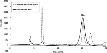
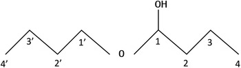
Five millilitres of butyl alcohol and 5 ml of butyl aldehyde were reacted at room temperature with 0·01 g of p-toluenesulphonic acid and 0·03 g of hydroquinone. After 24 h, BBA was separated by a Sephadex LH-20 column (35 × 700 mm) at a constant flow rate (1 ml/min) with a mobile phase of methanol, dichloroethane and water (9 : 2 : 1, v/v), and monitored by absorbance at 275 nm using TOSOH UV-8010 (TOSOH, Tokyo, Japan). The fractions of BBA were collected and the mobile phase was evaporated by a vacuum evaporator (conditions: 31 hPa; temperature of water bath, 30 °C). The retention time of the synthesized BBA on high performance liquid chromatography (HPLC) under the conditions previously reported (Mahfudz et al. Reference Mahfudz, Nakashima, Ohtsuka and Hayashi1997) was the same as that of BBA extracted from SDBP (Fig. 1). The molecular weight of the synthetic compound was determined by GC-MS (Polaris Q, Thermo Finnigan, Tokyo, Japan) equipped with a capillary column (DB-WAXetr, 60 m length, 0·25 mm I.D., 0·25 μm film-thickness, J&W Scientific Inc., Folsom, USA). The 1H and 13C NMR spectra were obtained by an INOVA-300 spectrometer (Varian, Palo Alto, USA) equipped with an Oxford 300/58 magnet, and chemical shifts were expressed on a δ (ppm) scale with tetramethylsilane (TMS) as an internal standard. The solvent used for NMR measurements was chloroform-d (CDCl3) and TMS (100 : 3, v/v). The chemical structure of the synthesized BBA is illustrated in Fig. 2.
HPLC chromatogram of natural BBA from SDBP and synthesized BBA.

The structure of BBA.

Animal experimental design
Two studies were conducted. The experimental protocol was approved by the Animal Care and Use Committee of Kagoshima University and Tohoku University. Body weight and feed intake were recorded every 3 days. At 27 days of age, all the chickens were killed by decapitation under carbon dioxide anaesthesia, and tissues were collected. In Study 1, excrement was collected and pooled daily for 3 days (from 24 to 27 days of age), and stored at −30 °C until analysis. In Study 2, plasma and muscle sample were taken and stored at −80 °C until analysis.
Study 1
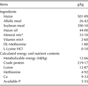
One hundred male broiler chicks (Ross 308) were obtained from a local commercial hatchery (Kagoshima Kumiai Chicken Foods Co., Ltd., Kagoshima, Japan) at 0 day of age. They were housed in a brooder and provided water and a commercial diet (220 g crude protein/kg and 12·55 MJ metabolizable energy/kg) ad libitum. After 12 days, 16 birds of similar body weight (c. 300 g) were selected and placed in individual aluminium cages. They were then fed the basal diet (Table 1), comprising mainly maize and soybean meal, until 15 days of age, after which they were divided randomly into two groups: control and BBA. In the experimental diet, 30 mg of BBA was mixed with 1 kg of basal diet, 2·5 ml of ethanol and 5 g of sucrose. The control diet was also mixed with similar amounts of ethanol and sucrose. These diets were then given for 12 days from 15 to 27 days of age. The experiment was undertaken in a temperature-controlled room (24 ± 1 °C) with a 12-h light and dark cycle.
Diet components and calculated contents of the basal diet

* Amounts per kg diet: CaHPO4·2H2O, 20·86 g; CaCO3, 6·62 g; NaCl, 5·06 g; MnSO4·5H2O, 0·22 g; ZnSO4·7H2O, 0·13 g; FeSO4·7H2O, 0·20 g; CuSO4·5H2O, 7·71 mg; NaIO3, 0·56 mg; Na2SeO3, 0·27 mg.
† Amounts per kg diet: retinol acetate, 1·57 mg; cholecalciferol, 0·01 mg; riboflavin, 6·50 mg; thiamine hydrochloride, 2·60 mg; pyridoxine hydrochloride, 1·30 mg; cyanocobalamin, 0·03 mg; calcium pantothenate, 10·40 mg; nicotinic acid, 26·00 mg; 2-methyl-1,4-naphthoquinone, 0·65 mg; folic acid, 0·52 mg; choline chloride, 0·78 mg; biotin, 0·07 mg; DL-α-tocopherol acetate, 6·50 mg; sucrose, 2·54 g.
Study 2
Forty male broiler chicks (Ross 308) were purchased from a commercial hatchery (Matsumoto Poultry Farms & Hatcheries Co., Ltd., Miyagi, Japan) at 0 day of age, and divided randomly into two groups: control and BBA. They were then housed in groups in electrically heated batteries under continuous light for 7 days, while being provided water and experimental diets (219 g crude protein/kg and 12·66 MJ metabolizable energy/kg) ad libitum. The composition of the experimental diets was the same as in Study 1. At 7 days of age, pairs of chicks were placed in wire-bottomed cages for 2 days. From 9 to 27 days of age, eight representative chicks from each group were placed in individual cages under controlled temperature (9–12 days of age, 26 °C; 12–15 days of age, 25 °C; 15–27 days of age, 24 °C) and continuous light.
Measurement of N τ-methylhistidine
The rate of skeletal muscle protein degradation was estimated by measuring the concentration of N τ-methylhistidine in the excrement and plasma as described previously (Hayashi et al. Reference Hayashi, Maeda, Toyomizu and Tomita1987; Kamizono et al. Reference Kamizono, Nakashima, Ohtsuka and Hayashi2010). The excrement sample was hydrolysed with 6 mol/l hydrochloric acid for 20 h at 115 °C in an autoclave. The hydrolysates were then cooled and passed through filter paper. After hydrochloric acid was removed by evaporation under reduced pressure, the hydrolysates were dissolved in water and again evaporated. The residue was dissolved in 0·2 mol/l pyridine and applied to a cation-exchange column (7 × 60 mm, Dowex 50W-X8, 200–400 mesh, pyridine form). After most of the acidic and neutral amino acids were washed out with 0·2 mol/l pyridine, N τ-methylhistidine was eluted with 1 mol/l pyridine and collected. The solvent was evaporated and the residue was dissolved in the mobile phase (15 mmol/l sodium 1-octanesulfonate in 20 mmol/l potassium dihydrogen phosphate). An aliquot was injected into an HPLC (LC-6A, Shimadzu, Kyoto, Japan) equipped with an Inertsil ODS-80A column (4·6 × 250 mm, 5 μm, GL Sciences, Tokyo, Japan). The column was attached to an oven at 50 °C. A fluorescence detector (RF-535, Shimadzu, Kyoto, Japan) set at an excitation wavelength of 365 nm, and an emission wavelength of 460 nm was used to monitor the fluorescence derived from the reaction with o-phthalaldehyde. Plasma was mixed with 200 g/l sulphosalicylic acid and centrifuged at 9600 g for 5 min. The supernatant was recovered and evaporated under reduced pressure. The residue was dissolved in 0·2 mol/l pyridine, after which the remainder of the treatments were the same as those for excrement.
Measurement of mRNA levels
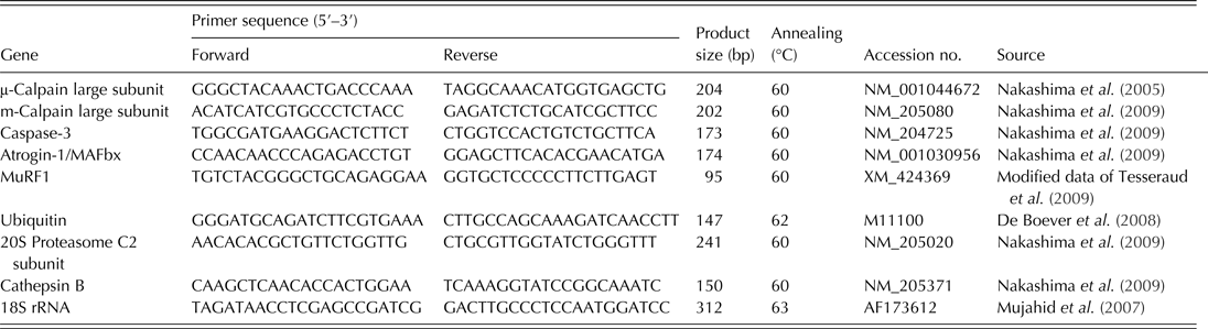
Standard molecular biological techniques were used, essentially as described by Sambrook et al. (Reference Sambrook, Fritsch and Maniatis1989). Total RNA was extracted from 100 mg pectoralis profundus muscle samples using TRIzol® Reagent (Life Technologies, Tokyo, Japan), according to the manufacturer's protocol. The RNA concentration and purity were determined spectrophotometrically using A260 and A280 values in a photometer (NanoDrop 2000c, Thermo Fisher Scientific, Yokohama, Japan). The ratio of A260/280 of all samples was between 1·8 and 2·0. Four micrograms of total RNA was reverse transcribed using a mixture of oligo(dT)12–18 and random primers (99 : 1, v/v), and M-MLV reverse transcriptase (Life Technologies, Tokyo, Japan). Two microlitres of each reverse transcription reaction product served as a template in a 20 μl polymerase chain reaction (PCR) containing 2 mmol/l magnesium chloride, 0·5 μmol/l of each primer, 0·5 U TaKaRa Ex taq (TaKaRa, Shiga, Japan) and 0·5 × SYBR Green master mix (Bio Whittaker Molecular Applications, Rockland, USA). Gene expression was measured by real-time PCR using the CFX ConnectTM Real-Time PCR Detection System (Bio-Rad Laboratories, Hercules, USA). The fluorescence of SYBR Green was monitored at the end of each cycle as the PCR product formed during that cycle. Melting curve profiles were recorded at the end of each run. The primers and annealing temperatures for analysis of mRNA expression related to proteolysis (i.e. μ- and m-calpain large subunits, caspase-3, atrogin-1/MAFbx, MuRF1, ubiquitin, 20S proteasome C2 subunit and cathepsin B) and internal standard (i.e. 18S rRNA) are shown in Table 2. The mRNA level of 18S rRNA as the internal standard was not significantly different between the control and BBA groups. The mRNA levels in the control were arbitrarily set to 1·0.
Sequences for real-time PCR primers
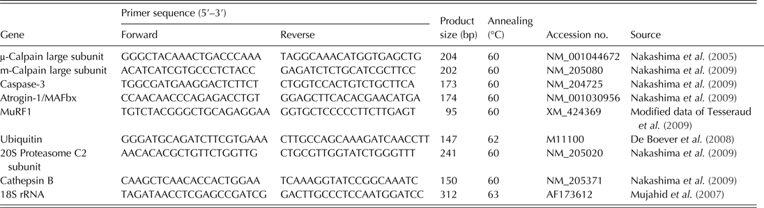
MAFbx, muscle atrophy F-box; MuRF1, muscle RING finger 1.
Statistical analysis
Values are presented as means with their standard errors (s.e.). Data were analysed with the Student's t test. P < 0·05 were considered statistically significant.
RESULTS
Identification of chemical structure of butoxybutyl alcohol
Butoxybutyl alcohol was synthesized and its chemical structure (Fig. 2) was determined by GC-MS, 1H and 13C NMR, after confirming the synthesized BBA was identical to the natural BBA by HPLC (Fig. 1). The results of GC-MS and NMR are shown below. Electron ionization mass spectrometry (EI-MS) (by GC-MS) m/z 145 [M-H]+, 127 [M-H-H2O]+, 73 [C4H9O]+, 57 [C4H9]+. 1H NMR (CDCl3) δ: 0·92 (6H, t, J = 7·2 Hz, CH3 × 2, H-4, H-4′), 1·41 (4H, m, CH2 × 2, H-3, H-3′), 1·53 (4H, m, CH2 × 2, H-2, H-2′), 3·41 (1H, dt, J = 9·6, 6·6 Hz, H-1′), 3·58 (1H, dt, J = 9·6, 6·6 Hz, H-1′), 4·47 (1H, t, J = 5·7 Hz, H-1). 13C NMR (CDCl3) δ: 14·0 (CH3 × 2, C-4, C-4′), 18·1 (CH2 × 2, C-3 or C-3′), 19·4 (CH2 × 2, C-3′ or C-3), 31·9 (CH2 × 2, C-2′ or C-2), 35·6 (CH2 × 2, C-2 or C-2′), 65·1 (C-1′), 103·0 (C-1).
Growth performance
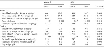
Data on final body weight, feed intake, feed efficiency and muscle weights (pectoralis superficialis muscle, pectoralis profundus muscle and leg) from Studies 1 and 2 are presented in Table 3. The final body weight was not significantly increased by feeding BBA in Study 1 but was significantly increased in Study 2 (P < 0·05), although feed intake did not change. As a result, BBA significantly increased feed efficiency in both studies (P < 0·05). The weight of the pectoralis superficialis muscle was either significantly (Study 1; P < 0·05) or slightly (Study 2; P = 0·059) increased by a diet containing BBA. Furthermore, the weights of the pectoralis profundus muscle and leg were significantly increased by BBA in Study 2 (P < 0·01). These results show that feeding chickens BBA improves their growth performance, and in particular their skeletal muscle growth.
Initial and final body weights, feed intake, feed efficiency and muscle weights of controls and chickens receiving BBA (30 mg/kg basal diet) in Studies 1 and 2 (mean values with their s.e., n 7–8 per group)

Control, chickens not receiving BBA; BBA, chickens receiving BBA (30 mg/kg basal diet); SEM, standard error of the mean.
* P values were obtained by Student's t test.
† Feeding periods of experimental diets were from 15 to 27 days and 0 to 27 days of age, for Studies 1 and 2, respectively.
‡ Leg, thighs+drumsticks.
Protein degradation of skeletal muscle
Nτ-Methylhistidine
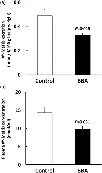
Data on excrement and plasma N τ-methylhistidine concentration from both studies are presented in Fig. 3. Butoxybutyl alcohol significantly decreased the N τ-methylhistidine concentration in both excrement (Fig. 3(a)) and plasma (Fig. 3(b)), which serve as indices of the rate of skeletal muscle protein degradation (P < 0·05 in both cases). These results demonstrate that BBA suppresses skeletal muscle protein degradation.
Changes of N τ-methylhistidine (N τ-MeHis) concentration of (a, Study 1) excrement and (b, Study 2) plasma of controls (□) and chickens receiving BBA (30 mg/kg basal diet, ■). Feeding periods of experimental diets were from 15 to 27 days and 0 to 27 days of age, for Studies 1 and 2, respectively. Values are means, with s.e. represented by vertical bars (n 7–8 per group). P values are shown above the columns.

Gene expression
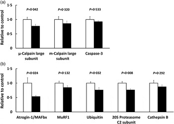
Then, in pectoralis profundus muscle, the effects of BBA on the expression of genes related to four proteolytic systems: (1) the calpain, (2) the caspase, (3) the lysosomal and (4) the ubiquitin–proteasome systems were examined (Fig. 4). In the first two systems (Fig. 4(a)), the mRNA level of the μ-calpain large subunit was significantly decreased by feeding BBA (P < 0·05), whereas the expression of the m-calpain large subunit and caspase-3 did not change. In the third system (Fig. 4(b)), the expression of cathepsin B was unchanged. In the fourth system (Fig. 4(b)), the mRNA level of atrogin-1/MAFbx was markedly decreased in response to BBA (P < 0·05), whereas that of MuRF1 was not statistically different. The expression of ubiquitin and the 20S proteasome C2 subunit were also significantly decreased by feeding BBA (P < 0·05 and P < 0·01, respectively). These results indicate that the reduction in the rate of proteolysis in skeletal muscle can be attributed to gene expression related to the calpain and ubiquitin–proteasome systems.
Changes of mRNA levels of (a) μ- and m-calpain large subunits and caspase-3, and (b) atrogin-1/muscle atrophy F-box (MAFbx), muscle RING finger 1 (MuRF1), ubiquitin, 20S proteasome C2 subunit and cathepsin B in pectoralis profundus muscle of controls (□) and chickens receiving BBA (30 mg/kg basal diet, ■) in Study 2. The experimental diet was administered from 0 to 27 days of age. The mRNA level of each target gene was normalized to the 18S rRNA mRNA level and expressed relative to the control level, which was arbitrarily set to 1·0. Values are means, with s.e. represented by vertical bars (n 6–7 per group). P values are shown above the columns.

DISCUSSION
In order to increase the productivity of edible meat from domestic animals, it is reasonable to focus on protein metabolism in skeletal muscle and regulate its turnover. One can propose the efficient usage of un-utilized feed materials capable of down-regulating protein degradation in skeletal muscle. In previous studies, it has been reported that feeding broiler chickens SDBP (Sultana et al. Reference Sultana, Kamizono, Furusono and Hayashi2011) or its hexane extracts (Kamizono et al. Reference Kamizono, Nakashima, Ohtsuka and Hayashi2010) promotes their growth due to the suppression of skeletal muscle proteolysis. It was speculated that BBA could be the active component of SDBP that produces these outcomes. Therefore, in the present study, BBA was synthesized and its chemical structure was identified. Then, two experiments were conducted to investigate the effects of feeding the synthesized BBA to broiler chickens.
Under the experimental conditions of the present study, a diet containing synthesized BBA promoted growth performance, especially skeletal muscle growth. Furthermore, the BBA diet led to a lower concentration of N τ-methylhistidine in the birds’ excrement and plasma. N τ-Methylhistidine is a component of the skeletal muscle proteins actin and myosin (Asatoor & Armstrong Reference Asatoor and Armstrong1967; Johnson et al. Reference Johnson, Harris and Perry1967). When skeletal muscle protein is degraded in rats, N τ-methylhistidine is excreted in the urine, and is not reused for protein synthesis due to the lack of N τ-methylhistidine tRNA (Young et al. Reference Young, Alexis, Baliga, Munro and Muecke1972). Thus, a quantitative change in the level of N τ-methylhistidine in excrement or plasma was used as an index of the degradation rate of skeletal muscle protein in the present study. The decrease in N τ-methylhistidine observed in the excrement and plasma of the BBA group suggests that the growth promotion due to an increase in skeletal muscle mass which occurred in response to dietary BBA might be caused by a decrease in the rate of protein degradation.
In order to investigate the underlying mechanism in more detail, the mRNA levels related to four proteolytic systems (i.e. calpain, caspase, lysosomal and ubiquitin–proteasome systems) in the pectoralis profundus muscle were measured. The calpain and caspase systems, which can initiate turnover of myofibrillar proteins, cleave titin and nebulin (Goll et al. Reference Goll, Thompson, Li, Wei and Cong2003), and actomyosin and actin (Du et al. Reference Du, Wang, Miereles, Bailey, Debigare, Zheng, Price and Mitch2004), respectively. Thereafter, the resultant cleavage fragments can be degraded by the lysosomal and/or ubiquitin–proteasome systems (Goll et al. Reference Goll, Neti, Mares and Thompson2008). Based on these findings, the present results were considered in two phases. Phase 1 (i.e. the initial stage of proteolysis) was represented by the μ- and m-calpain large subunits and caspase-3, with the mRNA level of the μ-calpain large subunit being significantly decreased by BBA supplementation, whereas the mRNA levels of the m-calpain large subunit and caspase-3 were unchanged. These results, which are in agreement with those of a previous study on the feeding of hexane extracts of SDBP (Kamizono et al. Reference Kamizono, Nakashima, Ohtsuka and Hayashi2010), suggest that the down-regulation of the calpain system can be attributed to a decrease in the initiation of protein degradation during BBA feeding. In both the present study and a previous one (Kamizono et al. Reference Kamizono, Nakashima, Ohtsuka and Hayashi2010), the effect of BBA on the mRNA level of the μ-calpain large subunit was larger than that for the m-calpain large subunit. The finding is in agreement with the results of Saleh et al. (Reference Saleh, Eid, Ebeid, Ohtsuka, Yamamoto and Hayashi2012), who reported that the mRNA level of the μ-calpain large subunit was more effective in increasing skeletal muscle size and reducing proteolysis than that of m-calpain in broiler chickens fed the fungi, Koji. Thus, the μ-calpain may play a more important role than the m-calpain in the system of skeletal muscle proteolysis of chickens. Phase 2 (i.e. the late stage of proteolysis) was represented by atrogin-1/MAFbx, MuRF1, ubiquitin, 20S proteasome C2 subunit and cathepsin B, with the mRNA level of atrogin-1/MAFbx being significantly decreased by BBA supplementation, whereas that of MuRF1 was not. Atrogin-1/MAFbx and MuRF1 are both ubiquitin ligases, which provide specificity to the ubiquitin–proteasome system. In particular, gene expression of atrogin-1/MAFbx has been suggested to play a critical role in the development of skeletal muscle proteolysis and muscle mass in broiler chickens (Nakashima et al. Reference Nakashima, Ishida and Katsumata2009; Kamizono et al. Reference Kamizono, Nakashima, Ohtsuka and Hayashi2010; Ohtsuka et al. Reference Ohtsuka, Kawatomi, Nakashima, Araki and Hayashi2011). The mRNA levels of ubiquitin and the 20S proteasome C2 subunit were decreased by BBA supplementation, but that of cathepsin B did not change, suggesting that the ubiquitin–proteasome system is involved in the decrease in protein degradation that occurs in the late stage of proteolysis during BBA feeding. This conclusion is supported by the proposal of Goll et al. (Reference Goll, Neti, Mares and Thompson2008) that, in skeletal muscle, the calpain and ubiquitin–proteasome systems play major roles in protein degradation. However, the mechanisms underlying the BBA-down-regulated calpain and ubiquitin–proteasome systems are, as yet, not clear, but may be associated with (1) hormone secretion (e.g. corticosterone and triiodothyronine related to proteolysis); (2) the prevention of the inflow of endotoxin from the intestinal tract, or (3) other unidentified metabolic pathways. It is also important to examine the transport of dietary BBA in the intestine and its metabolism to determine how BBA and its metabolites affect growth performance in chickens. Taken together, these results indicate that the reduction in the rate of proteolysis in skeletal muscle observed in the present study can be attributed to gene expression related to both the calpain and ubiquitin–proteasome systems.
The effects of dibutoxybutane (DBB) on growth and protein degradation in in vitro experiment using chicken muscle cells have been reported previously (Kamizono et al. Reference Kamizono, Ohtsuka, Hashimoto and Hayashi2013). dibutoxybutane is presumed to be formed by an addition of butyl alcohol to BBA, and is also included in SDBP. The result of a decreased atrogin-1/MAFbx mRNA level in the previous study in which DBB was used (Kamizono et al. Reference Kamizono, Ohtsuka, Hashimoto and Hayashi2013) is in agreement with that of the present study. In contrast, the mRNA levels (i.e. decreased MuRF1 and unchanged μ-calpain, ubiquitin and 20S proteasome C2 subunit) were not in agreement with those of the present study. These findings suggest that BBA and DBB may work by different mechanisms to improve skeletal muscle cell growth by decreasing the rate of protein degradation.
In conclusion, the present study provides the first evidence that feeding synthesized BBA to broiler chickens improves their growth performance due to a decrease in the protein degradation of skeletal muscle via the calpain and ubiquitin–proteasome systems. This highlights the potential dietary value of synthesized BBA, a finding that warrants further investigation.
The authors are grateful to Kagoshima Kumiai Chicken Foods Co., Ltd. (Kagoshima, Japan) for supply of broiler chickens and Kenya Hayasaka (Itochu Feed Mills Co., Ltd., Tokyo, Japan) for supplying the soybean meal. The present work was supported by Grant-in-Aid for Japan Society for the Promotion of Science (JSPS) Fellows (T. K., grant number 23·2096), and by Grant-in-Aid for Scientific Research (B) from JSPS (M. T., grant number 24380147).