




No CrossRef data available.
Article contents
The distribution of genotypes of the trypanosome parasite, Crithidia bombi, in populations of its host, Bombus terrestris
Published online by Cambridge University Press: 06 August 2004
Abstract
This study reports the distribution of parasite genotypes for the trypanosome Crithidia bombi across individual units (the colonies) in host populations of a social insect, the bumble bee Bombus terrestris. A number of microsatellite primers were developed and several of them were found to be polymorphic in our samples. Furthermore, a simple algorithm was used to identify the likely multi-locus genotypes present in multiply infected host individuals. The results demonstrated a remarkably high degree of genetic diversity among infections. A first sample from 1997 could only use a low resolution with 2 loci and showed a total of 11 different genotypes of C. bombi from 12 colonies. The sample from 2000 was analysed at 6 polymorphic loci and contained data from 8 colonies that were infected by 27 different C. bombi genotypes. Roughly 16% of all individual bees but half of all colonies (2000 sample) were infected with more than 1 genotype. The infections in the different colonies were also genetically distinct from each other, and the parasite population as a whole was in linkage disequilibrium and deviated from Hardy–Weinberg expectations. The highly structured and genetically diversified population of C. bombi is likely to result from strong genotypic host–parasite interactions.
Information
- Type
- Research Article
- Information
- Copyright
- 2004 Cambridge University Press
INTRODUCTION
The genetic structure of pathogen populations in their host populations has profound effects on many unsolved problems concerning the ecology and evolution of host–parasite interactions, such as the evolution of virulence (Nowak & May, 1994; Van Baalen & Sabelis, 1996; Grigg & Suzuki, 2003), the design of efficient strategies of intervention (Gupta, Ferguson & Anderson, 1997; Jelinek et al. 1999; Read & Taylor, 2001), or the possible role of parasites in maintaining genetic diversity and sexual reproduction in their hosts (Hamilton, Henderson & Moran, 1981; Kirzhner, Korol & Nevo, 1999; Lively, 1996; Lively & Apanius, 1995; Stahl et al. 1999). It has in fact long been recognized that most aspects of host–parasite co-evolution is based on genotype–genotype interactions (Thompson & Burdon, 1992; Antonovics, 1994; Wakelin & Walliker, 1996; Tibayrenc, 1999; Carius, Little & Ebert, 2001; Schmid-Hempel, 2003; Schmid-Hempel & Ebert, 2003). Despite the significance of these issues, comparatively little is known about the actual genetic structure of pathogen populations, and virtually all knowledge is restricted to important human pathogens such as Plasmodium, Trypanosoma, Leishmania, Toxoplasma or Cryptosporidium (Tibayrenc, 1995; Oliveira et al. 1998; Abderrazak et al. 1999; Jelinek et al. 1999; Tibayrenc & Ayala, 1999; MacLeod et al. 2000; Rich & Ayala, 2000; Leclerc et al. 2002; Cui et al. 2003; Grigg & Suzuki, 2003; Mallon et al. 2003).
What kind of population structure is found in pathogens is a controversial topic. Researchers disagree on whether micro-parasite populations (e.g. protozoans) in general are clonal or not, or whether there is partial clonality, that is, clonal population structure interrupted by occasional genetic exchange between lines (Thompson & Burdon, 1992; Tibayrenc, 1995; Dwyer, Elkinton & Buonaccorsi, 1997; Gibson & Stevens, 1999; Tibayrenc & Ayala, 1999, 2002; Awadalla et al. 2001). However, just as important is the question of how parasite genotypes distribute themselves over different host genotypes in a natural population regardless of how the types arose. For example, strong and specific immune selection (Gupta & Anderson, 1999; Lythgoe, 2002) should lead to parasite populations being well structured with respect to the host genotypes they can infect. Non-structured populations, in turn, could result from weak interactions, lack of clonality, or from frequent genetic exchange between lines that inhibit the appearance of persistent genetic entities. Whatever the exact processes, the investigation of the parasite population genetic structure is a crucial first element in the analysis of host–parasite co-evolution.
Here, we investigate pathogen population structure in a field situation, using the model system of the bumble bee, Bombus terrestris L., and its intestinal parasite, Crithidia bombi Lipa & Triggiani, 1980 (Trypanosomatidae) (Schmid-Hempel, 2001) to address this empirical question. Bumble bees, Bombus spp., are eusocial insects. The fertilized queens emerge from hibernation and start their colony in spring. When the first workers hatch, social life in the colony begins with workers raising brood and the queen laying eggs. Colonies can contain anything from a few dozen to many hundreds of workers. Bumble bees are host to a wide variety of parasites (Macfarlane, Lipa & Liu, 1995; Schmid-Hempel et al. 1990). C. bombi, in particular, is a flagellated protozoan that resides in the hind gut and is contracted by ingestion of cells on flowers (Durrer & Schmid-Hempel, 1994), by contact with infected nest mates, honey stores, or nest material. It resides in the intestines. A major effect of C. bombi is to constrain infected spring queens severely from successfully founding a colony, thus lowering fitness by 40% or more (Brown, Schmid-Hempel & Schmid-Hempel, 2003). Hence, C. bombi is likely to exert the necessary strong effects on its host that is required for a parasite-driven cycling of host and parasite genotype frequencies (Hamilton, 1980).
Earlier studies provided evidence that the interaction between C. bombi and its host, B. terrestris, is affected by their genotypes. For example, experimental transmission between colonies is affected by the identities of both, infected and susceptible colony and is associated with the genotype of the parasite (Schmid-Hempel et al. 1999; Schmid-Hempel & Schmid-Hempel, 1993). Although not clonal, colonies of social Hymenoptera (the ants, bees and wasps) typically consist of unusually closely related individuals. Colonies of B. terrestris, in particular, typically represent full sister groups (Schmid-Hempel & Schmid-Hempel, 2000) so that workers, on average, have unusually similar genotypes. Here, we will consider groups of workers from the same colony as representing host groups with similar genotypes as compared to other such worker groups from other, unrelated colonies that have a different genotypic background.
MATERIALS AND METHODS
Sources of bumble bees and of C. bombi
Emerging queens of B. terrestris were collected in the spring of two years of study, 1997 and 2000, in suitable areas north of Zurich, Switzerland. All 5 collection sites of 1997 (Rafz, Steinmaur, Buchberg, Warth, and Buchs) are separated flowering meadows within 20 km of one another. The sites of the 2000 collection were in the same geographical region but at different localities. The collected queens were kept in the laboratory and allowed to raise a colony according to standard protocols (e.g. Plowright & Jay, 1966). The colonies were kept in the laboratory and could complete their life-cycle under standard conditions (22 °C, 18[ratio ]6 L[ratio ]D cycle, 60% R.H.). Typically, a proportion of the queens that emerge from hibernation are naturally infected by C. bombi. As the queens transmit the infection to their workers, the colony becomes infected, too. These infections can then be typed by sampling the workers. In this way, a total of 163 individual workers from 12 infected colonies became available for the 1997 sample, and a total of 80 individuals from 8 colonies for 2000.
After our laboratory colonies had grown to mature size, we removed a random sample of workers and screened them for the presence of C. bombi. This can readily be detected as cells of the parasite appear in the host's faeces within a few days of infection (Schmid-Hempel & Schmid-Hempel, 1993). When at least 15 infected workers had been collected (n=8 in one case), we made whole-sample preparations by freeze-killing the workers and extracting C. bombi from the gut (where the parasite is most prevalent). These extracts served as our test samples. For the development of microsatellite primers, 2 clones of C. bombi, prepared by the method of Wu (1994) and stored in liquid nitrogen were used.
Development of microsatellite primers
Microsatellites for the 1997 samples.
Protocols followed those described by Glenn (2001). The total DNA of C. bombi was extracted with phenol–chloroform and digested overnight with Sau3 and Hae3. Products were separated on agarose (1997: 1·5%; 2000: 2%) and the DNA products in the range 200–700 bp were removed. Using different vectors (PTZ19, Gibco; pBluescript2, KSM13(+) plasmid, Stratagene; pGem-3Zf+, Promega; cutting with BamH1 and Hinc2), Crithidia DNA was incubated overnight at 16 °C followed by a further 4–6 h at 4 °C to ensure as many successful ligations as possible. The products were transformed into E. coli by electroporation (XL1-blue colonies, treated with CaCl2). Positive clones were then removed, further grown on Luria Broth (LB) plates and finally transferred by membrane lifting, and prepared for hybridization.
We screened the clones for the presence of microsatellite repetitive sequences using a range of di- and tri-nucleotide motifs. Oligolabelling was done either by labelling (Promega Labelling System) with alpha-33P or gamma-33P labelling with T4-Kinase (Pharmacia). The hybridized membranes were then exposed on X-ray film to identify clones with microsatellite repeats. Positive clones were removed from the plates and then grown on agar plates for a second hybridization step. This served to doubly check on positive clones. Positive clones were isolated from the microtitre plate and cultivated (at 37 °C) overnight in liquid LB-medium. Inserts of positive clones were directly amplified with forward/reverse primers (PUC/M13) with a standard PCR protocol, using DNA-Template, buffer and Super-Taq (denaturing for 5 min at 94 °C, followed by 35 cycles; 1 min at 94 °C, annealing at 52 °C (1 min), extension at 72 °C (1 min), followed by extension at 72 °C for 10 min). The products were purified using the Wizard™ PCR Preps DNA Purification System (Promega) and cycle sequenced (Promega Cycle Sequencing Kit).
Microsatellites for the 2000 samples.
During the project, novel techniques to develop primers became available (provided by B. Gautschi, personal communication). We used the Dynabeads enrichment library to develop more efficiently further microsatellite primers. Again, DNA was extracted with phenol–chloroform and digested overnight with Tsp509 I (Promega) at 37 °C and then separated on an agarose gel (2%). A 200–700 kb fragment was cut out and eluted in several steps under low- and high-salt buffers over DEAE–cellulose membranes, followed by chloroform–isoamyl alcohol/ethanol precipitation. For the cloning, the DNA was ligated with linkers TspAD short and TspAD long (12–16 h at 16 °C) and then amplified by PCR. Then, the 13-mer oligonucleotides CA and GA were marked at the 3′-end with biotin (Boehringer) with the help of terminal transferase (Roche). The targeted microsatellite fragments were subsequently retrieved with magnetic beads M-280 (Dynal) in a biotin–streptavidin reaction. The so-retrieved oligonucleotides were hybridized with C. bombi DNA fragments (5 h, 65 °C), purified, and amplified by PCR in several steps to enrich the targeted sequences. Finally, the products were transformed into E. coli by heat shock using TOP10F cells (30 s, 42 °C). Positive clones were removed and grown overnight in a microtitre plate in liquid LB medium with Ampicilin. Subsequently, the positive clones were again transferred onto a Hybond N+ membrane and prepared for hybridization. The oligonucleotides were labelled with fluorescein-11-dUTP by means of terminal transferase (1 h, 37 °C) (ECL nucleic acid labelling and detection system, Amersham Pharmacia). After several steps (hybridization, washing and detection) the membrane was exposed for 30 min to ECL radiofilters and then isolated from the microtitre plate and cultivated (at 37 °C) overnight in liquid LB medium. Plasmids were purified from the E. coli cells with the GFX Micro Plasmid Prep Kit (Amersham Pharmacia). Sequencing of the insert was performed by the Microsynth company (Zürich) after amplification.
For both years, primers were designed with program MacVector 4.1 based on the obtained sequences and synthesized by Microsynth. The primers were then tested against DNA obtained from the original clones. In addition, the primers were checked for functionality and variability in the products by using a number of faeces samples and homogenized tissue (gut) from, arbitrarily selected, infected workers, using the Chelex extraction method. Those primers found to produce variable products were used to screen the genotypes of infections in the laboratory colonies mentioned above. The PCR conditions were optimized such that amplification was maximized while minimizing mispriming. The products were sized with a size-marked (SequaMark, Research Genetics) electrophoresis run on the same gel. Sizes (length of repeat in bp) denote alleles. For the 1997 samples, polyacrylamide gels were used. For 2000, electrophoresis was carried out with the Spreadex System® (Elchrom).
Not all of the obtained sequences were suitable to design primers. Some repeats were too short in length, or the repeats were too close to the vector sequence. After eliminating such impractical cases, we were able to generate primers for 20 loci in the first round. Of those, 7 did not amplify in our test samples, 11 showed no variation in their products. But two of the primers (Cri 4 and Cri 16) reliably generated polymorphic products and were used to analyse the 1997 samples. For 2000, an additional 15 primers were found, 4 of which were polymorphic and easy to read (Cri 1.A5, 1.B6, 4.G9, 2.F10). The remaining ones did not amplify correctly, thus making available a total of 6 primers for the 2000 samples (Table 1). Primers amplified a product in most cases, but some probes could not be read and are entered as missing values. Therefore, sample sizes vary slightly among locations, colonies or loci (see figures given in Tables 2 and 4). However, previous studies show that the methods used here are sensitive enough to detect very low concentrations of different C. bombi strains in mixed infections (detection level is less than 80 cells in a probe (Schmid-Hempel et al. 1999)). Hence, we should have been able to detect also rare strains in mixed infections.

Table 2. Alleles found in workers naturally infected by Crithidia bombi in 1997 (Entries are allele lengths in bp.)


Table 4. Synopsis of alleles and inferred multi-locus genotype for Crithidia bombi infections (Missing values indicate failure to amplify product.)

Population genetic analysis
For the population genetic analysis of the data we used the web-based program GENEPOP (Raymond & Rousset, 1995) and GenAlEx for Excel (Peakall & Smouse, 2001). Colonies were treated as separate populations and each identified multi-locus genotype in the colony entered as one independent data point. The procedures follow standard population genetic methods and the details of computation can be taken from software described by Raymond & Rousset (1995) and Peakall & Smouse (2001). Analyses were run separately for singly and multiply infected animals.
RESULTS
Algorithm to identify C. bombi strains
Our data consist of allele patterns found at the different loci in any given bee. An allele combination at a single locus defines a single-locus genotype. The single-locus genotypes can be combined into a multi-locus genotype which, by definition, is the genotype of the infecting C. bombi strain. In the 1997 sample, the strain genotype therefore refers to the allele combinations at 2 loci, and in the 2000 sample to those at 6 loci. When several alleles are found at each of the loci, the number of different multi-locus genotypes that can be constructed, compatible with a given pattern of alleles, is potentially very large. In cases of such multiple infections we identified the minimum number of strain genotypes that can explain the pattern of alleles found, using the following procedure.
Based on previous knowledge, we assumed that populations of C. bombi consist of diploid lines (‘strains’) (Wu, 1994). This implies that with only one or two alleles found at a given locus, there is only one single-locus genotype present at this locus. Let a be the number of different alleles found at this single locus. Counting all possible homozygous infections (a) and all possible combinations for heterozygotes, there is a maximum of

possible strains. Similarly, the minimum number of independent infections, nmin, is determined by a. For a=1, there is 1 (homozygous) strain, for a=2 there is at least 1 (heterozygous) strain, and for a>2, the minimum number of independent infections is the value of (a/2) rounded up to the next integer (i.e. a=3 yields 2 strains, etc.). We thus calculated the values of nmin for all single loci in a given bee. Finally, we assumed that the most parsimonious estimate for the minimum number of independent strains infecting a given bee is the largest value of nmin over all loci, i.e. the maximum possible minimum number of strains estimated for this bee. With this number at hand, the following algorithm was used to find the multi-locus type of the infecting strains.
First, the allele combinations at each single locus for all bees in the entire sample were read and screened for instances where a single strain was infecting the bee – by definition where nmin=1 for all loci. In these cases, the combination of the respective single-locus genotypes defined the multi-locus type of the infecting strain. These single strains were then stored as reference strains in a library for later use. Then, the library was ranked according to the frequency with which the reference strains were found in the entire sample with the most frequent type ranking highest.
Second, the data were again screened to identify the combination of strains that would account for all observed alleles in a given bee. Bees with single infections (nmin=1) were the same as before and thus were simply assigned their respective single strain (multi-locus) genotypes. Typically, the majority of all bees fell in this category and could thus readily be assigned an infection. If more than 1 strain was present (nmin>1), we used a simple algorithm to identify what combinations of strains were most likely to make up the infection as follows.
All possible single-locus types for each locus were first defined in the entire sample. Then, all multi-locus types that can be constructed by combining the single-locus types in all possible ways were constructed (a potentially very large number; in practice, we found it to be around 35000 different variants). We then ranked these multi-locus types in the following way. First, the already identified reference strains (which had also been ‘constructed’ in the process) were ranked ahead of all other types (while preserving their respective ranking from before). We ranked all other multi-locus types based on the ranking of their constituent single-locus types. The single-locus types in turn were first ranked by always giving precedence to single-locus types that resulted from either a single observed allele (i.e. ‘true’ homozygotes; e.g. allele 123 giving the homozygote 123,123) or a combination of 2 alleles that were observed to be different (i.e. ‘true’ heterozygotes; e.g. alleles 123,125 yielding 123,125) over all other possible combinations. Within this ranking, single-locus types found more frequently inside a given colony were given precedence over those found less frequently. Third, single-locus types more prevalent in the population were given precedence over those less common in the population. This procedure resulted in the ranking of all multi-locus types in the entire sample. We considered this ranking to reflect the likelihood of the occurrence of strains based on plausibility (real observed types more likely than ‘constructed’ ones), frequency in a given colony and in the population at large.
For the multiply infected bees (nmin>1), we thus searched for the combination of strains that accounted for all observed alleles in a given bee and, at the same time, represented the highest possible ranking of its multi-locus types based on this list. For this, we first picked the nmin top-ranking multi-locus types (typically containing 1 or several reference strains) and checked whether this combination accounted for all alleles at all loci that were found in the bee. If this was the case, this combination of strains was considered to be most likely pattern of multiple infections. If the chosen combination did not account for all alleles, we picked the next combination of multi-locus types until the criterion of completeness of alleles was fulfilled. In picking the types, we first retained the highest-ranking strain while systematically exchanging the rest (nmin−1) of the strains to make up the total of infections. Hence, if necessary, the last (lowest ranking) type of this nmin−1 combination was replaced by the next lower ranking type from the list and alleles again checked. In this case, this was again swapped for the next lower type from the list, before stepping back and replacing the second-to-last (better-ranked) strain by its lower-ranking counterpart from the list to start another loop. Loops were carried out to the iteration depth (here set at 10 steps, but varying this value did not drastically affect the results). In the case where the first picked type was a reference strain, it was always kept in the combination. The algorithm quickly and efficiently assigned a plausible combination of likely strains for the multiply infected bees in the 2000 samples. The pattern of the 1997 sample was simple enough to assign infections by hand.
The samples of 1997
At locus Cri 4 all workers showed 2 alleles with the following exceptions (Table 2). All of the workers in colony no. 64 showed 3 alleles, and one worker in colony no. 39 also showed 3 alleles. The pattern at locus Cri 16 was similar. All workers showed 2 alleles with the following exceptions. All workers of colony no. 70 showed 1 allele, all workers of colony no. 64 showed 3 alleles. A total of 7 different alleles were found at locus Cri 4, and 3 different alleles at locus Cri 16. The pattern of alleles is consistent with the suggestion that C. bombi is diploid (Wu, 1994).
Judged by the multi-locus genotypes (loci Cri 4 and Cri 16), workers of most colonies were infected by only 1 genotype of C. bombi (Table 3). Workers of colony no. 64 were infected by at least 2 genotypes, similar to one worker in colony no. 39. Most colonies had their own, unique genotype, but genotypes nos 2 and 3 occurred in more than one colony (Table 3). However, as the data from the 2000 sample showed (see below), this coincidence is most likely due to the limited resolution achieved with two loci. There was no clear association between collection site and the genotypes found in the colonies.
The samples of 2000
Infections from 8 colonies (FF26 to FF228) were analysed. There was a maximum of 4 alleles found per locus and individual, but, typically, only one or two alleles per locus were present. The analysis yielded a total of 27 independent infections (i.e. multi-locus genotypes) as defined by particular combinations of alleles at the 6 loci (Table 5). None of the strains occurred in more than one colony. For half of the colonies (FF91, FF124, FF178, FF228) the procedure suggested a single infection. In the other half, the number of independent infections was estimated to be 5 (FF127, FF133) or 6 (FF26, FF112) (Table 5). In these colonies, 2–4 of the 10 tested bees were multiply infected, but with no more than a maximum of 2 genotypes (Table 4). Hence, a vast majority of bees (66 out of 79 where strains could be assigned; products of bee no. 4 in FF127 did not amplify) in our sample carried a single infection, while a minority (13 bees, 16·5%) carried 2 infections.

Population genetic analysis
The so identified multi-locus genotypes of C. bombi in different colonies were used to analyse the overall population genetic structure of the infections with the aid of the web-based program GENEPOP (Raymond & Rousset, 1995). Colonies were treated as separate populations and each identified multi-locus genotype in the colony entered as one independent data point. First, the analysis was run separately for the single-infected and for all animals (i.e. including the multiple-infected bees).
Not surprisingly, we found that the C. bombi populations in different colonies were highly significantly separated from one another, regardless of whether the analysis included only single-infected (for all loci: range of values for pairwise Fst: 0·107–0·578; for pairwise Rhost: 0·010–0·410) or all animals (for all loci: range of values for pairwise Fst: 0·108–0·577; for pairwise Rhost: 0·010–0·290). This would also be suggested by a visual inspection of Table 4. Similarly, for the entire population, the F-values indicated a strongly structured population across colonies for, both, single-infected (FST=0·237; FIS=−0·527) and all infected animals (FST=0·285; FIS=−0·631).
Furthermore, for each locus, as well as for all loci together, the population was highly significantly deviating from Hardy–Weinberg equilibrium (all P[Lt ]0·01; Fisher's method), again regardless of whether only single- or multiple-infected animals were included. Of all 15 combinations of 2 loci across all populations, 12 had allele frequencies that were in linkage disequilibrium (i.e. significant deviations P<0·02). Locus Cri 4.G9 was in equilibrium with locus Cri 16 (P=0·194) and locus Cri 1.B6 (P=0·173), and the latter with locus Cri 4 (P=0·098).
Within all colonies analysed separately, we found non-zero FIS-values regardless of whether only single-infected animals (for colony FF26: FIS=0·075; all other colonies FIS<0 with range −0·433 to −1·0) or all animals (for colony FF26: FIS=0·050; all other colonies FIS<0 with range −0·203 to −1·0) were included; none of the within-colony populations was in Hardy–Weinberg equilibrium (all at least P<0·021), irrespective of whether only single-infected or all animals were included, indicating a highly structured population for C. bombi.
DISCUSSION
At present, methodology does not allow the unambiguous identification of the kind and number of independent infection genotypes in naturally infected hosts, particularly those infected by several strains. However, based on the screening of alleles at several different loci, it is possible to infer a likely combination of strains with a reasonable algorithm such as we have done here. With this proviso, the most remarkable results of our study are, first, the remarkable degree of diversity of different parasite genotypes (11 genotypes in 12 colonies in 1997; and 27 genotypes in 8 colonies in 2000), and, secondly, the close association of distinguishable parasite genotypes with different colonies such that no strain is found in more than 1 colony, as particularly seen in the 2000 sample even though many alleles are shared. The overlap between colonies in 1997 was most likely due to a low resolution. Note that the different colonies used here represent different genotypic backgrounds (containing genetically similar workers) and thus the clear sorting of parasite genotypes according to colony gives evidence for a close host–parasite association.
The genetic evidence for the host–parasite association found here complements and confirms the existence of strong host–parasite genotypic interactions found in previous infection experiments in the same study system (Baer & Schmid-Hempel, 1999, 2003; Schmid-Hempel, 2001; Shykoff & Schmid-Hempel, 1991). The previous studies also strongly suggested that colonies differ in the ‘bandwidth’ of susceptibility to incoming infections. Workers of some colonies can become infected by transmission from many different sources while others are only susceptible to one parasite genotype (Schmid-Hempel & Schmid-Hempel, 1993). Similarly, in the current study, colonies vary in the number of infections they have.
Our results provide insight into the population structure of natural infections by a trypanosome parasite in an insect host. As indicated here, the degree of resolution in population genetic studies depends on the number of loci and the number of alleles that can be identified. In the case of the 1997 samples, 2 loci with 7 and 3 alleles, respectively, would define 168 distinguishable parasite genotypes (homo- and heterozygotes). Of those, 11 were realised in the 134 workers that were screened. An additional 47 genotypes could theoretically be represented by the allele pattern in the doubly infected colony no. 64. The 2000 samples revealed a potentially much larger number of different genotypes mainly due to the higher number of loci that were screened (potentially many tens of thousands in some cases). In some cases, the genotypes differed by only 1 allele among all 6 loci. Similarly, not one single genotype was the same in the 1997 and 2000 samples even though the samples were from the same geographical region. The combined evidence therefore suggests that the total diversity of multi-locus genotypes potentially present in a population of C. bombi is very large. On the other hand, the number of genotypes actually found in a given individual host, colony and year is much smaller.
The formal population genetic analysis of the 2000 samples confirmed that C. bombi populations in different colonies are highly separated. As a whole, and within each colony, the parasite population was far from Hardy–Weinberg equilibrium and most pairs of loci in linkage disequilibrium. In the present system, parasite transmission between colonies is frequent during the summer months (Durrer & Schmid-Hempel, 1994; Imhoof & Schmid-Hempel, 1998). Hence, it is likely that the strong genotypic interactions of C. bombi with its host B. terrestris (Schmid-Hempel et al. 1999; Shykoff & Schmid-Hempel, 1991) leads to the segregation of parasite genotypes across different host lines and maintains linkage equilibria due to associated epistatic interactions; only a subset of the entire population of C. bombi can then be sampled in any one colony (see Tibayrenc, 1996).
The ploidy number of trypanosomes and of the genus Crithidia is not generally known. Different parts of the genome may even have different levels of ploidy. For example, using allozymes and karyotype analyses Gibson, Garside & Bailey (1992) found that Trypanosoma brucei is diploid for tubulin and phospholipase C but triploid for a number of other important genes. The triploid state appears to coexist stably with the diploid state in other trypanosomes (Gibson & Stevens, 1999). However, the currently accepted working hypothesis for the related genera Trypanosoma and Leishmania is diploidy (Tait, 1980; Zajicek, 1991; Tibayrenc, 1995, 1996; Oliveira et al. 1998; Gibson & Stevens, 1999; Tibayrenc & Ayala, 1999; Gaunt et al. 2003). Votypka, Ray & Lukes (2001) also confirmed diploidy for C. fasciculata. For C. bombi, allozyme typing, pulsed-field gel electrophoresis (Wu, 1994) as well as the current and previous micro-satellite studies strongly indicate that C. bombi is diploid, too. Cases where more than 2 alleles are found per locus can readily be explained by multiple infections with different parasite genotypes as done here and does not require triploidy.
Our study was not explicitly designed to answer the question of whether C. bombi is clonal or whether genetic exchange occurs. Nevertheless, the remarkable contrast of similarity in the alleles at any one locus among all identified infections versus the clear divergence when multi-locus genotypes are considered, together with the known strong genotypic interactions established in experiments (Schmid-Hempel, 2001), tentatively suggests that genetic exchange may occur but that its products could be canalized by strong selection. Hence, the major process of genotypic diversification in C. bombi presumably is recombination rather than mutation. To our knowledge, C. bombi is monoxenic, i.e. has only 1 host in its life-cycle and cannot survive long outside its host (Schmid-Hempel et al. 1999). If any genetic exchange occurs, it would thus have to happen when a single host is infected by at least 2 different genotypes, as is the case for about 16% of the individual bees is our samples. The frequency of co-infections depends on the parameters of the epidemic of C. bombi, i.e. the force of infection, the transmission parameters, and so forth. C. bombi may thus show ‘epidemic clonality’ (Maynard Smith et al. 1993) where clones of the parasite only persist for some time until an opportunity for genetic exchange arises – this opportunity being dependent on the epidemicity of the disease. Further studies are in progress to clarify this question for C. bombi and for the dynamics of host–parasite co-evolution in the field.
We thank Mark Brown, Regula Schmid-Hempel and an anonymous reviewer for comments. Financially supported by the Swiss National Science Foundation (no. 3100-667733.01 to P.S.H.).
References
REFERENCES
ABDERRAZAK, S. B.,
OURY, B.,
LAL, A. A.,
BOSSENO, M. F.,
FORCE, B. P.,
DUJARDIN, J. P.,
FANDEUR, T.,
MOLEZ, J. F.,
KJELLBERG, F.,
AYALA, F. J. &
TIBAYRENC, M.
(1999).
Plasmodium falciparum: population genetic analysis by multilocus enzyme electrophoresis and other molecular markers.
Experimental Parasitology 92, 232–238.CrossRefGoogle Scholar
ANTONOVICS, J.
(1994).
The interplay of numerical and gene-frequency dynamics in host–pathogen systems. In
Ecological Genetics
(ed. Real, L.), pp. 129–145.
Princeton University Press,
Princeton NJ.
AWADALLA, P.,
WALLIKER, D.,
BABIKER, H. &
MACKINNON, M.
(2001).
The question of Plasmodium falciparum population structure.
Trends in Parasitology 17, 351–353.CrossRefGoogle Scholar
BAER, B. &
SCHMID-HEMPEL, P.
(1999).
Experimental variation in polyandry affects parasite loads and fitness in a bumblebee. Nature, London 397, 151–154.CrossRefGoogle Scholar
BAER, B. &
SCHMID-HEMPEL, P.
(2003).
Bumblebee workers from different sire groups vary in susceptibility to parasite infection.
Ecology Letters 6, 106–110.CrossRefGoogle Scholar
BROWN, M. J. F.,
SCHMID-HEMPEL, R. &
SCHMID-HEMPEL, P.
(2003).
Strong context-dependent virulence in a host–parasite system: reconciling genetic evidence with theory.
Journal of Animal Ecology 72, 994–1002.CrossRefGoogle Scholar
CARIUS, H. J.,
LITTLE, T. J. &
EBERT, D.
(2001).
Genetic variation in a host–parasite association: potential for coevolution and frequency-dependent selection.
Evolution 55, 1136–1145.CrossRefGoogle Scholar
CUI, L.,
ESCALANTE, A. A.,
IMWONG, M. &
SNOUNOU, G.
(2003).
The genetic diversity of Plasmodium vivax populations.
Trends in Parasitology 19, 220–226.CrossRefGoogle Scholar
DURRER, S. &
SCHMID-HEMPEL, P.
(1994).
Shared use of flowers leads to horizontal pathogen transmission.
Proceedings of the Royal Society Biological Sciences, Series B 258, 299–302.CrossRefGoogle Scholar
DWYER, G.,
ELKINTON, J. S. &
BUONACCORSI, J. P.
(1997).
Host heterogeneity in susceptibility and disease dynamics: tests of a mathematical model.
American Naturalist 150, 685–707.CrossRefGoogle Scholar
GAUNT, M. W.,
YEO, M.,
FRAME, I. A.,
STOTHARD, J. R.,
CARRASCO, H. J.,
TAYLOR, M. C.,
SOLIS MENA, S.,
VEAZEY, P.,
MILES, G. A. J.,
ACOSTA, N.,
ROJAS DE ARIAS, A. &
MILES, M. A.
(2003).
Mechanism of genetic exchange in American trypanosomes.
Nature, London 421, 936–939.CrossRefGoogle Scholar
GIBSON, W.,
GARSIDE, L. &
BAILEY, M.
(1992).
Trisomy and chromosome size changes in hybrid trypanosomes from a genetic cross between Trypanosoma brucei rhodesiense and T. b. brucei.
Molecular Biochemistry and Parasitology 52, 189–200.CrossRefGoogle Scholar
GIBSON, W. &
STEVENS, J.
(1999).
Genetic exchange in the Trypanosomatidae.
Advances in Parasitology 43, 1–46.CrossRefGoogle Scholar
GLENN, T. C.
(2001).
Microsatellite Manual, Version 6. Laboratory of Molecular Systematics, MRC 534, MSC, Smithsonian Institution, Washington DC.
GRIGG, M. E. &
SUZUKI, Y.
(2003).
Sexual recombination and clonal evolution of virulence in Toxoplasma.
Microbes and Infection 5, 685–690.CrossRefGoogle Scholar
GUPTA, S. &
ANDERSON, R. M.
(1999).
Population structure of pathogens: the role of immune selection.
Parasitology Today 15, 497–501.CrossRefGoogle Scholar
GUPTA, S.,
FERGUSON, N. M. &
ANDERSON, R. M.
(1997).
Vaccination and the population structure of antigenically diverse pathogens that exchange genetic material.
Proceedings of the Royal Society London, B 264, 1435–1443.CrossRefGoogle Scholar
HAMILTON, W. D.
(1980).
Sex versus non-sex versus parasite.
Oikos 35, 282–290.CrossRefGoogle Scholar
HAMILTON, W. D.,
HENDERSON, P. A. &
MORAN, N. A.
(1981).
Fluctuation of environment and coevolved antagonist polymorphism as factors in the maintenance of sex. In
Natural Selection and Social Behavior
(ed. Alexander, R. D. &
Tinkle, D. W.), pp. 363–381.
Chiron Press, New York.
IMHOOF, B. &
SCHMID-HEMPEL, P.
(1998). Colony success of Bombus terrestris and microparasitic infections in the field.
Insects Sociaux 46, 223–238.Google Scholar
JELINEK, T.,
KILIAN, A. H. D.,
WESTERMEIER, A.,
PROELL, S.,
KABAGAMBE, G.,
NOTHDURFT, H. D.,
VON, S. F. &
LEOSCHER, T.
(1999).
Population structure of recrudescent Plasmodium falciparum isolates from Western Uganda.
Tropical Medicine and International Health 4, 476–480.CrossRefGoogle Scholar
KIRZHNER, V. M.,
KOROL, A. B. &
NEVO, E.
(1999).
Abundant multilocus polymorphism caused by genetic interaction between species on trait-for-trait basis.
Journal of Theoretical Biology 198, 61–70.CrossRefGoogle Scholar
LECLERC, M. C.,
DURAND, P.,
DE, M. T.,
ROBERT, V. &
RENAUD, F.
(2002).
Genetic diversity and population structure of Plasmodium falciparum isolates from Dakar, Senegal, investigated from microsatellite and antigen determinant loci.
Microbes and Infection 4, 685–692.CrossRefGoogle Scholar
LIVELY, C. M.
(1996).
Host–parasite coevolution and sex – Do interactions between biological enemies maintain genetic variation and cross-fertilization?
BioScience 46, 107–114.Google Scholar
LIVELY, C. M. &
APANIUS, V.
(1995).
Genetic diversity in host–parasite interactions. In
Ecology of Infectious Diseases in Natural Populations
(
ed. Grenfell, B. T. &
Dobson, A. P.
), pp. 421–449.
Cambridge University Press,
Cambridge.CrossRef
LYTHGOE, K. A.
(2002).
Effects of acquired immunity and mating strategy on the genetic structure of parasite populations.
American Naturalist 159, 519–529.CrossRefGoogle Scholar
MACFARLANE, R. P.,
LIPA, J. J. &
LIU, H. J.
(1995).
Bumble bee pathogens and internal enemies.
Bee World 76, 130–148.CrossRefGoogle Scholar
MacLEOD, A.,
TWEEDIE, A.,
WELBURN, S. C.,
MAUDLIN, I.,
TURNER, C. M. R. &
TAIT, A.
(2000).
Minisatellite marker analysis of Trypanosoma brucei: reconciliation of clonal, panmictic, and epidemic population genetic structures.
Proceedings of the National Academy of Sciences, USA 97, 13442–13447.CrossRefGoogle Scholar
MALLON, M.,
MacLEOD, A.,
WASTLING, J.,
SMITH, H.,
REILLY, B. &
TAIT, A.
(2003).
Population structures and the role of genetic exchange in the zoonotic pathogen Cryptosporidium parvum.
Journal of Molecular Evolution 56, 407–417.CrossRefGoogle Scholar
MAYNARD SMITH, J.,
SMITH, N. H.,
O'ROURKE, M. &
SPARTT, B. G.
(1993).
How clonal are bacteria?
Proceedings of the National Academy of Sciences, USA 90, 4348–4388.Google Scholar
NOWAK, M. A. &
MAY, R. M.
(1994).
Superinfection and the evolution of parasite virulence.
Proceedings of the Royal Society of London, B 255, 81–89.CrossRefGoogle Scholar
OLIVEIRA, R. P.,
BROUDE, N. E.,
MACEDO, A. M.,
CANTOR, C. R.,
SMITH, C. L. &
PENA, S. D. J.
(1998).
Probing the genetic population structure of Trypanosoma cruzi with polymorphic microsatellites.
Proceedings of the National Academy of Sciences, USA 95, 3776–3780.CrossRefGoogle Scholar
PEAKALL, R. &
SMOUSE, P. E.
(2001).
GenAlEx V5: Genetic Analysis in Excel. Population Genetic Software for Teaching and Research.
Australian National University,
Canberra, Australia.
PLOWRIGHT, R. C. &
JAY, S. C.
(1966).
Rearing bumble bee colonies in captivity.
Journal of Apicultural Research 5, 155–165.CrossRefGoogle Scholar
RAYMOND, M. &
ROUSSET, F.
(1995).
GENEPOP (version 1.2): population genetics software for exact tests and ecumenicism.
Journal of Heredity 86, 248–249.Google Scholar
READ, A. F. &
TAYLOR, L. H.
(2001).
The ecology of genetically diverse infections.
Science 292, 1099–1102.CrossRefGoogle Scholar
RICH, S. M. &
AYALA, F. J.
(2000).
Population structure and recent evolution of Plasmodium falciparum.
Proceedings of the National Academy of Sciences, USA 97, 6994–7001.CrossRefGoogle Scholar
SCHMID-HEMPEL, P.
(2001).
On the evolutionary ecology of host–parasite interactions – adressing the questions with bumblebees and their parasites.
Naturwissenschaften 88, 147–158.CrossRefGoogle Scholar
SCHMID-HEMPEL, P.
(2003).
Variation in immune defence as a question of evolutionary ecology.
Proceedings of the Royal Society London, B 270, 357–366.CrossRefGoogle Scholar
SCHMID-HEMPEL, P. &
EBERT, D.
(2003).
On the evolutionary ecology of specific immune defence.
Trends in Ecology and Evolution 18, 27–32.CrossRefGoogle Scholar
SCHMID-HEMPEL, P.,
MÜLLER, C.,
SCHMID-HEMPEL, R. &
SHYKOFF, J. A.
(1990).
Frequency and ecological correlates of parasitism by conopid flies (Conopidae, Diptera) in populations of bumblebees.
Insectes Sociaux 37, 14–30.CrossRefGoogle Scholar
SCHMID-HEMPEL, P.,
PUHR, K.,
KRÜGER, N.,
REBER, C. &
SCHMID-HEMPEL, R.
(1999).
Dynamic and genetic consequences of variation in horizontal transmission for a microparasitic infection.
Evolution 53, 426–434.CrossRefGoogle Scholar
SCHMID-HEMPEL, P. &
SCHMID-HEMPEL, R.
(1993).
Transmission of a pathogen in Bombus terrestris, with a note on division of labour in social insects.
Behavioural Ecology and Sociobiology 33, 319–327.CrossRefGoogle Scholar
SCHMID-HEMPEL, R. &
SCHMID-HEMPEL, P.
(2000).
Female mating frequencies in social insects: Bombus spp. from Central Europe.
Insectes Sociaux 47, 36–41.Google Scholar
SHYKOFF, J. A. &
SCHMID-HEMPEL, P.
(1991).
Parasites and the advantage of genetic variability within social insect colonies.
Proceedings of the Royal Society London, B 243, 55–58.CrossRefGoogle Scholar
STAHL, E. A.,
DWYER, G.,
MAURICIO, R.,
KREITMANN, M. &
BERGELSON, J.
(1999).
Dynamics of disease resistance polymorphism at the Rpm1 locus of Arabidopsis.
Nature, London 400, 667–671.CrossRefGoogle Scholar
TAIT, A.
(1980).
Evidence for diploidy and mating in trypanosomes.
Nature, London 237, 536–538.CrossRefGoogle Scholar
THOMPSON, J. N. &
BURDON, J. J.
(1992),
Gene-for-gene coevolution between plants and parasites.
Nature, London 360, 121–125.CrossRefGoogle Scholar
TIBAYRENC, M.
(1995).
Population genetics of parasitic protozoa and other microorganisms.
Advances in Parasitology 36, 47–115.CrossRefGoogle Scholar
TIBAYRENC, M.
(1996).
Towards a unified evolutionary genetics of microorganisms.
Annual Review of Microbiology 50, 401–429.CrossRefGoogle Scholar
TIBAYRENC, M.
(1999).
Toward an integrated genetic epidemiology of parasitic protozoa and other pathogens.
Annual Review of Genetics 33, 449–477.CrossRefGoogle Scholar
TIBAYRENC, M. &
AYALA, F. J.
(1999).
Evolutionary genetics of Trypanosoma and Leishmania.
Microbes and Infection 1, 465–472.CrossRefGoogle Scholar
TIBAYRENC, M. &
AYALA, F. J.
(2002).
The clonal theory of parasitic protozoa: 12 years on.
Trends in Parasitology 18, 405–410.CrossRefGoogle Scholar
VAN BAALEN, M. &
SABELIS, M. W.
(1996).
The dynamics of multiple infection and the evolution of virulence.
American Naturalist 146, 881–910.Google Scholar
VOTYPKA, J.,
RAY, D. S. &
LUKES, J.
(2001).
Crithidia fasciculata: a test for genetic exchange.
Experimental Parasitology 99, 104–107.CrossRefGoogle Scholar
WAKELIN, D. &
WALLIKER, D.
(1996).
Genetics of host and parasite – implications for immunity, epidemiology and evolution.
Parasitology 112, S1–S4.Google Scholar
WU, W.
(1994).
Microevolutionary studies on a host–parasite interaction. Ph.D. thesis, Phil.-Naturwiss. Fakultät, University of Basel, Basel.
ZAJICEK, P.
(1991).
Trypanosoma cf. carassii: the combination of maloc enzyme patterns supports the theory of diploidy in trypanosomes.
International Journal for Parasitology 21, 753–755.Google Scholar

Table 1. Primers for variable microsatellite loci found in Crithidia bombi
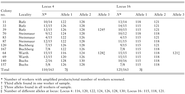
Table 2. Alleles found in workers naturally infected by Crithidia bombi in 1997

Table 3. Genotypes of Crithidia bombi found in 1997

Table 4. Synopsis of alleles and inferred multi-locus genotype for Crithidia bombi infections

Table 5. Genotypes of Crithidia bombi found in 2000
 You have
Access
- 67
- Cited by