Myocardial growth during fetal life is characterized by hyperplasia, which is strongly governed by the genetic programme, but which can be modulated by extrinsic factors.Reference Bishop1, Reference Saiki, Konig, Waddell and Rebeyka2 For example, an increase in cardiac workload induced by constriction of the outflow tract in the embryonic chick heart,Reference Clark, Hu, Frommelt, Vandekieft, Dummett and Tomanek3 or banding of the ascending aorta in the fetal guinea pig,Reference Saiki, Konig, Waddell and Rebeyka2 have been shown to increase ventricular growth by myocytic hyperplasia. Conversely, a decrease in left ventricular mechanical loading induced by ligation of the left atrial appendage in the embryonic chick heart was associated with decreased myocytic hyperplasia without affecting apoptosis.Reference Sedmera, Hu, Weiss, Keller, Denslow and Thompson4 The signalling mechanisms governing hyperplasia, or hypertrophy in postnatal stages, are not well defined. Studies using cell culture models, nonetheless, have suggested a role for various growth factors.Reference Hefti, Harder, Eppenberger and Schaub5, Reference Pasumarthi and Field6 Manipulation of growth factors, therefore, is an area of great interest in the investigation of the regulation of cell cycles as a possible basis for treating congenital cardiac disease. The primary players are angiopoietins, vascular endothelial growth factors, placental growth factors, platelet-derived growth factors, and fibroblast growth factors.Reference Sylven7
The chick embryo has a long and well-established history as a model system in developmental biology. Use of this model has contributed major concepts to many other disciplines.Reference Stern8 The recent release of the first draft of the sequence of its genome will further advance the chick as a model system.Reference Antin, Fallon and Schoenwolf9 In addition, the chick embryo is well suited to study experimental haemodynamic perturbations such as left atrial ligation.Reference Sedmera, Pexieder, Rychterova, Hu and Clark10, Reference Tobita and Keller11 By ligation of the left atrium, it is possible to produce a phenotype similar to hypoplastic left heart syndrome as seen in the human.Reference Sedmera, Cook, Shirali and McQuinn12 In this model, the normal pattern of flow of blood is redistributed from the developing left ventricle towards the right ventricle. Remodelling of ventricular myocardial architecture can be observed as soon as 2 days after ligation, and left ventricular myocardial volumes are significantly reduced after 4 days.Reference Sedmera, Pexieder, Rychterova, Hu and Clark10 These changes in myocardial architecture have been linked to alterations in the proliferative structure of the embryonic ventricle, as well as expression of fibroblast growth factor-2.Reference Sedmera, Hu, Weiss, Keller, Denslow and Thompson4
To date, 23 fibroblast growth factors have been identified in organisms ranging from nematodes to humans. The various factors vary in size from 17 to 34 kDa in vertebrates, and up to 84 kDa in Drosophila.Reference Itoh and Ornitz13 Members of the human family share a high affinity for heparin and heparin sulfate proteoglycans, as well as between 30 and 60% homology within a central core domain of 120 amino acids. This central core folds into twelve antiparallel ß-strands arranged into a triangular array necessary for receptor interaction.Reference Ornitz14 Fibroblast growth factors are widely expressed in developing and adult tissues. Their biological activities have been studied both experimentally and in life, and include roles in cellular proliferation, differentiation, and migration during embryonic development and tissue repair, wound healing and tumour angiogenesis in the adult organism.Reference Eswarakumar, Lax and Schlessinger15 In the settings of myocardial ischaemia, adenovirus-based delivery of fibroblast growth factor-5 was shown to be beneficial to recovery of myocardial function, both through stimulation of angiogenesis and direct effects on myocytes.Reference Lynch, Lee, Fallavollita, Canty and Suzuki16, Reference Suzuki, Lee, Fallavollita and Canty17 When mutated or improperly expressed, fibroblast growth factors can cause diverse abnormalities, ranging from morphogenetic disorders to cancer.Reference Demiroglu, Steer and Heath18
The fibroblast growth factors bind to fibroblast growth factor receptors located on the cell surface. These receptors are members of the receptor tyrosine kinase superfamily. The first fibroblast growth factor receptor was isolated in the mid-1980s from the chicken cDNA of a receptor capable of binding fibroblast growth factor-1.Reference Lee, Johnson, Cousens, Fried and Williams19 Since then, four such receptors have been characterized, named 1 through 4, which share between homologies of up to 72% at the protein level.Reference Powers, McLeskey and Wellstein20
Fibroblast growth factor-2 is known to modulate numerous cellular functions in a large panel of cells and tissues, including cellular proliferation and differentiation.Reference Detillieux, Sheikh, Kardami and Cattini21 Fibroblast growth factor-2 can be produced by cardiac myocytes and non-myocytes. It may act, therefore, in a paracrine or autocrine fashion in the heart.Reference Speir, Tanner, Gonzalez, Farris, Baird and Casscells22 Many studies implicate this second factor as the major functional fibroblast growth factor in the developing and adult myocardium.Reference Speir, Tanner, Gonzalez, Farris, Baird and Casscells22, Reference Lavine, Yu and White23 During the phase of rapid growth through hyperplasia, fibroblast growth factor-2, -9, and fibroblast growth factor receptor-1 are the only fibroblast growth factors and receptors that were shown to be expressed in mouse and chick ventricles.Reference Colvin, Feldman, Nadeau, Goldfarb and Ornitz24, Reference Pennisi, Ballard and Mikawa25 It was reported that fibroblast growth factor-2 is highly expressed during early cardiac morphogenesis in chick embryos,Reference Parlow, Bolender, Kokan-Moore and Lough26 and when inhibited by antisense oligonucleotides, a decrease in cellular proliferation was observed.Reference Sugi, Sasse and Lough27 This is in accord with experimental studies that have shown fibroblast growth factor-2 exerts a regulatory effect on cell proliferation and differentiation.Reference Jimenez, Sheikh and Jin28–Reference Velez, Aranega, Melguizo, Fernandez, Prados and Aranega30 Living studies, where beads soaked with fibroblast growth factor-2 were implanted adjacent to the heart of chick embryos at stages 18 through 24 showed a significant increase in myocytic proliferation.Reference Franciosi, Bolender, Lough and Kolesari31 Similarly, results from our laboratory have shown a localized decrease of fibroblast growth factor-2 in experimental models of embryonic ventricular hypoplasia.Reference Sedmera, Hu, Weiss, Keller, Denslow and Thompson4
Myocardial cells respond to fibroblast growth factor signalling via cognate receptors. Retrovirus-mediated suppression of fibroblast growth factor receptor-1, applied early during heart development, has been shown to result in significantly diminished myocardial proliferation after 8 to 10 days of further growth in the egg.Reference Mima, Ueno, Fischman, Williams and Mikawa32 Proliferation of cardiomyocytes is regulated at least in part by fibroblast growth factor signalling during the first week of development in the chick, but was thought subsequently to become independent of these factors.Reference Mikawa33 Other studies have shown that fibroblast growth factor-2 can signal in a receptor-independent manner, via heparin sulfate proteoglycans, and act directly on the cell nucleus, probably through its nuclear localization sequence.Reference Kardami, Liu, Kishore, Pasumarthi, Doble and Cattini34 Regulation of the number of receptors on the cell surface plays an important role in such signalling. This was demonstrated in neonatal myocyte cultures, in which overexpression of fibroblast growth factor receptor-1 led to an increase in myocytic proliferation, suggesting the continued importance of signalling during later development.Reference Sheikh, Fandrich, Kardami and Cattini35 In the chick model of hypoplastic left heart syndrome, there is a decrease in fibroblast growth factor-2, coupled with an upregulation of fibroblast growth factor receptor-1.Reference Sedmera, Hu, Weiss, Keller, Denslow and Thompson4 We thus set to verify the experimental relevance of fibroblast growth factor-2 signaling for myocytic proliferation during fetal stages, and the applicability of its modulation in the setting of experimental left heart hypoplasia.
Methods
Embryo selection
Fertile white Leghorn chicken eggs were purchased from Sunkist hatchery in Sumter, South Carolina, and delivered via courier in a temperature controlled manner in order to ensure viability and quality. The eggs were incubated blunt end up in a forced-draft constant-humidity incubator at 37.5°C, with continuous rocking, and studied at Hamburger-Hamilton stage 24, representing 4 days of incubation, stage 31 representing 7 days, and stage 35 equivalent to 9 days of an overall period of 46 stages or 21 days.Reference Hamburger and Hamilton36 Embryos that were dysmorphic at stage 24 were excluded from further study.
Ligation of left atrial appendage
At stage 24, the eggs were positioned under a dissecting microscope, and the eggshell and its membrane removed to expose the embryo. The embryo was gently turned over using an L-shaped fine glass hook to expose the left side, and microforceps were used to make a slit-like opening in the thoracic wall. A loop of 10–0 nylon suture was placed around the left atrial appendage and tightened.Reference Sedmera, Pexieder, Rychterova, Hu and Clark10 The embryo was then gently repositioned to its original right-side-up position. The opening in the egg was sealed with electrical tape, and the embryo returned to the incubator for reincubation without rocking until stage 31, equivalent to day 7. Normal embryos were windowed, but unoperated.
Microinjection of adenovirus
At stage 31, equivalent to day 7, and 3 days after ligation of the left atrial appendage, the embryos and their controls were aseptically microinjected with a volume of 8 microlitres of a viral suspension with a concentration of 109–1010 infectious particles per millilitre. The embryos were gently lifted from the eggs by their necks using a fine glass hook. Microinjection into the left ventricular myocardium was then performed via a pedal-controlled pressure injector and a hand-held needle. A volume of 3 microlitres of adenovirus expressing green fluorescent protein under the control of human cytomegalovirus promoter was used to visualize proper injection, while the remaining volume was used to deliver virus expressing the human fibroblast growth factor-2 coding sequence under the control of human cytomegalovirus promoter with simian virus 40 polyadenylation signal.Reference Nesbit, Nesbit and Bennett37 The fibroblast growth factor-2 virus was a kind gift from Dr Meenhard Herlyn at The Wistar Institute, Philadelphia, Penn. and custom propagated by Qbiogene, Montreal, Canada. Needles for microinjection were prepared from thin borosilicate glass using a vertical pipette puller. After resealing with electrical tape, the eggs were returned to a dedicated, humidified incubator set at 37.5°C and incubated with no rocking for an additional 48 hours prior to bromodeoxyuridine labelling and sampling. Intact controls were only windowed and vector controls were injected with the green fluorescent protein vector only. Additional controls included injection with solvent only. Fibroblast growth factor-2 protein was injected in the same manner on day 8. Only hearts expressing green fluorescent protein in the left ventricle, microscopically visible under blue illumination, were used for further analysis. All adenoviral microinjections and subsequent incubation and sampling were performed in an approved Biosafety Level 2 facility.
Bromodeoxyuridine pulse labelling and immunohistochemistry
Two hours prior to sacrifice, the embryos were labelled with a saturating dose of 50 micrograms of 5-bromodeoxyuridine in 200 microlitres of Tyrode’s saline applied directly over the vascular bed.Reference Sedmera, Hu, Weiss, Keller, Denslow and Thompson4 Only the hearts with a distinct phenotype for hypoplastic left heart syndrome were used for further analysis. The embryos were fixed in Dent’s fixative, comprising 80% Methanol and 20% dimethylsulfoxide, and processed into paraffin. Serial transverse sections were cut at 8 micrometers using a rotary microtome, and mounted on silane-coated slides. Histological analysis was performed using a triple staining protocol for sarcomeric actin as a myocytic marker, bromodeoxyuridine for S-phase nucleuses, and DRAQ5 for all nucleuses (Fig. 1).
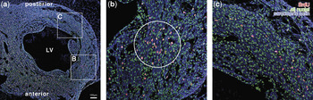
The hot spot of increased bromodeoxyuridine labelling after injection with recombinant fibroblast growth factor-2 protein. Immunohistochemical analysis of bromodeoxyuridine incorporation (a) shows increased abundance of S-phase nucleuses (red/yellow) in the injected area within wall of embryonic day 8 hypoplastic left ventricle (LV; panel b, circled) compared with the non-injected region (panel c). Nucleuses are shown in green, and sarcomeric actin, a myocytic marker, in blue.

Microscopy and image processing
Serial tissue sections with triple staining for bromodeoxyuridine, visualized by a cyanine-2 coupled secondary antibody, sarcomeric actin by cyanine-3, and DRAQ5 were examined on a Leica TCS SP2 AOBS confocal microscope. Fields from the ventricular free wall were recorded using a 40× oil immersion lens at 1024 by 1024 pixel resolution. The final images were made as maximum intensity projections of 7 optical sections, 1 micrometre apart, collapsed using Leica software to produce a single projection image. Cells were counted in Adobe Photoshop 7.0, where black dots were placed in separate layers over the nucleuses, then transferred for automated counting to ImageJ. The percentage of BrdU-labelled myocyte nucleuses of total myocytic nucleuses was then calculated.Reference Sedmera, Hu, Weiss, Keller, Denslow and Thompson4
Immunostaining for apoptosis
Immunofluorescence was performed on serial sections using a rabbit polyclonal antibody, anti-active caspase 3 detected by cyanine-3 coupled secondary. The sections were then counterstained with DRAQ5 nuclear dye, dehydrated, and cover-slipped with Depex mounting medium. Tissue sections were examined by Leica TCS SP2 AOBS microscopy as described above. Label intensity was determined by summing the corresponding pixel values in a 1024 by 1024 pixel image and dividing by the area covered by cells.
Single channel quantitative immunofluorescence
Sister sections were stained with rabbit polyclonal antibodies against green fluorescent protein, fibroblast growth factor-2, and fibroblast growth factor receptor-1, the latter both diluted by 1 in 50, followed by cyanine-5 and cyanine-3 coupled secondary antibodies, respectively. The sections were then counterstained with Hoechst 33342 nuclear dye. An additional set of sister sections was stained with anti-myosin heavy chain antibody followed by cyanine-2 coupled secondary antibody and Propidium Iodide nuclear counterstain.Reference Sedmera, Hu, Weiss, Keller, Denslow and Thompson4 Control slides included unstained sections to assess tissue autofluorescence, and secondary antibody only for control of nonspecific binding. Slides were subsequently dehydrated and cover-slipped with Depex mounting medium. For quantification, single optical section images were taken sequentially on a Leica TCS SP2 confocal system using a low power objective to minimize photobleaching. Negative controls with no primary antibody and tissue autofluorescence were used for background subtraction to determine specific signal.Reference Kajstura, Rota and Whang38 Gray level intensity was measured in region of interest positioned over carefully matched areas using ImageJ. The values are reported after background subtraction as mean plus or minus standard error. Black equals zero, and white, the maximum, equals 255 in an 8-bit image.
Statistical analysis
All data are shown as mean plus or minus standard error of the mean. Statistical comparison of differences between groups was performed after analysis of variance using an unpaired two-tailed Student’s t-test. For comparison of regions within the same heart, a paired t-test was used. Results were considered significant at 5% probability level.
Results
Optimization of mode of delivery
To determine the effect of fibroblast growth factor-2 on myocytic proliferation in the normal and hypoplastic chick heart, we delivered 2 micrograms of fibroblast growth factor-2 recombinant protein dissolved in 10 microlitres of sterile phosphate buffer saline by microinjection directly to the left ventricular myocardium on embryonic day 8. In the injected region, verified by tracer amount of MitoTracker Green, we observed 24 hours later a hot spot of increased bromodeoxyuridine labelling (Fig. 1), indicative of a localized increase in myocytic proliferation. Few embryos, however, survived until the time of sampling. It was previously reported that fibroblast growth factor-2 acts as a vasoconstrictor,Reference Zhou, Sutliff and Paul39 thereby raising the possibility that the embryos may be dying as a result of vasoconstriction. To test this hypothesis, we intravenously injected embryonic day 8 chick embryos in the shell-less culture with human recombinant fibroblast growth factor-2 protein and phosphate buffer saline alone, video recording the embryos every hour for 8 hrs. As time progressed, vasoconstriction was apparent, as well as a slowing heartbeat, until the death eventually occurred within 6 hours. We concluded that a mode of delivery independent of this complication was required.
Non-specific adenoviral infection does not influence survival or proliferation
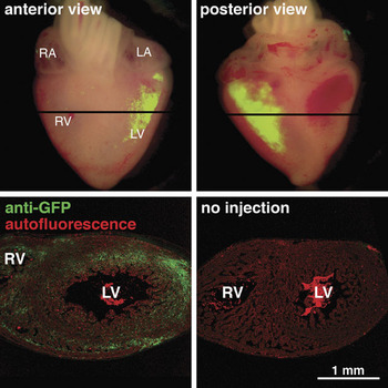
To circumvent vasoconstriction induced by fibroblast growth factor-2 following its introduction into the circulation, we used replication-deficient adenovirus as an alternative. This viral-based mode of delivery enabled sustained and localized delivery of fibroblast growth factor-2 to the embryonic heart. All embryos were injected with adenoviruses at embryonic day 7 and sampled at embryonic day 9. There was no difference in survival after viral infection in comparison with injection of the same volume of buffer. By using tracer amounts of a green fluorescent protein expressing adenovirus to target the left ventricular myocardium via microinjection, we were able to visualize a positive injection into the left ventricle via blue illumination (Fig. 2). Detection using a rabbit anti-green fluorescent protein antibody confirmed expression throughout the myocardium in tissue paraffin sections (Fig. 2).
Targeted gene delivery in the embryonic chick heart. Embryonic day 9 heart injected with green fluorescent protein-expressing adenovirus to the left ventricular myocardium shows intense staining within the wall. The expression is confirmed by anti-green fluorescent protein immunohistochemistry on paraffin sections, and extends throughout the wall. LA, left atrium, LV, left ventricle, RA, right atrium, RV, right ventricle.

To account for any non-specific effects of viral infection, we examined the effect of the green fluorescent protein expressing adenovirus on myocytic proliferation and apoptosis. Since the green fluorescent protein virus was used solely for visualization of positive left ventricle injection, it was important to rule out possible effects from injection and viral infection and overexpression of foreign protein. Embryos were pulse-labelled with bromodeoxyuridine 2 hours before sampling. No significant difference in proliferation between normal left ventricle and green fluorescent protein injected left ventricle was seen (19.8 plus or minus 1.2% versus 21.1 plus or minus 1.3%, statistically non-significant at 5% level). Additional samples were treated in a similar fashion and stained with anti-active Caspase 3 antibody in order to assess changes in apoptosis. No significant difference in apoptosis was found (data not shown).
Co-infection with fibroblast growth factor-2 adenovirus increases myocytic proliferation in the injected region
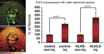
We utilized co-infection of green fluorescent protein and fibroblast growth factor-2 expressing adenoviruses as a means of verifying correct tissue targeting and induction of myocytic proliferation respectively. Immunohistochemistry was used on serial sections to identify the injected regions. Co-expression of the two proteins was seen using rabbit anti-green fluorescent protein and rabbit anti-fibroblast growth factor-2 antibodies on adjacent sections (Fig. 3). This region of co-expression was the region used for subsequent measurements of myocytic proliferation. The level of fibroblast growth factor-2 expression was determined in both control non-injected and control injected hearts and hypoplastic injected and hypoplastic non-injected hearts after 48 hours. A significant increase in fibroblast growth factor-2 expression in the injected region was seen in both control and hypoplastic hearts (Fig. 3).
Adenoviral co-expression and fibroblast growth factor-2 overexpression in the injected area. The panels on the left show on sister sections correlation between immunohistochemically detected expression of green fluorescent protein (in red) and fibroblast growth factor-2 (bottom panel, pseudocolor intensity scale) in the left ventricle (LV). The bar chart shows an average 2–3-fold increase in fibroblast growth factor-2 in the injected regions versus non-injected regions in the same heart.

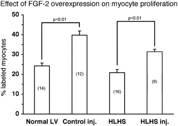
The effect of injection on myocytic proliferation was determined in control and hypoplastic hearts. There was a significant increase, of 45%, in proliferation in the normal hearts, and an increase of 39% in the hearts showing hypoplastic left heart syndrome (Fig. 4). These results demonstrate the mitogenic effect of fibroblast growth factor-2 in both normal and hypoplastic cultured hearts.
Fibroblast growth factor-2 overexpression increases myocytic proliferation. There is a significant increase in number of S-phase cells in both normal and hypoplastic (HLHS) left ventricles (LV) after treatment. The number of hearts analyzed in each group is given in parentheses.

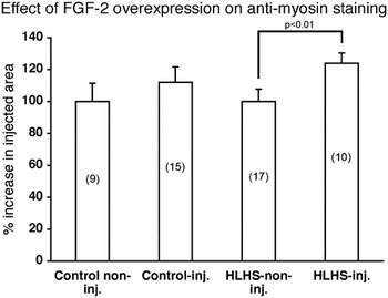
To determine the effect of fibroblast growth factor-2 overexpression on differentiation, anti-myosin immunostaining was performed on sister sections from hearts previously analyzed, in addition to already available anti-sarcomeric actin staining. The intensity of immunofluorescence was analyzed by fluorescent microscopy using low power objective to minimize photobleaching effects. As shown previously,Reference Sedmera, Hu, Weiss, Keller, Denslow and Thompson4 the hypoplastic left ventricle showed decrease in sarcomeric protein staining compared with normal. There was a significant increase in antimyosin expression in injected as opposed to non-injected hypoplastic hearts. No significant difference from normal left ventricles was observed in the control injected hearts (Fig. 5). The results were similar for the anti-sarcomeric actin staining (data not shown).
Fibroblast growth factor-2 overexpression increases myocyte differentiation. There is a significant increase in immunoreactivity against myosin heavy chains (MF20) in hypoplastic (HLHS) but not in the control left ventricles. Of note, the intensity of staining is significantly decreased in hypoplastic left heart syndrome compared with controls.

No structural anomalies are associated with viral injection after 5 days
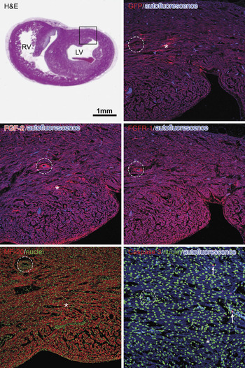
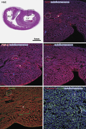
In order to determine the long-term effects of fibroblast growth factor-2 overexpression on chick hearts, various antibodies were used to investigate possible anomalies. Normal chick embryos were injected with green fluorescent protein/fibroblast growth factor-2 expressing adenoviruses at embryonic day 7, and sampled at embryonic day 12 instead of embryonic day 9. Morphological evaluation showed normal appearance of the ventricular wall in the injected region, and no increase in fibrous tissue. Antibodies staining for green fluorescent protein and fibroblast growth factor-2 showed continued presence at low levels in the viral injected region (Fig. 6). Fibroblast growth factor receptor-1 displayed no change in level following increases in exogenous fibroblast growth factor-2 ligand. Anti-myosin heavy chain staining showed normal alignment of the myofibrils, and anti-active Caspase 3 demonstrated lack of change in the number of apoptotic cells.
Long-term effects of fibroblast growth factor-2 overexpression on chick hearts. Normal embryos were injected with green fluorescent protein/fibroblast growth factor-2 adenoviruses on embryonic day 7 and sampled on embryonic day 12. Hematoxylin and Eosin staining shows the normal overall morphology of ventricular mid-section from which high-power views (boxed area, sister sections) were taken. A small coronary artery branch is circled for reference. Green fluorescent protein expression is still detectable (*), as well as small amount of fibroblast growth factor-2 over-expression (*); however, both fibroblast growth factor-2 and its major receptor are at this stage predominantly expressed in developing vascular structures. No myofiber disarray in the injected area (*) is visible in MF20 staining, and no significant active caspase 3 staining indicating apoptotic cells (arrows in last panel) is present. The last panel was taken at double magnification for increased resolution of single cells.
Discussion
There is growing evidence that fibroblast growth factor-2 is among a number of key growth factors that contribute to regulation of the cell cycle. Recently, platelet-derived growth factor-A has been identified as an epicardially-derived growth factor responsible for up to seven-tenths of the proliferative activity of the conditioned medium.Reference Kang, Gu, Li, Johnson, Sucov and Thomas40 Detailed dissection of relative contributions of such growth factors during normal development would be possible using specific inhibitors combined with bench or cultured experiments. The mitogenic effects of fibroblast growth factor-2 are mediated by binding to, and activation of, tyrosine kinase membrane receptors, among which the fibroblast growth factor receptor-1 isoform is predominant in cardiomyocytes at all developmental stages.Reference Sheikh, Hirst and Jin29 The importance of heparan sulfates in fibroblast growth factor signalling has been emphasized, albeit that the extent of the involvement of these molecules has yet to be resolved. Despite a wealth of data from bench experiments demonstrating that cardiomyoblasts proliferate in response to various fibroblast growth factors, including fibroblast growth factor-2, there is limited data that links fibroblast growth factor signaling to myocardial growth and development in life.
In this study, we sought to determine if the mitogenic activity of fibroblast growth factor-2 could be induced by microinjection of fibroblast growth factor-2 recombinant protein directly to the left ventricular myocardium of normal and hypoplastic left ventricles. We observed an area of increased bromodeoxyuridine labelling, indicative of a localized increase in myocytic proliferation, but only a few embryos survived until the time of sampling. Fibroblast growth factor-2 signaling network has recently increased in complexity by recognition of distinct developmentally regulated pathways for growth of myocardium and coronary vessels.Reference Lavine, Schmid, Smith and Ornitz41 Careful targetting of the intended cellular population, while avoiding undesirable effects of systemic administration, is thus pivotal. These results, as well as studies showing the vasoconstrictive action of fibroblast growth factor-2,Reference Zhou, Sutliff and Paul39 led us to the search for a mode of delivery that would have a more localized and sustained effect. Since retroviruses have low infection rates at these later embryonic stages, and potentially would lead to permanent expression, a replication-deficient adenovirus was employed. Adenovirus has high infection efficiency but is excluded from cells within a few weeks,Reference Liechty, Nesbit, Herlyn, Radu, Adzick and Crombleholme42 and our experiments with delayed sampling showed a steady decrease of the adenovirally delivered proteins after 5 days (Fig. 6). Adenovirus-mediated gene transfer has already proven successful in treating experimental conduction anomalies in a pig model.Reference Donahue, Heldman and Fraser43 Recently, fibroblast growth factor -5 delivered via adenovirus was shown to be beneficial in experimental myocardial ischaemia.Reference Lynch, Lee, Fallavollita, Canty and Suzuki16, Reference Suzuki, Lee, Fallavollita and Canty17 High-efficiency cardiac gene transfer was reported in mouse fetuses.Reference Christensen, Minamisawa, Gruber, Wang and Chien44 Although palliative in nature, such novel therapeutic strategies may prove usable in other severe congenital diseases.
We used adenoviruses expressing green fluorescent protein as a marker only, and fibroblast growth factor-2 in combination in order to visualize a positive injection into the left ventricle, and to induce myocytic proliferation, respectively. The effect of the green fluorescent protein expressing adenovirus alone on myocytic proliferation and apoptosis was analyzed in order to rule out any effects from this virus. No significant difference was found in either of these analyses. Levels of fibroblast growth factor-2 expression, and the effect of fibroblast growth factor-2 overexpression on myocytic proliferation, was determined in normal and hypoplastic left ventricles injected with the green fluorescent protein/fibroblast growth factor-2 adenoviruses. Fibroblast growth factor-2 expression was measured semi-quantitatively, and resulted in a significant increase in expression in both control and hypoplastic hearts. The increase was more pronounced in the hypoplastic hearts, where the baseline expression is significantly reduced in comparison to controls.Reference Sedmera, Hu, Weiss, Keller, Denslow and Thompson4 Likewise, the effect of fibroblast growth factor-2 overexpression on myocytic proliferation was measured in normal and hypoplastic hearts. An increase in proliferation of 45% was observed in the injected normal left ventricle, and of 39% in the injected hypoplastic left ventricle. These results are consistent with previous work demonstrating an increase in myocytic proliferation in response to exogenous fibroblast growth factor-2 at earlier embryonic stages.Reference Franciosi, Bolender, Lough and Kolesari31 These data also demonstrate the ability of fibroblast growth factor-2 to increase myocytic proliferation using fetal material in bench experiments without an increase in mechanical loading.
In the adult, viral infection would lead to both localized and global immune response, and contribution of recruited inflammatory cells to augmentation of tissue volume would be hard to exclude. In the embryo at this stage, in contrast, the immune system is immature, and no inflammatory reaction was previously reported by others,Reference Cheng, Litchenberg, Cole, Mikawa, Thompson and Gourdie45 or noted by us in response to viral injection. Similarly, the role of stem cells in maintenance of myocardial homeostasis and repair was recognized relatively recently.Reference Orlic, Kajstura and Chimenti46 Whatever the origin of stem cells in the heart, circulation-derived stem cells are not present in the myocardium prior to day 13 in the chick.Reference Zhang, Mustin and Reardon47 Thus, we attribute the observed proliferative effects of fibroblast growth factor-2 on myocytes to the protein itself, as corroborated also by experiments with direct injection of the recombinant protein, rather than to non-specific inflammatory response or stem cell activation, a known factor in adult heart injected with combinations of growth factors.Reference Linke, Muller and Nurzynska48
In addition to downregulation of fibroblast growth factor-2, anti-myosin staining was significantly reduced,Reference Sedmera, Hu, Weiss, Keller, Denslow and Thompson4 and myofibril organization delayed, in hypoplastic hearts.Reference Tobita, Garrison, Li, Tinney and Keller49 Normal and hypoplastic hearts injected with green fluorescent protein/fibroblast growth factor-2, therefore, were analyzed by immunostaining for MF20 following overexpression of fibroblast growth factor-2. No significant difference was observed in the normal hearts. On the other hand, a significant increase in myosin staining was observed in the hypoplastic hearts. There were similar changes in the intensity of staining of sarcomeric actin. Unfortunately, no other reliable cardiomyocytic-specific markers are presently available at this stage of development in birds. This data is consistent with previous work showing that fibroblast growth factor-2 plays a role in differentiation of cardiomyocytes during early stages of development.Reference Velez, Aranega, Melguizo, Fernandez, Prados and Aranega30
Our goal was to exploit the potential of fibroblast growth factor-2 as an inducer of myocytic proliferation, and apply it to an experimental model of hypoplastic left heart syndrome in the chick embryo. Previous studies have shown a downregulation of fibroblast growth factor-2 and upregulation of fibroblast growth factor receptor-1 in this experimental model.Reference Sedmera, Hu, Weiss, Keller, Denslow and Thompson4 It is likely that the depletion of myocardial growth factors is an effect of decreased loading, rather than a cause of hypoplastic left heart syndrome. Their addition, nonetheless, could have a positive effect, and result in sufficient improvement to allow postnatal loading of the left ventricle.
Further studies are necessary to understand the likely multifaceted effects exerted by fibroblast growth factor-2 in the heart. Among these, the long-term effects of fibroblast growth factor-2 should be analyzed, as fibroblast growth factor-2 has been implicated in certain cancers.Reference Dow and deVere White50, Reference Kumar-Singh, Weyler, Martin, Vermeulen and Van Marck51 We analyzed a set of green fluorescent protein/fibroblast growth factor-2 injected embryos at a later stage in order to assess any morphological or cellular abnormalities. No decrease in survival or gross morphological change was apparent. Our results demonstrated that both green fluorescent protein and fibroblast growth factor-2 were still expressed at these stages, although at lower levels. Caspase 3 staining showed no signs of abnormal apoptosis and myosin staining showed no disarray of myofibril alignment. These data are encouraging, albeit that even longer-term effects should be investigated.
One major limitation of this study is our inability to show, unlike with the surgical approachReference deAlmeida, McQuinn and Sedmera52 that the observed increase in myocytic proliferation will translate into increased myocardial mass. This would be an unrealistic expectation, since the injection was localized, providing a proof of principle rather than a direct attempt to provide a fully-formed therapy. While the survival of adenovirus-injected embryos was not decreased in comparison with saline-injected controls, longer term survival, past day 10, subsequent to ligation of the left atrial appendage results in selective loss of more severe phenotypes, providing an obstacle to unbiased interpretation of any changes in myocardial volumes. Future success of therapeutic strategies will depend on an increased understanding of the basic biology of fibroblast growth factor-2, and its diverse activities, as well as a more refined delivery system that enables spatial and temporal control of dosage.
Acknowledgements
We acknowledge Ms Chiffvon Stanley-Washington for excellent technical support, and Drs Rob Gourdie and Bobby Thompson for numerous helpful tips. Financial support from the National Institutes of Health (RR16434), March of Dimes (5-FY02–269), Ministry of Education of the Czech Republic (MSM 0021620806), and Academy of Sciences of the Czech Republic (AVOZ50450515, AV0Z5011059, GACR 304-08-0615, and Purkinje Fellowship) is gratefully acknowledged.