Introduction
Background
Daily changes in internal organ size and position can dramatically impact on tumour control and patient outcome.Reference Hector, Webb and Evans1, Reference Barker, Garden and Ang2 For this reason, on-treatment imaging verification has become the standard of care for many tumour sites as part of image-guided radiotherapy (IGRT).Reference Becker-Schiebe, Abac, Ahmad and Hoffmann3 Cone-beam computed tomography (CBCT) has become prominent among IGRT imaging methods largely due to the cross-sectional soft tissue imaging capability provided by the kilovoltage radiation.Reference Barney, Lee, Handrahan, Welsh, Cook and Sause4 Comparison of CBCT imaging data with the planned reference computed tomography (CT) images allows the relative positional and rotational discrepancies to be identified and an appropriate correction to be implemented. While this modality offers clear benefits to IGRT, image registration between the two datasets continues to be a clinical challenge in some tumour sites. The potential for real-time IGRTReference Schulze, Liang, Yan and Zhang5 has led to a surge in research into automated image-matching solutions.
Automated systems are capable of matching with millimetre accuracyReference Cui, Galvin and Straube6 but have shown a dependence on imaging dose, image quality and matching algorithms.Reference Barber, Sykes, Holloway and Thwaites7 Despite increasingly sophisticated automated matching algorithms and machine learning solutions, automated matches frequently require a trained clinical eye to approve or amend the proposed match. Time spent on manual interventions into these automated methods contributes significantly to the treatment time and clinician workload and directly affects the number of patients able to be treated per day as well as the amount of time a patient spends on the couch during treatment.8 Ultimately, this burdensome process limits how many IGRT cases each radiotherapy clinic can accommodate and how accessible IGRT is to the population in need.
To address this clinical challenge of time-intensive yet necessary clinical judgement during the image-matching phase, a novel Volumetric Image Matching Environment for Radiotherapy (VIMER) was developed. This unique application was developed by the authors to offer the clinician a more intuitive manual technique that promotes holistic volumetric matching. During a manual image match, the clinician currently is restricted to working with 2D orthogonal planes which can be time-intensive and limiting. The VIMER tool aims to improve the speed of this process by rendering both the primary and secondary patient CT datasets as a composite 3D model within a virtual reality (VR) environment such that anatomical borders are visualised as holistic volumes with maintained integrity in orientation rather than a collection of 2D slices. The aim of this initial study was to evaluate the potential value of VIMER for enhancing the clinical IGRT workflow.
Materials and Methods
Software development
A commercial VR headset presented a highly immersive and realistic interface readily available in VIMER. The application facilitates a dynamic cycle of inspecting matched patient dataset alignments and performing positional readjustments with intuitive confinements for optimal user control. The VIMER user interface replaces the traditional 2D orthogonal matching planar view with a novel 3D visualisation environment. Through the use of a pair of hand controllers, unique manipulation and anatomy slicing tools enable inspection of the matching boundaries between the two datasets at any location and in any orientation. Additional tools allow the user to perform intuitive six degree of freedom image-matching adjustments manually in the 3D virtual space. By doing so, the tool aims to visualise the relevant volumetric alignment information more efficiently when compared to the conventional 2D image-matching software.
Study design
Study participants were experienced therapy radiographers who also worked in an academic department feeding into IGRT training. As part of an early developmental phase, a pilot study was conducted to identify user training needs and preferences and inform the agile software development. A qualitative study was then conducted on a sample size of six participants in order to evaluate user experience when performing a 3D virtual image match using the VIMER tool. Qualitative methods were selected for several key reasons. First, as demonstrated by Kaplan’s research,Reference Kaplan9 one must take into consideration ‘cultural fit’. In addition to quantitative evidence-based benefits, the medical technology must align with the users’ cultural values for successful adoption into the clinical workflow. The exploration of cultural values which shape end-user acceptance cannot be informed by quantitative results and calls for qualitative research.
Qualitative methods allow descriptive findings to be disseminated and utilised to better one’s understanding of complex systems.Reference Heathfield, Pitty and Hanka10 The metrics of interest included user experience with using the tool, and overall user cognitive load experienced by the user during the image-matching process. The cognitive load and overall user experience within interactive applications are unique, subjective and complex. Accordingly, this qualitative approach was ideally suited for exploring and understanding the multiple facets that contribute to the users’ perceptions of VIMER.Reference Stewart and Shamdasani11 Therefore, an interview method was ideally suited for exploring and understanding the multiple facets that contributed to the users’ perception and experiences with this specific interactive application.Reference Naismith, Cheung, Ringsted and Cavalcanti12 Further down the road of VIMER development, clear quantifiable benefits in patient outcome must still provide the basis for the adoption of VIMER into the clinic.
Data collection
Data collection comprised of a 15-minute training session, a 15-minute practical image-matching session and a 15-minute individual interview with a sample group of six participants. Through an e-mail invitation, academic staff members who met the inclusion criteria of having significant CBCT image-matching experience were invited to participate. Individual interviews were chosen intentionally over a focus group format, so that each participant’s perception of the VIMER application was purely representation of his or her own formed opinions.Reference Tolley, Ulin, Mack, Robinson and Succop13
A training session took place with each participant prior to data collection in which subjects were given a brief demonstration of using the VIMER by the lead researcher. Following the demonstration, the participants were then introduced to a training application within the software and supported by ‘at-elbow’ guidance with regard to performing a manual image match. During training, participants were presented with two practice image-matching scenarios which allowed the user to gain control of the novel tools and techniques. This method of live demonstrations followed by user practice is common in medical technology user evaluation studies.Reference Eyal and Tendick14
Following training, the participants were challenged to complete two clinical image-matching cases which they had not seen before in the training modules (i.e., a parotid case and a bladder case). Two different anatomic sites were utilised to avoid site-based bias arising from the participants’ experiences. The first manual image-matching case contained a CT and CBCT male-torso dataset with a positional offset and poor image quality due to soft tissue contrast in the region of interest. The second manual image-matching case contained two head and neck datasets with a smaller positional offset but clearer image contrast. In each case, the participant was asked to perform an image match to the best of their ability. When users were satisfied with their image-matching solution, the experimental participant testing reached completion.
Directly after the user trial, each participant undertook an individual interview which lasted between 9 and 15 minutes to explore their perceptions of the VIMER application’s cognitive load and impact on image matching. A semi-structured approach comprised a series of questions based around the way in which VIMER presented the image-matching case, their interactive experiences and the learning process they adopted as first-time users. The interviews were audio-recorded and transcribed for further analysis.
Data analysis
A multi-stage qualitative thematic analysis process was used for the inductive analysis of the interview data. The purpose of thematic analysis was to identify patterns among the dataset and themes related to the viability and limitations of VIMER as a potential clinical matching tool.Reference Tolley, Ulin, Mack, Robinson and Succop13 An adapted six-step inductive thematic analysis and synthesis process was applied. Candidate themes were collated to formulate overarching themes which informed synthesis of conclusions.
Results
Emerging themes
Aside from user feedback concerning the software development, emerging candidate themes arising from the qualitative user evaluation were identified. From here, three major analytic themes surrounding the conceptual basis of this study were synthesised and further defined.
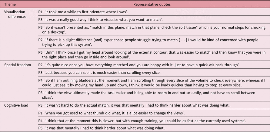
First, the clinicians found the immersive visualisation workspace to be different from their usual user interface but efficient for understanding patient anatomy and examining volumetric matching borders. Second, spatial freedom in view rendering was not always preferred, especially in positional adjustment tasks that required highly controlled movements on the order of millimetres. Users reported this freedom in spatial movement to be useful in accelerating the inspection process while promoting understanding of volumetric alignment. Lastly, although the different interface presented a significant barrier to the users, frustration and confusion decreased and user competency increased as the clinicians gained experience using the tool. It was noteworthy that users gained competence with the software after only 15 minutes of training. More substantial training is essential for future wide-scale implementation of the VIMER system. These resulting themes and representative quote are displayed in Table 1.
User feedback themes

Discussion
Spatial freedom
The development phase revealed radiographer preferences which were not otherwise evident in the literary review. More specifically, the radiotherapy users appeared to prefer viewing recognisable orthogonal surfaces. When presented with non-orthogonal viewing planes, the users struggled to recognise and spatially orient themselves with the anatomic data in VIMER. The unconventional views caused users to undertake unnecessary mental translations between the oblique view planes and the axial views and relationships they were accustomed to. While the software would allow for it, there was no reason or clinical motivation for unconventional rendering of anatomic extrusion in this situation. Therefore, the concept of unrestricted viewing geometry was limited down to a three-surface cubical shape which only rendered orthogonally normal-facing interior planes.
Likewise, the key motivation for introducing unrestricted spatial adjustments was to increase the user’s sense of control while manipulating the patient position. Unfortunately, the freedom to move patient anatomy freely through space appeared to be more of a drawback rather than a technological advancement. The radiotherapy user felt a lack of control over positional adjustments when they were granted complete freedom in movement of the secondary volumetric dataset. The user also felt a greater sense of motor control when seated compared to standing which caused anxiety and uncertainty in movement due to the loss of vision of the physical room surrounding the user. As a result, the users preferred button-controlled movements constricted to a range for adjusting patient position, but found the spatial freedom advantageous for inspecting matching borders all from a seated position. Thus, uniquely among VR-headset applications, VIMER is designed to operate from a seated workstation with confined positional adjustment tools. This maps well to the usual radiotherapy workplace environment.
VIMER advantages
Three distinct advantages can be identified from the user intervention with VIMER: volumetric visualisation, spatial freedom in inspection and a stereoscopic virtual viewing experience. First, the volumetric visualisation of corresponding 3D patient datasets presented the user with one merged object which displayed all relevant matching information in a manner which maintained orientation integrity, rather than three disconnect orthogonal views. Recent medical viewing technologies have capitalised on this computational voxel-rendering ability in diagnostics and surgical planning.Reference Wheeler, Deng and Toussaint15 Participants reported more intuitiveness in understanding spatial anatomic relationships as all the relevant data were presented ‘all at once’. This viewing modality eliminated the need to spatially reconstruct 3D models and could decrease intraobserver variability based on spatial ability in the long term.
Second, the spatial freedom experienced in slicing through the volumetric visualisation was preferred in parts of the match inspection process but not in others where fine-tuned precision was required. Previous studies have reported this ability to slice through anatomic models as a huge advantage for understanding spatial relationships of interior anatomy; however, no software has visualised two datasets in one space, a task specifically useful for image matching.Reference Seo, Smith and Cook16 The participants appreciated the ability to match volumetrically by inspecting the matching borders freely in 3D space. In the future, this method could lead towards more efficient pre-treatment verification of automated matches, which could ultimately lead towards shorter IGRT total treatment time.
Lastly, users reported advantages in the stereoscopic non-realistic viewing of multiple patient models in 3D space. The stereoscopic vision facilitated by the 3D immersive visualisation environment (IVE) has the primary function of depth perception. By presenting two concurrent models of the patient’s primary and secondary datasets, the viewer was able to gain a better spatial understanding of the relative patient positioning. In the long term, this tool may help facilitate more intuitive and rapid detection of gross setup errors or evaluating clinical target coverage during the planning stage.
Wider application
The applicability of this type of medical data visualisation demonstrated in VIMER extends beyond the image-matching uses explored in this study. Multiple medical technology companies have already released commercial VR applications which feature the ability to instantly visualise digital imaging and communication in medicine (DICOM) patient datasets as detailed 3D voxel-based models. The ability to segment and slice through these volumetric visualisations has already been released to the market. It is currently being used for surgical planning, anatomic training and intraoperative inspections of patient anatomy.Reference Shinomiya, Shindo and Kawanishi17 Within the past year, multiple other VR medical imaging companies have entered into the clinical field which is becoming increasingly competitive.
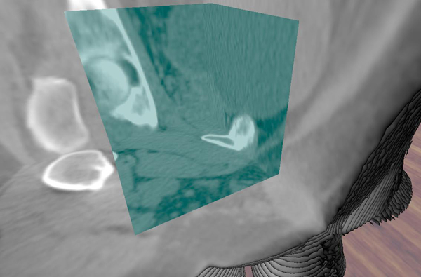
The VIMER addressed a niche area which has not yet been targeted by VR medical application companies. Certain aspects of the VIMER application, such as spatial freedom in volumetric inspection of matching borders, were found to be useful. Other features, such as full spatial freedom of movement for patient adjustments, were less useful and would suit more restricted controls. The VIMER may have a valuable role as an offline verification tool to assess the match quality between two already positioned datasets. The area of this fast-growing ‘VR in medicine’ market which VIMER has the ability to capitalise on most, however, is the dual-volume visualisation as seen in Figure 1.
Display of simultaneous visualisation of two separate dataset volumes within VIMER.

No other VR visualisation tool has rendered two 3D datasets in this composite volume fashion. Therefore, VIMER offers an innovative tool for comparing two sets of sequential patient datasets which might have slightly varying anatomical interior makeups. The potential impact of the application on clinical practice arises from the volumetric visualisation and the ability to view datasets in multiple orthogonal planes simultaneously. Combined with the unique interactive tools, this provides users with an intuitive image-matching platform that could speed up the manual matching process without delegating clinical decision-making to algorithms.
Limitations
The main limitation identified was insufficient participant training time which introduced a confounding learning variable into the user’s experience and overall opinion of VIMER. In 2015, a pilot study was conducted on an enhanced training method which provided instructional coaching to 28 radiotherapy students while they learned how to perform CBCT-based image matches on commercial software. Students were provided with general instructions, case details and image-matching protocols on 24 different cases over the course of a month.Reference Mohamoud, Ryan and Moseley18 When compared to precedential methods of training, it was clear that 15 minutes on a completely foreign image-matching technology with an assessment to follow on the same day did not provide the participants with sufficient training. For a fair assessment of the VIMER technology, the participants would have needed to undergo more substantial training sessions using a larger library of practice cases. This prolonged training requirement was not feasible with the timeline and resources devoted to this initial proof of principle project.
The disruptive change from the conventional image-matching user interface caused discomfort and frustration to the users which could deter new users. The radiotherapy workflow would not usually be best suited for a headset-based technology which prioritises mobility and takes the clinician away from the central control suite. The seated workstation interface, combined with adequate training should, however, overcome this challenge.
The other limitation of the study was the small sample size. While this was not an issue for this qualitative proof-of-principle phase, a quantitative assessment with a larger participant pool and more intensive training will be adopted for the next phases of preclinical testing.
Conclusions
The motivation behind the development of the VIMER was to deliver a more intuitive method of manual image matching to the clinical user. In an attempt to achieve this, the VIMER introduced volumetric visualisations of 3D patient datasets and novel tools within a 3D immersive VR environment alongside tools for manual volumetric image matching and match verification. Through a qualitative proof of principle approach, this study provided initial indications of the barriers and achievable benefits facilitated by the VIMER tool in image-matching procedures. Limitations include user discomfort with unfamiliar VR technology and substantial training requirements. Benefits included the novel interactive volumetric inspection of patient anatomy and border matching. While this matching tool was applied to CT/CBCT-based patient datasets, the technology could easily be transferred to MR-based patient datasets or to consecutive CBCT images over the course of a radiotherapy treatment course.
The volumetric inspection of an image match within a 3D IVE has clear benefits for integration into a radiotherapy clinical setting. The VIMER application provided the user with a holistic understanding of the image match quality between two 3D volumetric representations of anatomic patient datasets. The ability to volumetrically visualise two sets of 3D datasets in the same virtual space is a disruptive innovation which may transform the way that corresponding sets of imaging data are able to be inspected.
Author ORCID
P. Bridge 0000-0001-7704-9812
Acknowledgements
The authors gratefully acknowledge the support of the University of Liverpool Radiotherapy Department staff in completing this study. In addition, the datasets graciously provided by Simon Goldsworthy of the Taunton and Somerset NHS Foundation were pivotal for the development of this tool.
Financial Support
The research was supported financially by a scholarship provided by the US–UK Fulbright Foundation and additional funding from the University of Liverpool.
Conflict of Interest
The authors possess no conflicts of interest.
Ethical Standards
The authors assert that all procedures contributing to this work comply with the ethical standards of the UK Research Integrity guidelines on human experimentation and with the Helsinki Declaration of 1975, as revised in 2008, and has been approved by the Committee on Research Ethics with the School of Health Sciences at the University of Liverpool.