
Introduction
In Alzheimer's disease (AD), the accumulation of misfolded Aβ and pTau aggregates is accompanied by high levels of redox-active iron in senile plaque and neurofibril tangles.[Reference Bolt and Marchan1–Reference Zecca, Youdim, Riederer, Connor and Crichton3] These abnormal quantities of redox-active iron are in turn related to magnetite, a stable iron oxide that contains Fe+2 and has been found in iron-rich plaque core materials in human Alzheimer's brain[Reference Collingwood, Chong, Kasama, Cervera-Gontard, Dunin-Borkowski, Perry, Pósfai, Siedlak, Simpson, Smith and Dobson4, Reference Kirschvink, Kobayashi-Kirschvink and Woodford5] and in brain tissue sections from APP/PS1 transgenic mice.[Reference Gallagher, Finnegan, Grehana and Dobson6] Therefore, the redox-active form of magnetite may be transformed via the reductive effect of the iron–amyloid interaction because Fe+3 is favored over Fe+2 as the electronic state of isolated magnetite. Labile iron is free to participate in the Fenton reaction, resulting in the generation of reactive oxygen species, which go on to induce oxidative stress and neuronal damage.[Reference Smith, Harris, Sayre and Perry7, Reference Honda, Moreira, Liu, Siedlak, Zhu, Smith and Perry8] Based on x-ray magnetic circular dichroism examinations, ferrous iron formation following the co-aggregation of ferric iron and Aβ (1–42) peptide was accompanied by the formation of magnetite morphology, suggesting magnetite nanoparticles from ferrihydrites are generated by the reduction effect of Aβ.[Reference Everett, Céspedes, Shelford, Exley, Collingwood, Dobson, van der Laan, Jenkins, Arenholz and Telling9] Magnetite itself was shown to induce Aβ-fibril formation by direct interaction with the Aβ (1–42) peptide in vitro,[Reference Teller, Tahirbegi, Mir, Samitier and Soriano10–Reference Mahmoudi, Quinlan-Pluck, Monopoli, Sheibani, Vali, Dawson and Lynch12] in parallel with conversion into redox-active iron (II) as dominant redox status with a plaque-forming morphology of the iron–amyloid complex in vivo.[Reference Telling, Everett, Collingwood, Dobson, van der Laan, Gallagher, Wang and Hitchcock13] Therefore, magnetite in the iron–amyloid complex of AD plaque can be a therapeutic target to remove its neurotoxicity and the pre-existing senile plaque itself. Disruption of the magnetite-mediated iron–amyloid plaque or conversion of the redox-active iron into redox-inactive iron is a potential way to remove the neurotoxicity associated with AD plaque. The Coulomb nanoradiator (CNR) or x-ray photoelectric nanoradiator effect[Reference Kim, Seo, Kim, Kim, Chung, Kim and Ye14–Reference Kim, Titze, Schöffler, Trinter, Waitz, Voigtsberger, Sann, Meckel, Stuck, Lenz, Odenweller, Neumann, Schössler, Ullmann-Pfleger, Ulrich, Costa Fraga, Petridis, Metz, Jung, Grisenti, Czasch, Jagutzki, Schmidt, Jahnke, Schmidt-Böcking and Dörner17] produces burst emissions of fluorescent x-rays and low-energy electrons (LEEs) via Auger cascades and the interatomic/intermolecular Coulomb decay de-excitation pathway[Reference Gokhberg, Kolorenč, Kuleff and Cederbaum18, Reference Trinter, Schoeffler, Kim, Sturm, Cole, Neumann, Vredenborg, Williams, Bocharova, Guillemin, Simon, Belkacem, Landers, Weber, Schmidt-Böcking, Dörner and Jahnke19] from ionized atoms of high-Z nanoparticles under irradiation with a high-energy ion beam or synchrotron/conventional broad-band x-ray, respectively. Mechanism underlying gold nanoparticle-mediated therapeutic enhancement in radiotherapy[Reference Wolfe, Chatterjee, Lee, Grant, Bhattarai, Tailor, Goodrich, Nicolucci and Krishnan20, Reference Hainfeld, Dilmanian, Zhong, Slatkin, Kalef-Ezra and Smilowitz21] have been investigated also by other physical models.[Reference Taggart, McMahon, Currell, Prise and Butterworth22, Reference Shmatov23] Proton-irradiated magnetite has been shown to produce a CNR effect,[Reference Seo, Jeon, Han and Kim24, Reference Jeon, Han, Min, Seo, Ihm, Chang and Kim25] which give it therapeutic effects toward tumors as well as thrombus in animal models.[Reference Kim, Seo, Kim, Kim, Chung, Kim and Ye14, Reference Jeon, Han, Min, Seo, Ihm, Chang and Kim25] Importantly, traversing pristine Bragg-peak ion beam enables site-specific activation of dose-enhancement only at the target site with nanoparticles by a comparable dose with Bragg-peak (BPD) while depositing plateau dose (PD) in surrounding non-target, providing a distinctive opportunity for the treatment of diffusive target.[Reference Kim, Seo, Kim, Kim, Chung, Kim and Ye14, Reference Seo, Jeon, Han and Kim24, Reference Jeon, Han, Min, Seo, Ihm, Chang and Kim25]
In this study, we investigate the redox changes of iron and defibrillation of magnetite–amyloid complexes by traversing proton-induced CNR while not substantially impacting normal neuronal cells in an in vitro separate compartment model of neuronal cells and extracellular iron–amyloid complexes. Importantly, we found CNR-mediated redox changes from iron (II) to iron (III) and disruption of Aβ fibrils without impact to the neuronal cells when using a traversing proton beam, suggesting a potential application as a non-invasive Coulomb nanochelator therapy and amyloidolysis in AD plaque.
Experimental details
Primary culture of cortical neuron cells
Pregnant Sprague–Dawley rats were sacrificed by rapid cervical dislocation, and their embryos were removed and placed in sterile, ice-cold HBSS with 10 µg/mL gentamycin. Embryonic (days 18–19) rat cortices were gently removed from the medium, dissociated mechanically and plated on a series of single-well plates coated with coating buffer (1 mL of 10% PEI/1 L of borate). Cells were plated at a density of 500,000 cells/well in Neurobasal media with 2% B27, 0.5 mM L-glutamine, 1% penicillin–streptomycin (P/S) and 25 µM glutamate. Culture wells were incubated at 37°C with 5% CO2 and 95% humidity in plating medium for up to day in vitro (DIV) 2. In addition, the cells were incubated in Neurobasal media with 2% B27, 0.5 mM L-glutamine and 1% P/S (without glutamate) up to DIV 12 or 13 with periodic fluid replacement every 3 days.
Preparation of the Aβ/magnetite fibril complex
Magnetite was synthesized using a standard coprecipitation method. Aβ (1–42) peptide was obtained from a commercial manufacturer (AnyGen, AGP-8338, Seoul, Korea). The Aβ/magnetite complexes were prepared in a glove box under an N2 atmosphere to prevent changes in oxidation state of the iron. Monomeric synthetic Aβ (1–42) was dissolved in 0.1 M NaOH to afford a 220 µM Aβ-stock solution. This Aβ stock was left to stand for 30 min to ensure complete peptide dissolution prior to mixing with the 80 µg/mL magnetite solution. The Aβ/magnetite solutions were neutralized to pH 7 with 0.5 M hydrochloric acid. The final Aβ and metal concentrations were 110 µM and 40 µg/mL, respectively. The Aβ/magnetite solutions were incubated at 37°C for 144 h. A 10-μL aliquot of each Aβ/magnetite solution was pipetted onto carbon/Formvar-coated copper transmission electron microscopy (TEM) grids (TED PELLA Inc.), and excess liquid was removed to allow the deposition of Aβ/magnetite aggregates.
Proton irradiation and cell viability tests
A TEM grid with bound Aβ/magnetite fibril complexes was attached to the bottom of a single well, and cortical neuron cells were cultured in the well for 12 or 13 days. The culture medium was removed; however, 20 µL of the medium was left in the well to prevent the cells from drying prior to irradiation. Irradiation was performed at TR103 in KOMAC using a 100 MeV traversing proton beam with a plateau dose of 2 or 4 Gy (see Supplementary Materials Fig. S1). The dose rate was measured by 0.1 Gy/s in front of the sample position. After irradiation, Neurobasal media without glutamate was added to the cells, and the cells were incubated overnight. Cell viability was determined using the MTT (3-(4,5-Dimethylthiazol-2-yl)-2,5-diphenyltetrazolium bromide) assay. Disruption of the Aβ/magnetite fibrils was examined with Bio-TEM analysis and STXM imaging.
Scanning transmission x-ray microscopy/x-ray absorption spectroscopy (STXM–XAS) analyses
A 15-μL aliquot of each Aβ/magnetite solution (and their Aβ-free controls) was pipetted onto carbon/Formvar-coated copper TEM grids, and the excess liquid was removed. Sampling was performed under anoxic conditions after 30 min, 48 h, and 144 h of incubation at 37°C. The presence of the Aβ/magnetite fibrils was confirmed by thioflavin-T fluorescence measurement after 144 h of incubation,[Reference Cabrera, Mathews, Mezherichera, Beach, Deng, Neubert, Rostagnoa and Ghiso26] and the fibrils were investigated further by STXM/XAS and proton irradiation. STXM was performed to visualize the Aβ/magnetite complexes for the imaging and the elemental analysis of a given structure to a spatial resolution of 30 nm using beamline 10A at the Pohang Light Source (Pohang, Korea).
TEM grids containing the samples were mounted on stainless steel plates for STXM examination. Element-specific images displaying the nitrogen and iron contents of the amyloid aggregates were created by performing scans across the N K-edge (380–460 eV) and the Fe L 2,3-edges (770–730 eV).
The XAS spectra of Aβ/magnetite were recorded using the total electron yield method. To locate the sites of magnetite accumulation, two-dimensional Fe maps at a spatial resolution of 100 µm were obtained. Detailed XAS measurements across the Fe L 2,3 absorption-edge region (700–730 eV) of selected sites were conducted on the regions of Aβ/magnetite accumulation to study the Fe oxidation.
Results and discussion
The enhanced thioflavin-T fluorescence after 144 h of incubation indicated that the Aβ fibrils formed from the Aβ/magnetite complex (see Supplementary Materials Fig. S2).
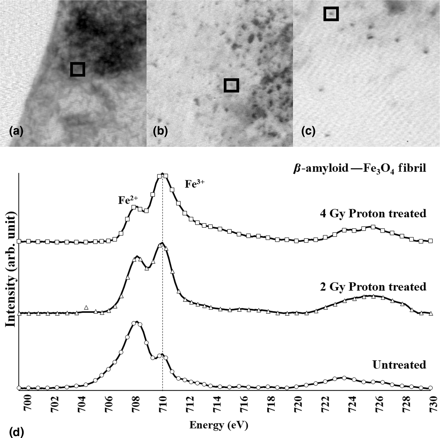
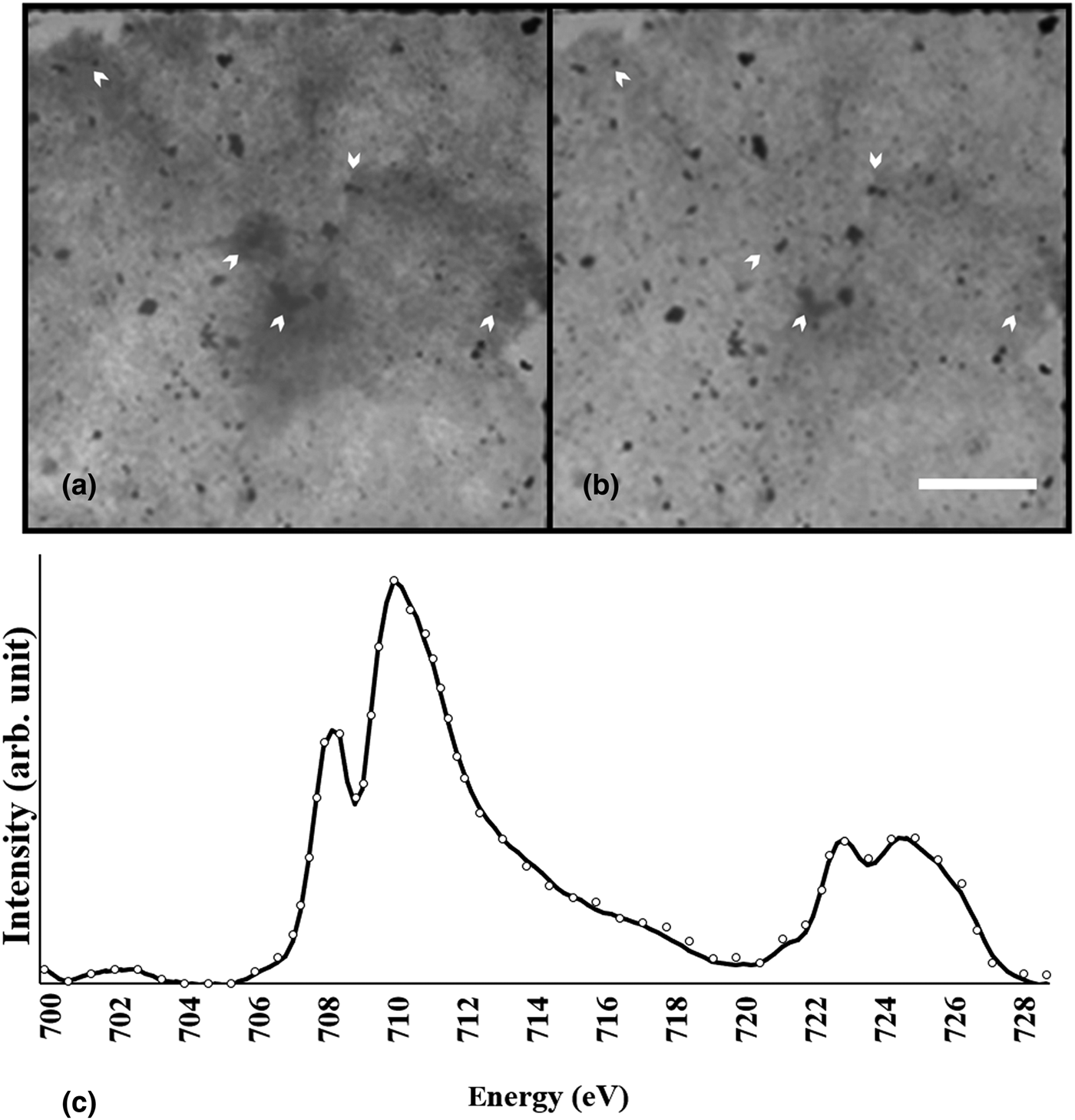
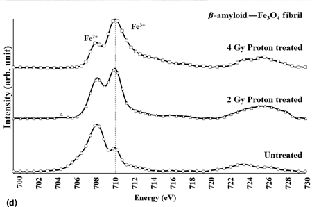
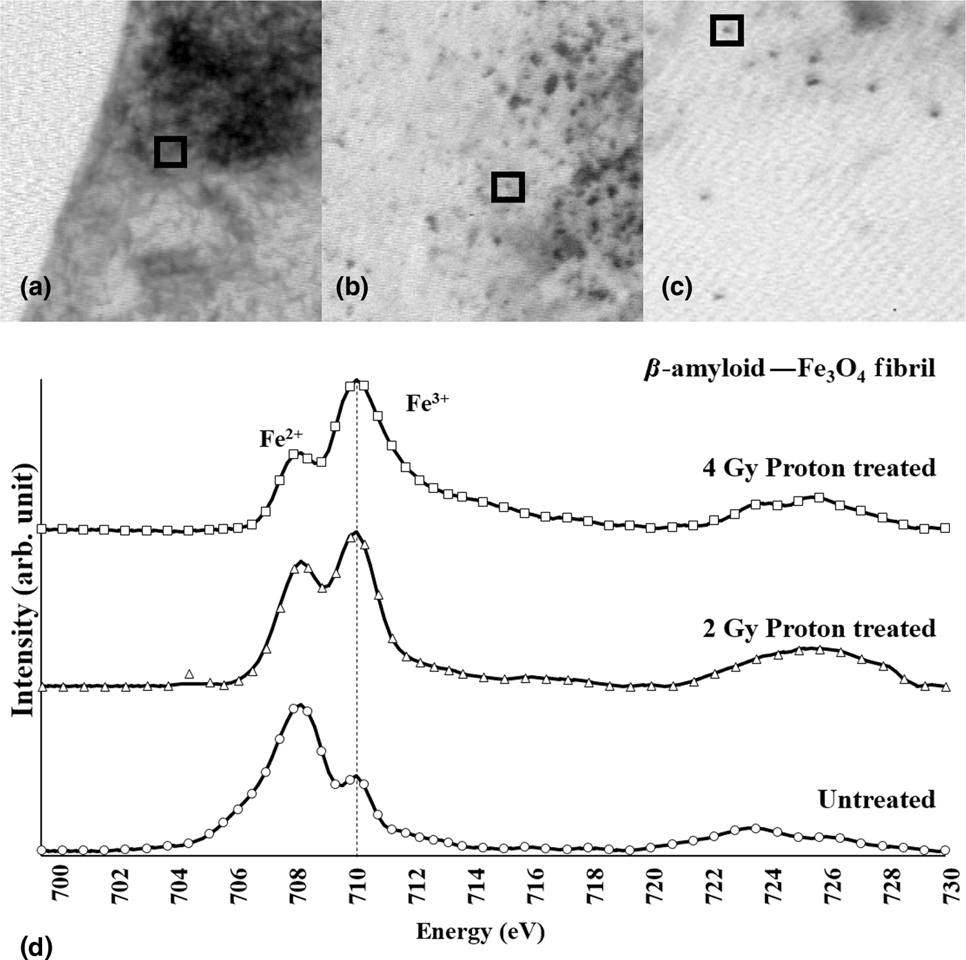
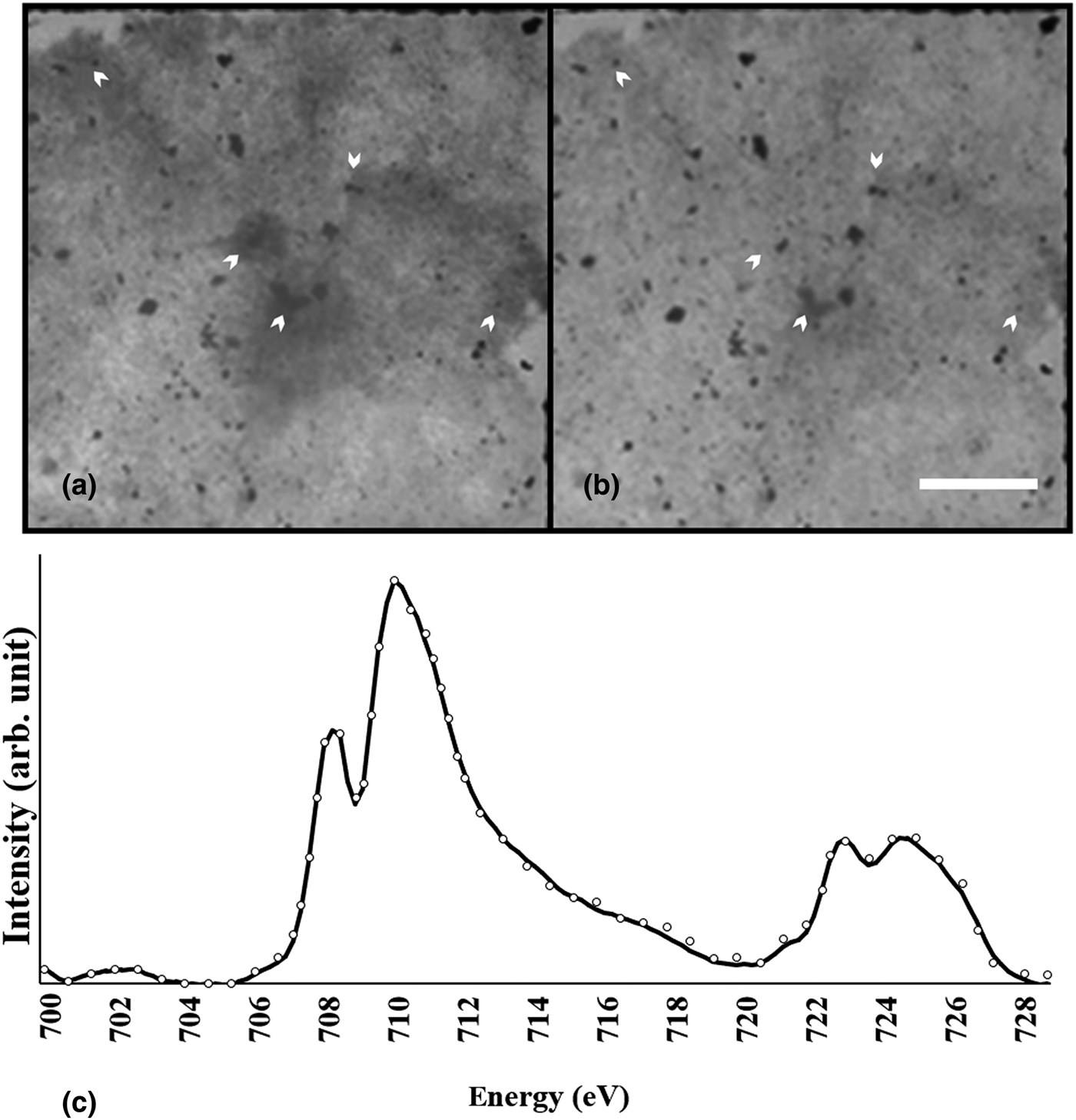
Ferrous iron [iron (II)] was observed in the STXM–XAS spectrum of the Aβ/magnetite fibrils after 144 h of incubation, as shown in Fig. 1. Because the preferred oxidation state of magnetite prior to the formation of aggregates with Aβ was ferric iron [Fe+2/Fe+3 = 0.5, dominant species is iron (III)], the presence of ferrous iron in the Aβ/magnetite fibrils indicates an increase in the Fe+2/Fe+3 ratio from the reduction effect of Aβ and stabilization of the magnetite. Aβ/magnetite fibrils exposed to a 100 MeV traversing proton beam showed a conversion of the iron to ferric iron in a dose-dependent manner. The relative portion of Fe+3 was higher in the Aβ/magnetite fibril subjected to proton irradiation at a dose of 4 Gy than that subjected to a dose of 2 Gy, as shown in Fig. 1. A nitrogen-STXM image in Fig. 2 showed multiple areas with reduced density in post-treatment image of Aβ/magnetite fibril compared with pre-treatment image where dominant ferric ion was detected, suggesting correlated disruption of peptide matrix in Aβ/magnetite fibril with redox change back to that of isolated magnetite.
X-ray absorption spectra measured across the Fe L 2,3 absorption edge (d) were obtained from magnetite deposit in the selected area (square box) of upper STXM image at 701 eV of untreated (a), proton irradiated 2 Gy (b), proton irradiated 4 Gy (c) of Aβ/magnetite complex samples. Fe2+ and Fe3+ were shown at 708.5 and 710 eV, respectively.
Nitrogen-mapping STXM image showing change in density (indicated by arrow) suggesting morphologic change in peptide matrix between untreated (a) and proton-treatment (b) where accompanied by redox change into dominant ferric ion (Fe+3, at 710 eV) in Fe L-edge XAS spectrum (c), presenting correlated Aβ fibril disruption with redox change of magnetite.
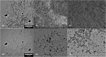
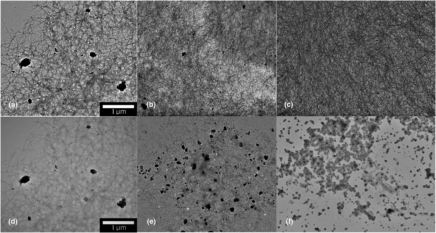
TEM imaging analysis of Aβ-fibril formation has been well-established technique distinguishing fibril and unformed fibril with a correlation of Thioflavin fluorescence.[Reference Teller, Tahirbegi, Mir, Samitier and Soriano10–Reference Mahmoudi, Quinlan-Pluck, Monopoli, Sheibani, Vali, Dawson and Lynch12] TEM images in this study showed typical feature of fibril strands in Aβ/magnetite [Figs. 3(a)–3(c)] and demonstrated various features of degraded fibril with a morphologic change in the damaged Aβ/magnetite under proton irradiation as shown in Figs. 3(d)–3(f). Faint or untangled fibril strands were obscured at a dose of 0.5–2 Gy possibly by CNR-mediated fibrinolysis. However, re-formation of Aβ fibril was possible reversibly by re-incubation of proton-irradiated dried Aβ/magnetite fibril in a well with a solution. More severe irreversible disruption of fibril was notable with remaining magnetite particles at more than 2-Gy traversing proton irradiation as shown in Fig. 3(f). Irreversible change of peptide matrix occurred depending on the local density of CNR-producing magnetite and proton dose. Proton irradiation was performed on the semi-dried sample (solvent was dried naturally in open air) while having a minimal surface hydration of the Aβ/magnetite complex at most in TEM grid and ruled out the potential damaging effect of primary radiolysis of bulk water solvent on Aβ/magnetite complex. Thus, this disruptive degradation of the Aβ/magnetite fibrils, effectively β-amyloidolysis, was attributed to primarily the CNR-mediated burst electron emission from the proton-irradiated magnetite.
TEM images of untreated (upper panels, a–c) and proton-treated (lower panel) Aβ/magnetite fibril complexes. Scale bar: 1 µm. All images were obtained with 100.0 kV and 8.0 µA. Depending on proton dose and local density of magnetite, obscured faint strands (0.5–1 Gy, d, e) or severe degradation (2 Gy, f) of Aβ fibril was notable.
Because magnetite-surface charge interacts with hydrophobic core (7–10 nm) of the Aβ peptide during fibrillation, and influence fibrillation and inhibition process,[Reference Mirsadeghi, Shanehsazzadeh, Atyabi and Dinarvand11, Reference Mahmoudi, Quinlan-Pluck, Monopoli, Sheibani, Vali, Dawson and Lynch12] CNR-electron emission at iron oxide nanoparticle (ION) site within Aβ fibril is able to damage fibril complex directly like LEE-mediated breaking DNA double-strand or alteration of protein structure.[Reference Boudaiffa, Cloutier, Hunting, Huels and Sanche27–Reference Rezaee, Hill and Jaffray30] This damage-inducing interaction would initiate electron emission upon nanoscaled energy deposition around magnetite and be transferred further via photoelectric absorption of fluorescent x-ray on surrounding iron atoms within directly proton-impact magnetite or surrounding magnetite as revealed from track analysis.[Reference McMahon, Hyland, Muir, Coulter, Jain, Butterworth, Schettino, Dickson, Hounsell, O'Sullivan, Prise, Hirst and Currell31, Reference Jeon and Kim32]
Since the emission of electrons from the CNR is followed by initial Coulomb ionization of the core–valence electrons of Fe atoms in the atomic nanocluster under proton irradiation, changes in the overall oxidation state of the magnetite are possible by rearrangement of Fe+2 and Fe+3 within individual magnetite nanocrystals. Therefore, changes in the crystallinity due to proton irradiation are possible, and detection studies of such changes are currently ongoing through either x-ray diffraction or electron diffraction analysis of the fibril-disrupted Aβ/magnetite aggregates.
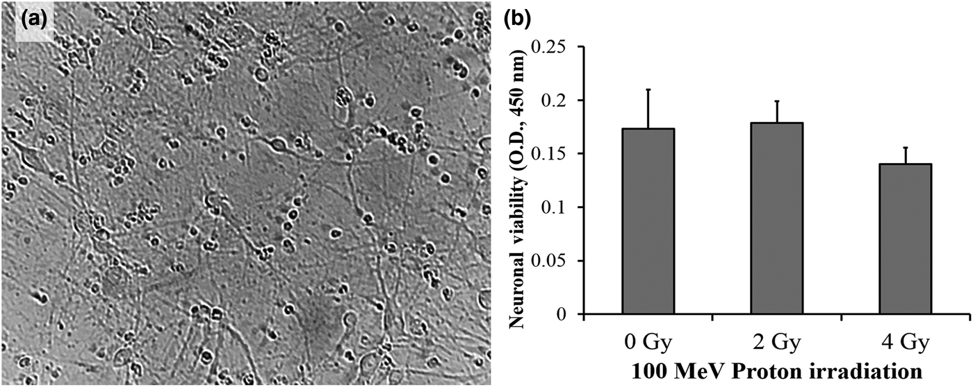
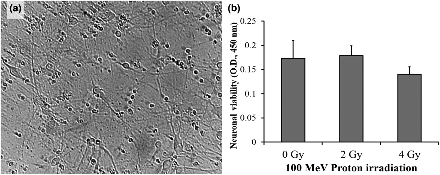
Because it is redox active, ferrous iron is known to induce neurotoxicity both in vitro and in vivo, and its conversion into redox-inactive ferric iron in CNR-treated Aβ/magnetite suggests a redox-change-based therapy for neurotoxic Aβ/magnetite fibrils or senile plaque of AD. In contrast, this traversing proton beam does not damage the neuronal cells in viability as shown in Fig. 4(b) while keeping neurite connection between neurons [Fig. 4(a)] that were exposed to PD of the traversing protons, suggesting redox inactivation by the Coulomb nanochelator without impacting the normal cells. 2–4 Gy is a typical dose of 1-day for fractionated proton therapy on brain tumor with total 40–50 Gy. However, single PD of 2–4 Gy is much less than accumulated dose in surrounding tissue after fractionated irradiation with total BPD of 40–50 Gy, suggesting potentially less damage than 3% reduction of hippocampal neurogenesis by a single 10-Gy dose of whole brain irradiation[Reference Monje, Mizumatsu, Fike and Palmer33] or other cognitive impairments by oncological radiotherapy.[Reference Robbins, Bourland, Cline, Wheeler and Deadwyler34, Reference Greene-Schloesser, Moore and Robbins35] Moreover, single plateau dose of traversing proton beam has normally much less linear energy transfer than that of BPD that is exploited in conventional oncological proton therapy,[Reference Robbins, Bourland, Cline, Wheeler and Deadwyler34, Reference Greene-Schloesser, Moore and Robbins35] suggesting a minimal effect on surrounding normal tissue. The prior study established removal of intravascular thrombus while saving vascular structure under same irradiation protocol as this study. A further safety study on cerebral microvessel is ongoing with transgenic mouse AD model.
Microscopic image showing neurite-connected neuronal cells 24 h after irradiation with a proton beam with a plateau dose of 2 Gy (a) and results of the MTT assay of the neuronal cells 24 h after traversing proton irradiation (b), indicating no or minimal effect on neuronal cell viability by 2–4 Gy plateau dose of 100 MeV traversing proton.
In conclusion, the traversing proton-mediated CNR effect induced the disruption of Aβ/magnetite fibrils, and the disruption was followed by the conversion of redox-active ferrous iron into redox-inactive ferric iron. The results of this study suggest a potential non-invasive Coulomb nanochelator therapy for the redox inactivation and β-amyloidolysis of neurotoxic senile plaque related to AD, and the applicability of such a therapy should be clarified with further in vivo animal model experiments.
Supplementary material
The supplementary material for this article can be found at https://doi.org/10.1557/mrc.2018.102
Acknowledgments
This work was performed with financial support from the National Research Foundation of Korea funded by the Ministry of Education, Science and Technology (grant numbers 2013M2B2B1075774 and 2015 M2A2A7A1045270).