Introduction
Olfactory hallucinations (OHs), or hallucinations of smell, occur in a range of conditions. These include temporal lobe epilepsy (Chen et al., Reference Chen, Shih, Yen, Lirng, Guo, Yu and Yiu2003), migraine (Fuller & Guiloff, Reference Fuller and Guiloff1987; Schreiber & Calvert, Reference Schreiber and Calvert1986), and schizophrenia (Kopala, Good, & Honer, Reference Kopala, Good and Honer1994). OHs in schizophrenia are of particular interest for several reasons. First, these symptoms are clinically significant with the vast majority of patients describing their OHs as intrusive and distressing (Kopala et al., Reference Kopala, Good and Honer1994), foul and unpleasant (Meats, Reference Meats1988). Stevenson, Langdon, and McGuire (Reference Stevenson, Langdon and McGuire2011) recently catalogued a wide range of OH experiences in schizophrenia. The OHs covered almost the entire spectrum of smells encountered in day-to-day life, with the vast majority being negatively valenced (e.g., smoky, decay, animal-like smells). Further attesting to their clinical significance, OHs in schizophrenia are associated with increased social anxiety and depression (Langdon, McGuire, Stevenson, & Catts, Reference Langdon, McGuire, Stevenson and Catts2011). OHs of smelling one's own body are of particular concern, being associated with self-deprecation (Langdon et al., Reference Langdon, McGuire, Stevenson and Catts2011). OHs are also indicative of poorer prognosis and more serious psychopathology. For example, Kwapil, Chapman, Chapman, and Miller (Reference Kwapil, Chapman, Chapman and Miller1996) found that premorbid OHs predict later onset of schizophrenia, as well as poorer global adjustment and social position. Research into “other” hallucinations in schizophrenia, which typically groups together OHs, with somatic, tactile, and gustatory hallucinations, also reports an earlier age-of-onset in people with hallucinations of this type (Lewandowski, DePaola, Camsari, Cohen, & Öngür, Reference Lewandowski, DePaola, Camsari, Cohen and Öngür2009). The relevance here is that earlier age-of-onset is indicative of increased illness severity (see, e.g., Delisi, Reference Delisi1992).
Despite their clinical significance, OHs in schizophrenia are under-researched and poorly understood. This has occurred, in part, because these symptoms are less prevalent than other hallucinations in schizophrenia. Auditory hallucinations are the most common, being present in 47–98% of patients with schizophrenia. Visual hallucinations are the second most common, being present in 14–69%, with the other modalities (including OHs) being present in only 4–25% (see, e.g., Langdon et al., Reference Langdon, McGuire, Stevenson and Catts2011, for a recent review). Prevalence rates vary, however, depending upon the samples (e.g., in-patients vs. out-patients), rating period (e.g., lifetime vs. current or past-month), and instrument used to probe presence. With regard to the latter, OHs are underrepresented in traditional diagnostic instruments and many symptom-rating scales, such as the widely used Positive and Negative Syndrome Scale (PANSS; Kay et al., Reference Kay, Opler, Spitzer, Williams, Fiszbein and Gorelick1991), rate all hallucinations on a single item. The implication here is that once hallucinations in any modality have been identified, typically auditory hallucinations, further probing may never occur. Recent studies that have more specifically probed OHs report a higher lifetime prevalence of up to 35% (Kopala et al., Reference Kopala, Good and Honer1994) and a past-month prevalence of 13–17% (Langdon et al., Reference Langdon, McGuire, Stevenson and Catts2011). Moreover, OHs can occur very frequently. Stevenson, Langdon, and McGuire (Reference Stevenson, Langdon and McGuire2011) found that OH patients self-report experiencing OHs up to 4 times per day.
OHs in schizophrenia are also theoretically important for several reasons. First, these symptoms are poorly explained by current cognitive models of hallucinations. Visual and auditory hallucinations are said to arise when imagery and/or involuntary sensory memories are externally misattributed (see Seal, Aleman, & McGuire, Reference Seal, Aleman and McGuire2004, for a review). However, olfactory imagery is relatively difficult to generate (Crowder & Schab, Reference Crowder and Schab1995; Herz, Reference Herz2000) and the capacity for involuntary olfactory memories is unknown, raising questions about the adequacy of such accounts. Neuropsychological understanding of OHs in schizophrenia is also limited. Consideration of the overlap between the neural underpinnings of OHs in other disorders and the neural basis of normal olfaction might advance neuropsychological understanding of OHs in schizophrenia.
With regard to OHs in other disorders, damage to the medial temporal lobes, especially the amygdala, appears to associate with OHs in temporal lobe epilepsy (see, e.g., Chen et al., Reference Chen, Shih, Yen, Lirng, Guo, Yu and Yiu2003; Chitanondh, Reference Chitanondh1966). Several researchers have also suggested that the predominance of negatively valenced OHs implicates abnormal amygdala function, associated with poor emotion processing, not only in temporal lobe epilepsy (Acharya, Acharya, & Luders, Reference Acharya, Acharya and Luders1998), but also in schizophrenia (Velakoulis, Reference Velakoulis2006).
The neural basis of normal olfaction (see Figure 1) also implicates amygdala dysfunction, as well as disruption to orbitofrontal cortex (OFC) functioning. Olfactory receptors within the nasal cavity have fibers that project to the olfactory bulb (OB) and then travel along the olfactory tract into the primary olfactory cortex (POC). Fibers from the POC then project into the OFC and secondary olfactory areas. Projections of olfactory tract fibers also connect the piriform cortex to the basolateral amygdala (Blumenfeld, Reference Blumenfeld2002; Hawkes & Doty, Reference Hawkes and Doty2009). Consistent with the latter claim, animal studies of the olfactory system in mice (Kang, Baum, & Cherry, Reference Kang, Baum and Cherry2009; Miyamichi et al., Reference Miyamichi, Amat, Moussavi, Wang, Wickersham, Wall and Luo2011) and the macaque monkey (Carmichael, Clugnet, & Price, Reference Carmichael, Clugnet and Price1994) have shown strong reciprocal connections between the amygdala and the OB.
Schematic representation of the direct (and indirect) pathways for normal odor processing within the olfactory system.

There is also extensive evidence of volume reductions in the amygdala and hippocampus in schizophrenia, as well as in other related structures, for example, the OFC (see Shenton, Dickey, Frumin, & McCarley, Reference Shenton, Dickey, Frumin and McCarley2001, for a review). Findings of this type accord with the proposal that disruptions to the amygdala, OFC and/or connections between these two regions might associate with OHs in schizophrenia. With regard to possible involvement of amygdala dysfunction, numerous studies have reported poor emotion recognition in schizophrenia, (see, e.g., Edwards, Jackson, & Pattison, Reference Edwards, Jackson and Pattison2002; Kohler & Brennan, Reference Kohler and Brennan2004). This impairment is particularly evident when patients view fearful facial expressions (Green, Williams, & Davidson, Reference Green, Williams and Davidson2003)—a prototypical test of amygdala function (see, e.g., Adolphs & Tranel, Reference Adolphs and Tranel2003). However, to the best of our knowledge, no studies have examined whether deficits of facial affect recognition might be more marked in people with schizophrenia and OHs.
Likewise, few studies have examined whether OFC dysfunction might associate with OHs in schizophrenia. There is overwhelming evidence that frontal brain regions are disrupted in schizophrenia, with numerous neuropsychological studies reporting evidence of executive deficits, in addition to generalized cognitive impairment (see, e.g., Heinrichs & Zakzanis, Reference Heinrichs and Zakzanis1998; Reichenberg & Harvey, Reference Reichenberg and Harvey2007, for reviews). However, neuropsychological studies have tended to focus more on tests of dorsolateral prefrontal cortex (DLFPC) than OFC function (see, e.g., Pantelis & Brewer, Reference Pantelis and Brewer1995, for discussion). The one notable exception is those studies that have used odor identification tests to assess the integrity of the OFC (and related semantic processing) in schizophrenia (see, e.g., Saoud, Hueber, Mandran, Dalery, & d'Amato, Reference Saoud, Hueber, Mandran, Dalery and d'Amato1998; Stedman & Clair, Reference Stedman and Clair1998). These studies have consistently found evidence of odor identification deficits in schizophrenia, but no evidence of an association with OHs. Specifically, both Kopala et al. (Reference Kopala, Good and Honer1994) and Stedman and Clair (Reference Stedman and Clair1998) found no correlation between odor identification deficits and OHs in schizophrenia.
However, odor identification is not the only function subserved by the OFC. The OFC is also critical for: processing reward associated with taste and odor; learning, including reversal learning, associated with both positive (i.e., rewarding) and negative (i.e., aversive) reinforcers; and social evaluations. Tasks that assess these other aspects of OFC functioning may yet reveal an association between the OFC and OHs in schizophrenia. One such task is the Object Alternation Task (OAT) from the Comparative Neuropsychological Tasks (CNT: Freedman, Reference Freedman1990; Oscar-Berman & Bardenhagen, Reference Oscar-Berman and Bardenhagen1998). This task was developed for use with the Delayed Alternation Task (DAT) to tease apart OFC from DLPFC functioning. The DAT is thought to tap into reward/punishment pathways, as well as measuring working memory, and the ability to shift set. As such, it is considered to tap both DLPFC and OFC function. In contrast, the OAT is said to measure the ability to inhibit responses, rather than merely shift set, while also tapping into reward/punishment pathways. It is, therefore, considered a more sensitive test of OFC function (see, e.g., Freedman, Black, Ebert, & Binns, Reference Freedman, Black, Ebert and Binns1998; Oscar-Berman & Bardenhagen, Reference Oscar-Berman and Bardenhagen1998). Of relevance here, Seidman et al. (Reference Seidman, Oscar-Berman, Kalinowski, Ajilore, Kremen, Faraone and Tsuang1995) found that patients with schizophrenia performed more poorly than controls on both the OAT and DAT. However, these authors did not consider whether OHs in schizophrenia might associate more specifically with performance deficits on the OAT.
We use two other, more experimental tests of OFC functioning. Both are variants of “theory of mind” task. Theory of Mind (ToM) refers to the capacity to infer the thoughts, feelings and intentions of others. Normal ToM function is subserved by a complex neural system involving the prefrontal cortex, superior temporal sulcus, and amygdala/temporopolar cortex (see, e.g., Gallagher & Frith, Reference Gallagher and Frith2003). Saxe and Kanwisher (Reference Saxe and Kanwisher2003) also highlight the role of the temporo-parietal junction. The ToM tests specifically selected for the present study involve evaluations of the social and emotional significance of inferred mental states. As such, these tests may rely more on OFC-amygdala connections than other, more classic ToM tasks, such as false-belief tasks (see Bell, Langdon, Siegert, & Ellis, Reference Bell, Langdon, Siegert and Ellis2010, and Langdon, Reference Langdon2005, for discussion of the taxonomy of ToM tasks, with a focus on schizophrenia research).
The first ToM task is the “Faux Pas” test, which requires participants to identify whether someone in a social vignette has said something that unknowingly upsets another character. This task was developed by Stone and colleagues to examine ToM deficits in patients with OFC lesions (Stone, Baron-Cohen, & Knight, Reference Stone, Baron-Cohen and Knight1998). The same task was later used to show ToM deficits in patients with bilateral amygdala lesions (Stone, Baron-Cohen, Calder, Keane, & Young, Reference Stone, Baron-Cohen, Calder, Keane and Young2003). The second ToM task is the “Reading the Mind in the Eyes Test” (RMET). When performing the RMET, participants are shown images of the eye regions of another person's face and must select a word that best describes the other person's likely thoughts and feelings. These other people's mental states typically have broader social significance, including being “accusing,” “suspicious,”or “flirtatious.” The RMET has been used to show ToM deficits in patients with fronto-temporal dementia. This is a condition that is associated with neuroanatomical and functional abnormalities of the OFC (see, e.g., Gregory et al., Reference Gregory, Lough, Stone, Erzinclioglu, Martin, Baron-Cohen and Hodges2002; Torralva et al., Reference Torralva, Kipps, Hodges, Clark, Bekinschtein, Roca and Manes2007). Stone et al. (Reference Stone, Baron-Cohen, Calder, Keane and Young2003) also used the RMET to show ToM deficits in patients with amygdala lesions, as have several other researchers (see, e.g., Adolphs, Baron-Cohen, & Tranel, Reference Adolphs, Baren-Cohen and Tranel2002; Shaw et al., Reference Shaw, Bramham, Lawrence, Morris, Baron-Cohen and David2005).
ToM deficits on tasks of this type have also been documented in schizophrenia (see, e.g., Bora, Yucel, & Pantelis, Reference Bora, Yucel and Pantelis2009; Langdon, Coltheart & Ward, Reference Langdon, Coltheart and Ward2006, Reference Langdon, Coltheart and Ward2010). However, no studies to date have considered whether ToM deficits are more severe in people with schizophrenia and OHs.
Study aims and hypotheses
Against this background, we compared the performances of patients with schizophrenia and OHs, patients with non-olfactory hallucinations (specifically, auditory-verbal hallucinations: AVHs), and healthy controls. We used a battery of neuropsychological tasks comprising: (a) standardized tests of odor identification and executive function; (b) the Object Alternation Task (OAT) and the Delayed Alternation Task (DAT) from the CNT, designed to tease apart DLPFC and OFC function; (c) an experimental test of facial affect recognition, used traditionally to index amygdala function; and (d) two ToM tasks (the Faux Pas test and the Reading the Mind in the Eyes Test: RMET), reported elsewhere to tap both OFC and amygdala function.
Consistent with previous research, we expected patients, as a whole, to perform more poorly than controls across all tasks, with no differences between OH and AVH patients on the standardized tests of odor identification and executive function. In contrast, it was hypothesized that, if amygdala dysfunction, as indexed by poor emotion recognition, contributes to OHs in schizophrenia, then OH patients will be more impaired than AVH patients on the facial affect recognition test. It was further hypothesized that, if OFC and related amygdala functioning, as indexed by our tests of ToM and reward/punishment processing, is more compromised in people with schizophrenia and OHs, then OH patients will also be more impaired on the two ToM tasks and the OAT, but not the DAT.
Methods
Participants
Potential clinical participants were recruited initially from participants who had taken part in a previous telephone survey of 51 patients who self-reported at least one OH experience in the previous 12 months (Stevenson et al., Reference Stevenson, Langdon and McGuire2011). Clinical recruitment was then augmented via community and hospital mental health centers across Sydney, and the Australian Schizophrenia Research Bank administered by the Schizophrenia Research Institute of Australia.
All clinical participants who took part in the study were currently symptomatic, had recently experienced an OH or AVH, but were not so acutely unwell as to be unable to complete testing. In total, 21 clinical participants with DSM-IV schizophrenia (n = 18) or schizoaffective disorder (n = 3) took part. The first author, DA, conducted all clinical interviews, using the Diagnostic Interview for Psychosis (DIP: Castle et al., Reference Castle, Jablensky, McGrath, Carr, Morgan, Waterreus and Farmer2006) to confirm diagnosis, and the Scales for Assessing Positive and Negative Symptoms of Schizophrenia (SAPS/SANS: Andreasen, Reference Andreasen1984a,b) to rate symptom severity. The SAPS hallucinations items, which probe all modalities, were also used to confirm the presence or absence of OHs and AVHs within the past 6 months and then across the lifetime. Patients were subsequently classified into an OH subgroup (n = 11) and a subgroup with AVHs and no lifetime history of OHs (n = 10). Given the relative lower prevalence of OHs, OH participants were not excluded if they also reported hallucinations in other modalities.
Eighteen age- and gender-matched healthy controls were also recruited from various general community sources (e.g., unemployment work-related Web sites, on-campus). Controls were screened using the affective, psychotic, and substance abuse screening modules from the Structured Clinical Interview for DSM-IV Axis 1 Disorders (SCID-I: First et al., 1998). Each patient was matched to at least one healthy control of the same gender and within 5 years of the same age. No attempt was made to match patients to controls on either years of education or IQ. This is because schizophrenia is conceived today as a neurodevelopmental disorder with neural changes believed to onset well before diagnosis, particularly compromising the adolescent stages of brain development and subsequently compromising intellectual achievement (see Gogtay, Vyas, Testa, Wood, & Pantelis, Reference Gogtay, Vyas, Testa, Wood and Pantelis2011; Pantelis et al., Reference Pantelis, Yucel, Wood, Velakoulis, Sun, Berger and McGorry2005, for discussion).
General inclusion criteria for all participants were 18–60 years of age, English speaking, more than 8 years of formal education, with no history of brain injury (operationalized as a loss of consciousness for no more than 1 hour) or central nervous system disease, and no substance abuse within the last 5 years according to DSM-IV criteria.
Ethical clearance for the current study was approved by the Ethics Committees of Macquarie University and the relevant Sydney Area Health Services. Informed written consent was obtained from participants before the commencement of testing.
Materials and Procedure
Tasks were administered in a fixed order comprising the Wechsler Test of Adult Reading (WTAR: Wechsler, Reference Wechsler2001) to approximate IQ, the University of Pennsylvania Smell Identification Test (UPSIT: Doty, Reference Doty1989), and a battery of standardized tests of executive function (see Table 1 for a list of the dependent variables: DVs). Another four, more experimental tasks, proposed to tap OFC-amygdala circuitry, were administered, as follows:
Standardized neuropsychological tasks and DVs used to compute a composite executive function score

(i) Facial Affect Recognition Task
Four male and 4 female faces, each displaying 6 emotional expressions (happy, sad, surprised, fearful, angry, disgusted), were taken from Ekman and Friesen's (Reference Ekman and Friesen1976) Pictures of Facial Affect series, and morphed with neutral faces to create expressions at 3 levels of intensity (50%, 75%, 100%). The facial affect images were presented randomly in the centre of the computer screen for 6 blocks of 24 trials (a total of 144 experimental trials), with participants given an opportunity to rest between blocks, and preceded by a block of six practice trials. Six emotion names were concurrently displayed across the bottom of the screen and participants were asked to press one of the six corresponding response keys to indicate their choice of correct emotion name. The DV was proportion correct for each emotional expression at each level of intensity.
(ii) Comparative Neuropsychological Tasks (CNT)
For both the Delayed Alternation Test (DAT) and the Object Alternation Test (OAT), the researcher and participant sat opposite each other, separated by a wooden frame (55 cm high × 60 cm wide) with a black adjustable felt curtain anchored to the frame. The curtain was partially raised on each trial to reveal a stimulus board (47 cm × 34 cm × 2 cm) containing two reinforcement cups (10 cm diameter) placed approximately 20 cm apart and each lined with black felt to mute any sounds of objects being placed inside. Each cup was covered with a black laminated cardboard cover (12 cm × 12 cm). For the OAT task, a small 3D object (e.g., green rectangle block; orange sphere) was also mounted on each cover. Only the examiner's hands and reinforcement cups were visible to participants on each trial. Participants were told that a 5 cent coin would be placed in one of the cups and they were to try to select the cup with the money. They were told that they could keep all monies collected during the tasks. Administration of the DAT and OAT was counterbalanced within and across groups.
On the DAT, participants had to learn that the cup with the coin was swapped after each correct response (e.g., on the first trial, a 5 cent coin was placed in each cup, and on the second trial the coin was placed in the cup opposite that chosen by the participant on the first trial). For all subsequent trials, a correction procedure was used (i.e., the coin remained on the same side until the participant made a correct response, thus completing a trial). On the OAT, participants had to learn that the cup with the coin was alternated after each correct response, and that this was independent of the location of the object on the cover. The location of the object was determined using Gellermann's (Reference Gellermann1933) random modified schedule. The DV was percent perseverative errors in each condition, where an error was coded as perseverative when there were two or more consecutive incorrect responses.
(iii) Faux Pas Test (Stone, Baron-Cohen, & Knight, Reference Stone, Baron-Cohen and Knight1998)
Participants read 20 stories, 10 of which contained a faux pas and 10 of which did not, and were asked to judge whether someone had said something that they should not have. The DV was sensitivity (d’).
(iv) Reading the Mind in the Eyes Test (Baron-Cohen, Wheelwright, Hill, Raste, & Plumb, Reference Baron-Cohen, Wheelwright, Hill, Raste and Plumb2001)
Participants were shown photographs of the eye regions of 18 male and 18 female faces and were asked to select which of four words best described how the person was thinking or feeling. The DV was number correct/36.
Statistical Analysis
Analyses were conducted using the Statistical Package for the Social Sciences (SPSS), version 17.0. Initial inspection of the data revealed one outlier among controls for the Trails Interference score. This outlier score was substituted with the next most extreme score. Since some data were not normally distributed, both nonparametric and parametric analyses were run initially. However, results were comparable, and so parametric results are reported. To reduce the number of tests, a composite executive score was computed by averaging the standardized Z-scores for DVs listed in Table 1.
Analyses of variance (ANOVAs) were used to analyze group results, with alpha set at .05 for individual tests. Whenever results were significant, nested planned comparisons compared patients, as a whole, to controls, and then between patient subgroups. Correlations were subsequently used to examine the contributions of estimated IQ and years of education to each DV. Whenever correlations were significant, partial correlations were used to establish whether one or other, or both of these measures, were unique predictors. Depending upon results, a subset of healthy controls was subsequently compared to patients in follow-up ANOVAs. This subset did not differ significantly from patients on estimated IQ or years or education, or both, depending on which of the latter measures were relevant.
Results
Demographics
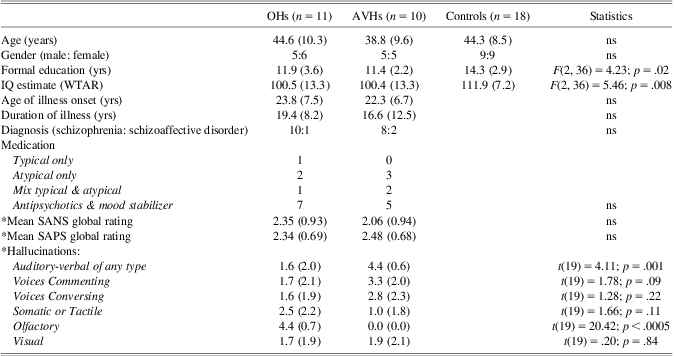
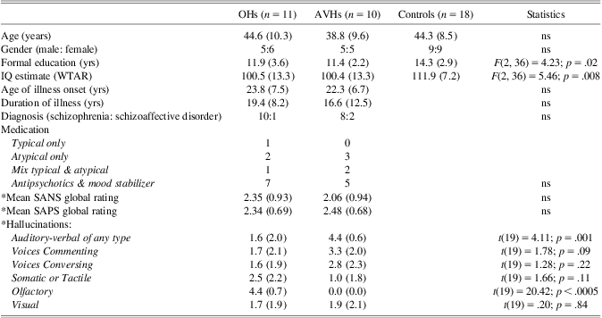
Table 2 summarizes results. Between group differences were not significant for years of age and gender ratio. There were, however, significant differences for years of education and IQ. Patients, as a whole, completed fewer years of education and had a lower estimated IQ than controls. However, neither years of education nor estimated IQ differed between patient subgroups.
Means (SDs) of demographic and clinical characteristics of participants
Note. WTAR = Wechsler Test of Adult Reading; OHs = olfactory hallucinations; AVHs = auditory-verbal hallucinations; SANS = Scale for the Assessment of Negative Symptoms of Schizophrenia; SAPS = Scale for the Assessment of Positive Symptoms of Schizophrenia (range 0 to 5).
* 0 = absent; 1 = questionable; 2 = mild; 3 = moderate; 4 = marked; 5 = severe.
The two patient subgroups were also similar with regard to diagnostic category, with the majority receiving a diagnosis of schizophrenia. Age of illness onset, duration of illness, medication type, and severity of symptoms, indexed by the global SAPS and SANS ratings, were also similar across patient subgroups. There were, however, significant differences in ratings of individual hallucinations items, as might be expected. The OH patients were rated as “marked,” on average, for OHs, “questionable” to “mild” for visual hallucinations, and “mild” to “moderate” for somatic (i.e., of the body) hallucinations. The slightly higher rating for somatic compared to visual hallucinations is to be expected, since OHs co-occur more often with somatic than other non-olfactory hallucinations (see, e.g., Langdon et al., Reference Langdon, McGuire, Stevenson and Catts2011). Nevertheless there were no significant differences between the patient subgroups on ratings of either somatic or visual hallucinations (see Table 2).
Experimental Tasks
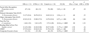
Table 3 summarizes results for the experimental tests believed to tap OFC-amygdala circuitry, as informed by previous lesion studies.
Means (SDs) on experimental tasks and standardized tests of odor identification (UPSIT) and executive function (composite score from DVs listed in Table 1)

Note. F-statistics and Cohen's d also provided.
OHs = olfactory hallucinations; AVHs = auditory-verbal hallucinations.
Data from the facial affect recognition task were analyzed using a (3 × 6 × 3) mixed design with 3 levels on the between factor group, 6 levels on the within factor emotion, and 3 levels on the within factor intensity. Of most interest was a main effect of group, F(2,36) = 3.45; p = .04; in contrast, the interaction of emotion by group was non-significant. Table 3 summarizes results collapsed across emotion. Patients, as a whole, performed more poorly than controls (p = .013), with no difference between the OH and AVH patients (p = .78).
With regard to the remaining experimental tasks, results revealed a non-significant group difference on the DAT (% perseverative errors). However, it is noted that patients, as a whole, did tend to perform more poorly than controls on the DAT (p = .04), with the two patient subgroups performing at a similar level (p = .56). This same pattern was seen on the Reading the Mind in the Eyes Test (RMET: number correct)—that is, patients, as a whole, performed more poorly than controls (p < .0005), with no difference between the OH and AVH patients (p = .69).
In contrast, two other tests of OFC function—the OAT (% perseverative errors) and the Faux Pas test (sensitivity)—showed significantly greater impairment in the OH patients. Not only did the patients, as whole, perform more poorly than controls on the OAT (p = .009), so too did the OH patients perform more poorly than the AVH patients (p = .03). Similarly, patients, as whole, performed more poorly than controls on the Faux Pas test, (p = .005), with the OH patients also performing more poorly than the AVH patients (p = .002).
UPSIT
Results for the UPSIT were similar to results for the facial affect recognition task, DAT and RMET. Patients, as a whole, performed more poorly than controls (p = .013), with no differences between the OH and AVH patients (p = .78).
Standardized Neuropsychological Tests of Executive Function
Table 3 also provides results for the composite executive score. Results mirrored those seen for the UPSIT. There was a significant main effect of group, with patients, as a whole, showing executive deficits compared to controls (p < .0005), and no differences between the patient subgroups (p = .16).
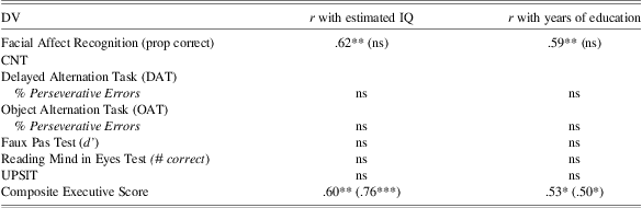
Influence of IQ and Years of Education
Table 4 lists the correlations of DVs with estimated IQ and years of education, within patients and within controls. The only significant relations were observed for proportion correct on the facial affect recognition task and the composite executive score. In each case, the relationship with years of education was no longer significant once the effect of estimated IQ had been partialled out. We, therefore, selected a subset of 13 controls who did not differ group-wise from patients on estimated IQ, F(2,31) = 2.11; p = .11. This control subgroup was then compared to the two patient subgroups on the composite executive score and the proportion correct score from the facial affect recognition test. While the main effect of group remained significant for the former, F(2,31) = 11.57; p < .0005, the group effect for the latter was no longer significant, F(2,31) = 1.48; p = .24.
Correlations within patients and within controls (in brackets) between the DVs listed in Table 3, estimated IQ, and years of education
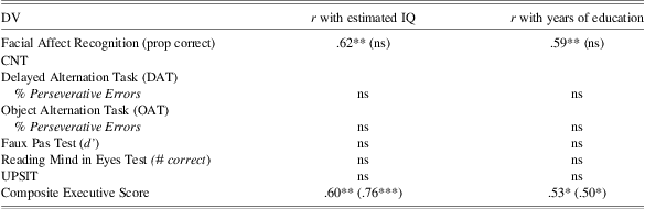
*p < .05; **p < .01; ***p < .005.
Relations Between OH Severity and DVs
Consistent with the between groups results, correlations within patients revealed a relation between severity of OHs and OAT % perseverative errors that approached significance, r = .41; p = .06, while the correlation of OH severity with Faux Pas sensitivity was significant, r = −.50, p = .022. No other correlations of task measures with OH severity reached significance.
Effect Sizes
Finally, we note that effect sizes (Cohen's d) for the observed differences between the OH and AVH patients on the OAT and the Faux Pas test were large: 1.04 and 1.32, respectively (see Table 3).
Discussion
The present study assessed neuropsychological functioning in people with schizophrenia experiencing OHs vs. those experiencing AVHs, and healthy controls. Tasks were selected to tap functions associated with the amygdala and OFC, as informed by previous lesions studies. These were administered in addition to the UPSIT and a battery of standardized executive tasks, commonly used in schizophrenia research.
The Comparative Neuropsychological Tasks (CNT) provided evidence suggestive of a modality-specific association between the presence of OHs (vs. AVHs) and perseverative errors on the OAT (but not the DAT). The implication here is that greater compromise to the OFC, rather than DLPFC, may contribute to OHs in schizophrenia. The OH patients also showed a similar pattern of impairment relative to the AVH patients on the Faux Pas test of theory of mind (ToM).
In contrast, performance on another test of ToM, the Reading the Minds in the Eyes Test (RMET), also believed to tap OFC-amygdala functioning, failed to reveal relatively greater impairment in the OH patients. Although both the Faux Pas test and the RMET are considered ToM tasks, they may tap ToM functioning in distinct ways. The Faux Pas test requires, not only the attribution of others’ mental states, but also the ability to make higher order social inferences about the consequences of those others’ mental states (i.e., inferring that one person will feel hurt by another person's unknowing words). In contrast, and as suggested by Baron-Cohen et al. (Reference Baron-Cohen, Wheelwright, Hill, Raste and Plumb2001), the RMET may only tap into lower-order ToM processing. This is despite that the mental states depicted on this task are fairly complex (e.g., “accusing”). In other words, the RMET may tap the ability to infer the presence of complex mental states but does not require any further inferencing about the social implications of those mental states. The implication here is that disrupted punitive feedback associated with aversive negative affect (e.g., the “cringe” associated with detecting a faux pas), and sustained by OFC/amygdala connections, may be implicated in the generation and/or maintenance of OHs in schizophrenia.
There is also debate in the literature concerning whether the RMET is best conceived as a test of “social perception” rather than “social cognition.” In other words, the RMET may be more similar to an emotion cue perception task than a classic ToM task (see, e.g., Bell et al., Reference Bell, Langdon, Siegert and Ellis2010). In the present study, there was no evidence that more basic amygdala processing, as required for simple facial affect recognition, associated specifically with OHs in schizophrenia. In contrast, and consistent with a large body of previous research (e.g., Edwards et al., Reference Edwards, Jackson and Pattison2002), patients, as a whole, showed poor emotion recognition. It is acknowledged, however, that the group difference was no longer significant when patients were compared to a subset of controls, matched group-wise on estimated IQ. Nevertheless, both the RMET and the facial affect recognition task showed a similar pattern of results, consistent with the view that the RMET may be more similar to an emotion perception task than a classic ToM task.
The executive battery revealed a similar pattern of results. In keeping with previous evidence of extensive executive dysfunction in schizophrenia (Heinrichs & Zakzanis, Reference Heinrichs and Zakzanis1998; Reichenberg & Harvey, Reference Reichenberg and Harvey2007), patients, as a whole, performed more poorly than controls on the composite executive measure, with no difference between the OH and AVH patients. A similar pattern was seen for the UPSIT. While the non-significant difference between the OH and AVH patients on the UPSIT is consistent with previous findings (Kopala et al., Reference Kopala, Good and Honer1994; Stedman & Clair, Reference Stedman and Clair1998), it is counter to expectations based on the location of the olfactory bulb within the OFC. Thus, while the ability to identify odors clearly involves the OFC, compromise of this ability appears insufficient to predict the presence of OHs. Instead, our findings suggest that it is compromise to the reward/punishment and socio-evaluative aspects of OFC functioning that may associate more specifically with OHs in schizophrenia. However, it is beyond the scope of this neuropsychological study to speculate about a functional model of how disrupted processing of reward/punishment signals and evaluation of social significance might contribute to OHs in schizophrenia.
Limitations of the present study include the relatively small clinical samples and the numbers of statistical tests, thus opening up the possibility of Type I and Type II errors. The relatively small clinical samples were an inevitable consequence of the requirements that patients be experiencing either OHs, which have relatively low current prevalence rates (e.g., 13–17%: Langdon et al., Reference Langdon, McGuire, Stevenson and Catts2011), or AVHs with no lifetime history of OHs, with lifetime prevalence of OHs estimated at up to 35% (Kopala et al., Reference Kopala, Good and Honer1994). At the same time, patients could not be so acutely unwell as to be unable to complete testing. Nevertheless, the reported large effect sizes for differences between patient subgroups on the OAT and Faux Pas task suggest that our results do not merely reflect chance. Replication of our findings with larger clinical subgroups is clearly warranted, however.
In conclusion, this is the first study to provide evidence of OH-specific neuropsychological impairments associated with reward/punishment decision-making and social evaluative aspects of OFC function in schizophrenia.
Acknowledgments
The authors received no specific grant from any funding agency, commercial or not-for-profit sector and have no conflicts of interest. The authors thank Sarah Jacek for her assistance with recruitment and Margery Aylett for her assistance with the initial planning stages of this study. Recruitment of participants for this study was also facilitated by the Australian Schizophrenia Research Bank (ASRB), which is supported by the National Health and Medical Research Council of Australia, the Pratt Foundation, Ramsay Health Care, the Viertel Charitable Foundation, and the Schizophrenia Research Institute.