Introduction
Of the 27 species of liverwort that are known from Antarctica, only one, the leafy liverwort Cephaloziella varians (Gottsche) Steph., occurs in the continental Antarctic (Bednarek-Ochyra et al. Reference Bednarek-Ochyra, Váňa, Ochyra and Smith2000). This species, which is the commonest and most widespread liverwort in Antarctica, is the only hepatic to occur at latitudes above 70°S. Only 22 native species of moss, and no higher plant species, have been recorded beyond this latitude (Ochyra et al. Reference Ochyra, Lewis-Smith and Bednarek-Ochyra2008). Terrestrial habitats beyond 70°S present some of the most hostile conditions for plant life on Earth, including very low winter temperatures, extended periods of ice- and snow-cover and darkness, extreme aridity, wide diurnal and annual variations in temperature and solar radiation exposure, and, in summer, frequent freeze-thaw cycles. However, despite the occurrence of C. varians in such extreme habitats, the reasons for its success in the Antarctic biome remain obscure.
This article has two aims. The first is to collate the available information from the literature on C. varians. A description of the hepatic and its reproduction is given, together with information on the habitats in which it occurs, its photosynthesis, pigments, water relations, responses to changing environmental conditions and its associated organisms. The second aim of the article is to attempt to identify the adaptations that have led to the success of C. varians in the Antarctic terrestrial biome.
The format of this paper is based upon that of the Biological Flora of the British Isles series published in Journal of Ecology, initiated by the late Professor Arthur Willis of the University of Sheffield.
Geographical distribution
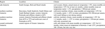
Cephaloziella varians occurs in the sub-, northern and southern maritime and continental Antarctic (Fig. 1a). Geographical definitions of each of these four regions are given in Table I. The species is widespread throughout the sub- and maritime Antarctic, occurring at Heard Island, South Georgia, Bouvetøya and the South Sandwich, South Orkney and South Shetland islands, and along the western Antarctic Peninsula to the eastern coast of Alexander Island (Fig. 1a & b). It is the only hepatic known from continental Antarctica, where it occurs at coastal locations in Victoria, Wilkes, Princess Elizabeth and Mac Robertson lands (Fig. 1a). Its southernmost known location is at 77°S, at Granite Harbour in Botany Bay, Victoria Land (Seppelt & Green Reference Seppelt and Green1998). The species usually occurs within 2 km of the coast and at low (< 350 m) altitudes, although it is present on Mount Melbourne in the continental Antarctic on heated ground at an altitude of 2700 m (Smith Reference Smith2007). For the precise locations of C. varians collections from the Antarctic, see Ochyra & Váňa (Reference Ochyra and Váňa1989a, Reference Ochyra and Váňa1989b) and Bednarek-Ochyra et al. (Reference Bednarek-Ochyra, Váňa, Ochyra and Smith2000).
The distribution of Cephaloziella varians in a. sub-, maritime and continental Antarctica, and b. the South Shetland Islands and Antarctic Peninsula. Data from the Antarctic Plant Database (http://www.antarctica.ac.uk/Resources/BSD/PlantDatabase/) and Bednarek-Ochyra et al. (Reference Bednarek-Ochyra, Váňa, Ochyra and Smith2000).

Regions of Antarctica in which C. varians occurs and their climates. Adapted from Smith (Reference Smith1984).

Outside Antarctica, C. varians is present in mountainous regions, including the Alps, Pyrenees and Carpathians ranges, and in New Zealand, Alaska, Canada, Greenland, Sweden, Iceland and the Russian Arctic (Schuster & Damsholt Reference Schuster and Damsholt1974, Schljakov Reference Schljakov1979, Söderström Reference Söderström1995, Konstantinova & Potemkin Reference Konstantinova and Potemkin1996, Bednarek-Ochyra et al. Reference Bednarek-Ochyra, Váňa, Ochyra and Smith2000). It is frequently recorded in the Arctic as C. arctica, which is considered to be synonymous with C. varians (Ochyra & Váňa Reference Ochyra and Váňa1989a). In agreement with this, Ochyra & Váňa (Reference Ochyra and Váňa1989b) note that C. varians, together with four other Antarctic hepatics (Anthelia juratzkana (Limpr.) Trevis., Barbilophozia hatcheri (Evans) Loeske, Lophozia excisa (Dicks.) Dumort. and Scapania obcordata (Berggr.) Arnell), belongs to a bipolar floristic element. They note that these taxa may have attained their southern range by long-range dispersal, rather than by fragmentation of their original ranges.
Species description
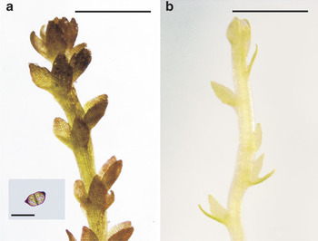
A detailed description of the species is given by Bednarek-Ochyra et al. (Reference Bednarek-Ochyra, Váňa, Ochyra and Smith2000), from which much of the following is derived. Plants very small and delicate, with stems 2–8 (-12) mm in length, usually creeping or semi-erect and sparsely or lateral-intercalary branched. Leaves 100–250 μm in length, plane or concave, barely overlapping, typically 1.5–2 times as wide as the stem, transversely or subtransversely inserted, distinctively bilobed to 0.5–0.7 of length (Fig. 2a & b). Lobes ovate to ovate-triangular, erect or incurved and 6–10 cells wide at their base. Leaf cell dimensions c. 12–15 × 10–13 μm. Leaves and stems smooth. Rhizoids hyaline, typically occurring at base of stem. Oil bodies frequent in cells (Post & Vesk Reference Post and Vesk1992).
Micrographs of a. sun-exposed, and b. shade-adapted forms of Cephaloziella varians. Bars are 500 μm. Inset in a. is a gemma. Bar in inset is 25 μm. Material collected from Léonie Island, Ryder Bay, south-western Antarctic Peninsula.

Leaves and stems slightly lustrous, especially in damp locations, and exceptionally variable in colour, ranging from deep violet to black, brown-green, green and yellow-green (Seppelt & Green Reference Seppelt and Green1998, Bednarek-Ochyra et al. Reference Bednarek-Ochyra, Váňa, Ochyra and Smith2000, Newsham et al. Reference Newsham, Geissler, Nicolson, Peat and Lewis-Smith2005). Considerable variation in pigmentation can occur within a given location: sun-exposed plants are deeply-pigmented (Fig. 2a), but plants growing in shaded habitats are uniformly green (Fig. 2b). Observations suggest that deeply-pigmented plants in the maritime Antarctic typically occur in unshaded habitats at the southern end of the species’ range (Bednarek-Ochyra et al. Reference Bednarek-Ochyra, Váňa, Ochyra and Smith2000, Newsham et al. Reference Newsham, Geissler, Nicolson, Peat and Lewis-Smith2005), possibly reflecting the more continental climate in this region, which is typified by extended periods of cloudless skies during summer (see Effects of solar radiation, below).
Reproduction
Autoecious or pseudodioecious. Male inflorescence terminal, becoming intercalary, bracts imbricate, monandrous and saccate at base, in 4–10 pairs. Female inflorescences on elongated shoots, bracts broadly ovate, and bracteoles, which are smaller than bracts, mostly bilobed. Perianths obloid to obloid-clavate, formed by thick-walled rectangular cells in the apical region. Capsule ovoid-elliptical, rounded at apices with bistratose walls. Epidermal cells of capsule with nodular thickenings and inner cells with semi-annular narrow bands. Spores 9–12 μm in diameter, delicately verruculose (Bednarek-Ochyra et al. Reference Bednarek-Ochyra, Váňa, Ochyra and Smith2000, Smith & Convey Reference Smith and Convey2002). Elaters free, bispiral and 7–8 μm in diameter. Gemmae two-celled, 15–25 μm in length, 10–15 μm in width (Fig. 2a, inset), borne on the margins of leaves and formed frequently in the natural environment. Vegetative reproduction also by branch fragmentation (Seppelt & Green Reference Seppelt and Green1998).
Although the species is almost entirely sterile in the Antarctic natural environment, sporophytes have been observed in populations on the South Shetland Islands, in the Marguerite Bay region and on Alexander Island in the southern maritime Antarctic since the mid 1990s, possibly as a consequence of climate warming in the maritime Antarctic (Smith & Convey Reference Smith and Convey2002). Mean spore counts per capsule from these locations are 23 750 and 14 000, respectively. Sporophytes are also occasionally found on plants in geothermally-heated habitats in the South Sandwich Islands (Convey et al. Reference Convey, Smith, Hodgson and Peat2000).
Chromosomes n = 18 (16 + 2 m), based on material from South Georgia and King George Island (Newton Reference Newton1980, Ochyra et al. Reference Ochyra, Przywara and Kuta1982). The smallest of the nine pairs of chromosomes are unique in being composed of two unequal m-chromosomes (Newton Reference Newton1980).
History
Cephaloziella varians was originally described as Jungermannia varians by Gottsche (Reference Gottsche1890) from material collected by H. Will at South Georgia during the German Polar Year Expedition in 1882–83. It was later assigned to Cephalozia varians by Stephani (Reference Stephani1901), based on material from the Gerlache Strait collected by E. Racovitza of the Belgian Antarctic Expedition in 1897–99. Stephani (Reference Stephani1905) subsequently assigned the species to Cephaloziella varians.
Taxonomic placement
There is considerable phenotypic plasticity between populations of C. varians from different locations in the Antarctic, which has led to the elevation of several different names for the taxon from the biome (e.g. Grolle Reference Grolle1972, Schuster & Damsholt Reference Schuster and Damsholt1974, Fulford Reference Fulford1976, Schuster Reference Schuster1980). The species has frequently been referred to in the literature as Cephaloziella exiliflora (Tayl.) Steph. (e.g. Seppelt Reference Seppelt1983, Williams et al. Reference Williams, Roser and Seppelt1994, Seppelt & Green Reference Seppelt and Green1998), but it is now accepted that all Antarctic Cephaloziella should be referred to C. varians, with C. exiliflora, a distinct species with larger leaves, vestigial underleaves and red-brown pigmentation, being restricted to Australasia (Ochyra & Váňa Reference Ochyra and Váňa1989a, Bednarek-Ochyra et al. Reference Bednarek-Ochyra, Váňa, Ochyra and Smith2000).
Nucleotide sequence data for a specimen of C. varians from New Zealand are deposited in GenBank under accession numbers AY608222, DQ439689, AY608074 and AY607953 (Davis Reference Davis2004, Forrest et al. Reference Forrest, Davis, Long, Crandall-Stotler, Clark and Hollingsworth2006). These nucleotide sequences are of 26S large subunit ribosomal RNA, the ribulose biphosphate carboxylase (rbcL), small ribosomal protein (rps4) and the CD122 photosystem II 32 kDa protein (psbA) genes, respectively. Based on these data, Forrest et al. (Reference Forrest, Davis, Long, Crandall-Stotler, Clark and Hollingsworth2006) place C. varians in a clade of leafy liverworts including C. hirta Schuster and species of Cephalozia, Stenorrhipis, Scapania, Diplophyllum, Tritomaria, Anastrophyllum, Tetralophozia, Herzogobryum, Nowellia, Schiffneria, Odontoschisma and Adelanthus.
Microhabitat
Substratum
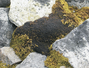
Cephaloziella varians forms dense compact mats (Fig. 3), often of a few square metres in area, on the surface of moribund mosses, or more usually intertwined with the shoots of other bryophytes. The species occurs predominantly in moist to wet habitats, normally on acidic substrata with pH values of 4.9–6.9 (Table II). Davis (Reference Davis1986), Wynn-Williams (Reference Wynn-Williams1988) and Smith (Reference Smith1990) similarly report pH values of 4.8–5.1 for mixed bryophyte communities in which C. varians occurs on Signy Island in the South Orkney Islands, at Rothera Point on Adelaide Island and on Bailey Peninsula in Wilkes Land, continental Antarctica. However, the species also occurs on more alkaline soils, such as those derived from marble on Signy Island, with pH values of up to 8.2 (Smith Reference Smith1972). The substrata on which C. varians occurs are variable in their organic matter content, with between 24% and 95% loss on ignition (LOI) for material from Bird Island to Moutonnée Valley on Alexander Island, with more mineral substrata being encountered at more southerly locations (Table II). Roser et al. (Reference Roser, Melick, Ling and Seppelt1992) and Melick & Seppelt (Reference Melick and Seppelt1992) report 95% and 88% LOI for the substratum under C. varians from Bailey Peninsula, whilst Wynn-Williams (Reference Wynn-Williams1988) reports 91% and 87% LOI for peat under C. varians at Signy and Adelaide Islands, respectively. LOI values of between 3.3% and 96.4% are reported by Smith (Reference Smith1972) for soils on which C. varians occurs at Signy Island. Cephaloziella varians is also present on geothermally-heated ground, where soil surface temperatures reach 25–35°C, on Mount Melbourne in Victoria Land, on Deception Island, and on Leskov, Visokoi, Candlemas and Bellingshausen islands in the South Sandwich Islands (Longton & Holdgate Reference Longton and Holdgate1979, Broady et al. Reference Broady, Given, Greenfield and Thompson1987, Convey et al. Reference Convey, Smith, Hodgson and Peat2000, Smith Reference Smith2005, Convey & Smith Reference Convey and Smith2006).
A darkly pigmented colony of sun-exposed Cephaloziella varians growing amongst shoots of the moss Sanionia uncinata at Rothera Point on Adelaide Island. The colony is c. 100 mm in width.

pH and organic matter content of substrata on which C. varians occurs at a range of maritime and sub-Antarctic locations. Data are means of 5–8 replicates ± SD.

1550°C for 2 h, 2South Orkney Islands
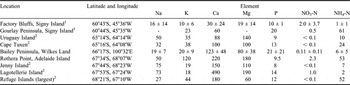
Concentrations of extractable elements in substrata on which C. varians occurs are highly variable (Table III), and, in broad terms, not limiting for plant growth (Allen Reference Allen1989). For the nine locations for which data are available, moderate to high concentrations of sodium, potassium, calcium, magnesium, phosphorus and ammonium have been recorded in soil on which the species occurs (Table III). Low concentrations of nitrate (< 0.2 mg 100 g-1 dry weight; Allen Reference Allen1989) have been recorded in soil at five of the nine locations for which data are available (Table III).
Extractable elements (mg 100 g-1 dry weight) in substrata on which C. varians occurs. Data for Factory Bluffs and the Bailey Peninsula are means of 5–7 replicates ± SD. Other data are single measurements.

Data, which were all analysed by the same analytical chemistry facility, are from Holdgate et al. (Reference Holdgate, Allen and Chambers1967), Wynn-Williams (Reference Wynn-Williams1988) and Smith (Reference Smith1990, Reference Smith1996). 1South Orkney Islands. 2Sites for which soil chemical data are reported by Smith (Reference Smith1996) for Polytrichum strictum, Sanionia uncinata and Bryum pseudotriquetrum/Brachythecium austrosalebrosum, but at which C. varians also occurs (R.I.L. Smith, personal communication 2009). Dashes indicate missing data.
Topography
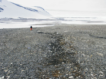
Cephaloziella varians usually occurs on level or gently-sloping ground, commonly along rills, meltwater flushes, seepage channels and in sheltered depressions that are irrigated with meltwater during summer (Smith Reference Smith1990, Bednarek-Ochyra et al. Reference Bednarek-Ochyra, Váňa, Ochyra and Smith2000). It often occurs on north-facing slopes, which are exposed to more solar radiation than south-facing areas, and where meltwater channels are more frequent (Fig. 4). The species is also found in drier habitats, such as on rock ledges or in crevices and fissures.
Cephaloziella varians growing in meltwater channels with mixed mosses on the north-facing slopes of Moutonnée Valley on Alexander Island (71°S), close to the southernmost limit of the species’ range in the maritime Antarctic.

Climate and plant temperature
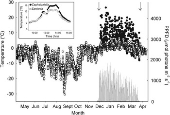
Cephaloziella varians occurs in a wide range of Antarctic habitats, in which mean monthly air temperature can exceed freezing point for several months each year, but can fall to -25°C or below in winter (Table I). Precipitation in these environments varies widely between c. 100 mm and > 900 mm water equivalent per annum (Table I). Rainfall, particularly at more southerly latitudes, can be very rare, with any precipitation usually falling as snow. The temperature within a mat of C. varians varies widely, and often on a diurnal basis: at Rothera Point in the southern maritime Antarctic, mat temperature can reach 27°C at solar noon during the summer, and often exceeds air temperature by 20°C, but frequently falls to below freezing point at night (Fig. 5). After the first snowfalls of autumn, mat temperature declines to near freezing point, closely tracking air temperature, and can fall to -30°C during winter (Fig. 5). The high temperatures attained by sun-exposed shoots during the summer relative to air temperature are apparently due to their purple-black pigmentation, which enables efficient heat absorption: under clear skies, the temperature of C. varians mats can be up to 4°C higher than that of nearby Sanionia uncinata (Hedw.) Loeske shoots (Fig. 5, inset), which are light green in colour (Fig. 3).
Temperature of (●) sun-exposed Cephaloziella varians and (○) air (left axis) and (—) photosynthetic photon flux density (PPFD; right axis) between May 2006 and April 2007 at Rothera Point on Adelaide Island. Data were recorded every 3 h. Plant temperature was measured by button loggers (SL51 Smart Button Logger, Status Instruments, Tewkesbury, UK) inserted into mats and air temperature was measured in the shade by two platinum resistance thermometers (PT100; Labfacility Ltd., Teddington, UK). PPFD was measured with a Skye SKP 215 sensor (Skye Instruments Ltd., Llandrindod Wells, UK). Arrows mark the period when plants were free from snow and ice. Inset shows temperatures of sun-exposed C. varians and the moss Sanionia uncinata at Rothera Point between 09h00 and 16h00 on 27 November 1999. The two colonies were separated by c. 0.2 m.

Light environment
Cephaloziella varians experiences wide fluctuations in its light environment in the Antarctic. At Signy Island, the length of the growing season, when plants are not covered by snow and ice, is approximately six months (Wynn-Williams Reference Wynn-Williams1980), whereas at a more southerly site, Rothera Point, the season rarely lasts for more than four months. Plants at the latter site become covered with snow and ice in the autumn, with photosynthetic photon flux density (PPFD) incident on the mats falling to close to zero when the depth of snow over them exceeds c. 200 mm. This depth of snow is usually maintained for approximately eight months. Mild spells, when air temperatures can reach freezing point, do occur during winter (Fig. 5), but mats usually remain covered with snow and ice. In contrast, after snowmelt has occurred during the late spring, PPFD often reaches 1500 μmol photons m-2 s-1 at solar noon (Fig. 5), and plants are exposed to almost continual daylight. However, low solar elevation angles during the early hours of the morning in the summer cause PPFD to fall to 20–40 μmol photons m-2 s-1 (Snell et al. Reference Snell, Convey and Newsham2007).
The exposure of plants to UV-B radiation (280–315 nm) can also vary widely, both as a consequence of changes in solar elevation angles and, in recent decades, anthropogenic depletion of ozone in the stratosphere. The daily mean biologically effective dose of UV-B radiation in the southern maritime Antarctic during late spring and early summer, when plants emerge from melting snow and ice, is c. 3 kJ m-2 under undepleted ozone columns (c. 350 Dobson Units, DU). However, when depletion of the gas occurs in the stratosphere to 200 DU, the daily dose of biologically effective UV-B radiation to which plants are exposed can reach 10.3 kJ m-2 (Newsham et al. Reference Newsham, Hodgson, Murray, Peat and Smith2002), which is three fold higher than the mean dose of UV-B received by plants in the region in midsummer, when ozone depletion does not occur. Cephaloziella varians is able to respond rapidly to these changes in UV-B exposure (see effects of solar radiation, below).
Photosynthesis, fluorescence measurements and net primary production
For C. varians growing in situ, the maximum quantum yield of photochemistry (F v/F m) is 0.4–0.6 (Snell et al. Reference Snell, Convey and Newsham2007, Reference Snell, Kokubun, Griffiths, Convey, Hodgson and Newsham2009), although values of this parameter can fall to 0.1–0.2 (Newsham et al. Reference Newsham, Hodgson, Murray, Peat and Smith2002). Given that F v/F m values are typically 0.7–0.8 for most plant species growing under optimal conditions, the low values of this parameter indicate that C. varians is probably under constant physiological stress in the natural environment. Non-photosynthetic quenching (qN), derived from fluorescence measurements, is 0.02–0.05 for the sun-exposed form of C. varians but 0.17 for the shade form of the species, which is attributable to increased heat dissipation from chloroplasts in cells lacking dark pigmentation (Snell et al. Reference Snell, Kokubun, Griffiths, Convey, Hodgson and Newsham2009).
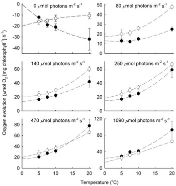
The shade-adapted and sun-exposed forms of C. varians also display different gas exchange characteristics. At low irradiances (80–250 μmol photons m-2 s-1), the shade form of the species evolves significantly (P < 0.05) more oxygen than the sun-exposed form at temperatures of between 5°C and 20°C (Fig. 6). In contrast, at a temperature of 20°C and at higher irradiances (470–1090 μmol photons m-2 s-1), the sun-exposed form evolves significantly (P < 0.05) more oxygen than the shade form (Fig. 6). Oxygen evolution of the shade form saturates at PPFD of 200–400 μmol photons m-2 s-1, whereas that of the sun-exposed form apparently does not saturate at < 1090 μmol photons m-2 s-1 (Post & Vesk Reference Post and Vesk1992, Snell et al. Reference Snell, Kokubun, Griffiths, Convey, Hodgson and Newsham2009). Gas exchange of C. varians is closely and positively associated with temperature, with sun-exposed and shade forms evolving up to 93 and 65 μmol O2 (mg chlorophyll-1) h-1 at 20°C, respectively (Fig. 6). At present, the reduction in the dark respiration rate of sun-exposed C. varians at higher temperatures, and the lower dark respiration rate at 7.5–20°C of sun-exposed, compared with shade-adapted plants (Fig. 6), remain anomalous.
Oxygen evolution of (●) sun-exposed and (○) shade-adapted forms of Cephaloziella varians as a function of temperature at irradiances of 0, 80, 140, 250, 470 and 1090 μmol photons m-2 s-1. Data for 20°C are means of measurements for December and February from Post & Vesk (Reference Post and Vesk1992). All other data are from Snell et al. (Reference Snell, Kokubun, Griffiths, Convey, Hodgson and Newsham2009). Bars are s.e.m.

Davis (Reference Davis1981) gives estimates of net primary production for C. varians of between 58 and 98 g dry weight m-2 yr-1 on Signy Island. However, these data should be regarded as approximations, as they are not based on empirical measurements.
Chloroplast ultrastructure
The chloroplasts of C. varians are spherical to slightly oval, and show little variation between different parts of the plant in size, shape or thylakoid content. Chloroplasts show marked differences between sun-exposed and shade-adapted forms of the species: in those of the former plants, the thylakoid system consists of sparsely-arranged elongated grana, usually comprising two or three thylakoids distributed evenly throughout the stroma. In those of shade-adapted plants, the thylakoid system is more extensive, with grana consisting of usually more than eight thylakoids (Post & Vesk Reference Post and Vesk1992). Thylakoids occupy 19% and 39% of the total chloroplast volume in sun-exposed and shaded plants, respectively (Post & Vesk Reference Post and Vesk1992).
Pigments
Chlorophylls
Roser et al. (Reference Roser, Melick, Ling and Seppelt1992) report a chlorophyll a concentration of 0.29 mg g-1 (fresh weight) in tissues of C. varians sampled from the Bailey Peninsula in Wilkes Land. Post & Vesk (Reference Post and Vesk1992) similarly report total chlorophyll (a and b) concentrations of 0.21 and 0.38 mg g-1 (fresh weight) respectively in the sun-exposed and shade-adapted forms of C. varians from the Clarke Peninsula, also in Wilkes Land. Similar values of 0.20 and 0.33 mg total chlorophyll g-1 (fresh weight) have been measured in sun-exposed and shade-adapted plants at Léonie Island in Ryder Bay, south-western Antarctic Peninsula, respectively (K.K. Newsham, unpublished data). Total chlorophyll concentrations in C. varians from Rothera Point of 1.2–3.3 mg g-1 (dry weight) are also reported by Newsham et al. (Reference Newsham, Geissler, Nicolson, Peat and Lewis-Smith2005) and Snell et al. (Reference Snell, Convey and Newsham2007, Reference Snell, Kokubun, Griffiths, Convey, Hodgson and Newsham2009). The chlorophyll a/b ratios of plants from sun-exposed and shaded habitats, which vary between 2.1 and 2.4, do not differ (Post & Vesk Reference Post and Vesk1992, Snell et al. Reference Snell, Convey and Newsham2007).
Carotenoids
The concentration of total carotenoids in C. varians tissues typically varies between 0.15 and 0.45 mg g-1 (dry weight) (Newsham et al. Reference Newsham, Hodgson, Murray, Peat and Smith2002, Snell et al. Reference Snell, Convey and Newsham2007, Reference Snell, Kokubun, Griffiths, Convey, Hodgson and Newsham2009), with increased concentrations of carotenoids in plants exposed to high levels of solar radiation (see Effects of solar radiation, below). HPLC analyses indicate that lutein is the most abundant carotenoid in the tissues of C. varians, forming 76% of the total mass of carotenoids (Newsham et al. Reference Newsham, Hodgson, Murray, Peat and Smith2002). Neoxanthin, zeaxanthin, antheraxanthin, β,β-carotene and violaxanthin are also relatively common, forming 10.3, 8.0, 1.4, 0.6 and 0.5% of the total mass of carotenoids, respectively. The carotenoids α- and β,ε-carotene and their derivatives are present in C. varians tissues at low (< 0.1%) concentrations (Newsham et al. Reference Newsham, Hodgson, Murray, Peat and Smith2002).
Non-photosynthetic pigments
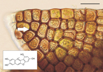
Post & Vesk (Reference Post and Vesk1992) isolated the dark pigment present in the tissues of C. varians sampled from Clarke Peninsula in Wilkes Land. The pigment was dark under ultraviolet light, had an R f value of 0.7 and similar mobility on thin layer chromatography plates to anthocyanins. Post & Vesk (Reference Post and Vesk1992) concluded that it was an anthocyanin-like pigment. The pigment, which is located in the walls of apical cells of sun-exposed leaves (Fig. 7), has a peak of absorption at 495 nm and minor peaks at 240, 281 and 330 nm. A later study (Snell et al. Reference Snell, Kokubun, Griffiths, Convey, Hodgson and Newsham2009) showed the pigment, which has a molecular weight of 285, to be riccionidin A (C15 H9 O6; Fig. 7, inset), a red to purple anthocyanidin found in the cell walls of Rhus javanica L., a vascular plant, and four other liverworts, including Ricciocarpos natans (L.) Corda and Marchantia polymorpha L. (Kunz et al. Reference Kunz, Burkhardt and Becker1994, Taniguchi et al. Reference Taniguchi, Yazaki, Yabu-uchi, Kawakami, Ito, Hatano and Yoshida2000). In the absence of an authenticated standard of riccionidin A, comparisons were made between the anthocyanidin extracted from Antarctic C. varians and that from north European R. natans and M. polymorpha. Snell et al. (Reference Snell, Kokubun, Griffiths, Convey, Hodgson and Newsham2009) found that the molecular masses, spectral profiles and elution times of the pigments from the three hepatics matched, and concluded that riccionidin A is present in C. varians tissues. The anthocyanidin is present at concentrations of c. 2 μmol g-1 (dry weight) in sun-exposed plants (Post & Vesk Reference Post and Vesk1992, Snell et al. Reference Snell, Kokubun, Griffiths, Convey, Hodgson and Newsham2009).
Micrograph of sun-exposed Cephaloziella varians leaf, showing dark pigmentation in cell walls (arrow). Bar is 20 μm. Inset shows molecular structure of riccionidin A.

Other flavonoids are also synthesized by C. varians. HPLC analyses have shown there to be at least six, and possibly as many as nine, other acidified methanol-extractable flavonoids within the tissues of C. varians, none of which have sugar attachments (T. Kokubun, personal communication 2009). In common with riccionidin A, these compounds tend to be localised in apical leaves, which have the highest exposure to solar radiation, but, unlike the anthocyanidin, are present in the cytosol, rather than the cell wall (Snell Reference Snell2007). The absolute concentrations of these pigments, which vary widely depending on exposure to solar radiation, are currently unknown, but total absorption of acidified-methanol extracts over the UV-B waveband is c. 3–6 mg-1 dry weight (Newsham et al. Reference Newsham, Geissler, Nicolson, Peat and Lewis-Smith2002, Reference Newsham, Hodgson, Murray, Peat and Smith2005, Snell et al. Reference Snell, Kokubun, Griffiths, Convey, Hodgson and Newsham2009).
Carbohydrates
Sucrose, trehalose and glucose are reported by Roser et al. (Reference Roser, Melick, Ling and Seppelt1992) to be the only sugars in the tissues of C. varians sampled from Bailey Peninsula. Concentrations of these carbohydrates are 3.3, 2.0 and 1.3 mg g-1 (dry weight), respectively (Roser et al. Reference Roser, Melick, Ling and Seppelt1992). A separate study, also on plants from Bailey Peninsula, reports concentrations of sucrose, trehalose, fructose and glucose of 4.3, 1.1, 0.2 and 0.2 mg g-1 (dry weight), respectively (Melick & Seppelt Reference Melick and Seppelt1992). Of the polyols, trace levels of arabitol and ribitol (total 0.5 mg g-1 dry weight) and higher levels of mannitol (1.7 mg g-1 dry weight) are present in C. varians tissues sampled from the field. Laboratory-grown plants contain 10.2 mg g-1 (dry weight) of mannitol (Roser et al. Reference Roser, Melick, Ling and Seppelt1992).
Tissue chemistry
Carbon, nitrogen, phosphorus and potassium concentrations of C. varians plants sampled from Bird Island and Signy, Adelaide and Alexander islands in the sub-, northern and southern maritime Antarctic vary between 31 and 48%, 1.2 and 2.5%, 0.009 and 0.026% and 0.14 and 0.46% (dry weight), respectively (Table IV). Reductions occur in the concentrations of the former three elements in C. varians tissues at more southerly locations (Table IV). Bokhorst et al. (Reference Bokhorst, Huiskes, Convey and Aerts2007) report a δ 15N value of 11.9 for C. varians growing on a soil with a δ 15N value of 14.0.
Carbon, nitrogen, phosphorus and potassium concentrations in C. varians tissues at a range of maritime and sub-Antarctic locations. See Table II for latitudes and longitudes of locations. Data are means of 5–8 replicates ± SD.

1Elementar Vario EL elemental analyser, 2Perkin Elmer DRC11 ICP-MS, 3South Orkney Islands
Water relations
Cephaloziella varians is poikilohydric, and is thus unable to control water loss from its tissues. The water content of tissues is high, owing to the damp environments that the species inhabits, with the substrate below C. varians becoming sufficiently waterlogged for decomposition to be inhibited and for methanogenesis to occur (Wynn-Williams Reference Wynn-Williams1980, Reference Wynn-Williams1988, Yarrington & Wynn-Williams Reference Yarrington and Wynn-Williams1985, Davis Reference Davis1986). Roser et al. (Reference Roser, Melick, Ling and Seppelt1992), Melick & Seppelt (Reference Melick and Seppelt1992) and Post & Vesk (Reference Post and Vesk1992) report water contents of 76–80% (fresh weight) of plants from continental Antarctic locations. Similarly, the water contents of plants at Signy and Adelaide islands are 68% and 88% (fresh weight), respectively (Davis Reference Davis1981, K.K. Newsham, unpublished data). Smith (Reference Smith1972) and Davis (Reference Davis1986) report water contents for substrata under plant communities in which C. varians occurs at Signy Island of between 16% and 1125% (dry weight). Water evaporation rates for vegetation in which C. varians occurs on Bailey and Clark peninsulas in continental Antarctica are 27 and 38 mg h-1 (Smith Reference Smith1990).
Response to environment
Effects of freeze-thaw cycles
Plants of C. varians subjected to a daily freeze-thaw cycle (between -15°C and 4°C) for 16 days lose 29% of low molecular weight carbohydrates after leaching with water, compared with plants not subjected to freeze-thaw events, which lose 10% of carbohydrates (Melick & Seppelt Reference Melick and Seppelt1992). This loss is largely accounted for by an increase in sucrose in the leachates. Changes in the freezing points of material subjected to freeze-thaw are not observed, however: after 16 days, those of the control and freeze-thawed material are -5.4°C and -5.3°C, respectively (Melick & Seppelt Reference Melick and Seppelt1992). These relatively high (> -9°C) freezing points suggest that C. varians does not avoid freezing, but rather tolerates the freeze-thaw process. The species is able to survive for long periods at temperatures below freezing point: Longton & Holdgate (Reference Longton and Holdgate1967) and Bednarek-Ochyra et al. (Reference Bednarek-Ochyra, Váňa, Ochyra and Smith2000) document the growth of C. varians after plants had been frozen at up to -20°C for three and 20 years, respectively.
Effects of snowmelt
Snell et al. (Reference Snell, Convey and Newsham2007) report a rapid increase from 0.18 to 0.45 in the maximum quantum efficiency of photosystem II (F v/F m), and an 88% increase in the concentrations of chlorophyll a and b, during the first 24 h after C. varians emerges from snow. However, there is little evidence of increased concentrations of photoprotective pigments in C. varians as it emerges from melting snow and ice at the end of spring (Snell et al. Reference Snell, Convey and Newsham2007).
Effects of solar radiation
Several studies demonstrate that the morphology and pigmentation of C. varians responds to solar radiation. In the sun-exposed form, apical leaves tend to be more closely spaced than in the shade form, and overlap each other slightly (Post & Vesk Reference Post and Vesk1992). The shade form of the species has elongated internodes and may have smaller leaves. Sun-exposed leaves have thick cell walls, particularly on the abaxial surface.
Post & Vesk (Reference Post and Vesk1992) report that apical leaves of C. varians from sun-exposed habitats accumulate higher concentrations of an anthocyanin-like pigment (riccionidin A; see non-photosynthetic pigments, above) than those from shaded habitats. They also show that sun-exposed plants have lower concentrations of chlorophyll and higher carotenoid/chlorophyll ratios than shade-adapted plants. The accumulation of riccionidin A in apical leaves is owing to exposure to solar UV-B radiation (Newsham et al. Reference Newsham, Geissler, Nicolson, Peat and Lewis-Smith2005): the pigment is lost within approximately three weeks when UV-B radiation exposure is reduced to 30% of that in sunlight, achieved by placing Mylar polyester screens over plants in the natural environment (Fig. 8). If the species is then exposed to an abrupt increase in solar UV-B exposure by removing the screens, the anthocyanidin is resynthesized within 48 h at a mean air temperature of 0.9°C (Snell et al. Reference Snell, Kokubun, Griffiths, Convey, Hodgson and Newsham2009), with a visible darkening of its tissues during this period. Snell et al. (Reference Snell, Kokubun, Griffiths, Convey, Hodgson and Newsham2009) estimate that a minimum of 1.85% of the carbon fixed in photosynthesis is used to synthesize riccionidin A during this period.
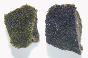
Mats of Cephaloziella varians exposed to reduced (left) or full (right) solar UV-B radiation for three weeks under Plexiglas screens covered with Mylar polyester or Plexiglas screens only, respectively. The green shoots in the right hand mat are of the moss Bryum pseudotriquetrum. Reproduced from Snell (Reference Snell2007).

Cephaloziella varians displays rapid responses to changes in UV-B exposure arising from ozone depletion, with concentrations of UV-B screening pigments (putative colourless flavonoids) tracking solar UV-B irradiances, implying that plants respond rapidly (within 24 h) to UV-B exposure (Newsham et al. Reference Newsham, Hodgson, Murray, Peat and Smith2002). Concentrations of carotenoids, notably neoxanthin, violaxanthin and lutein, are also positively associated with the flux of UV-B radiation in sunlight (Newsham et al. Reference Newsham, Hodgson, Murray, Peat and Smith2002).
Associated organisms
Bryophytes and vascular plants
Cephaloziella varians is frequently associated with many other bryophyte species in the Antarctic (Ochyra et al. Reference Ochyra, Lewis-Smith and Bednarek-Ochyra2008). It is a common member of the short moss cushion and turf sub-formation, the tall moss turf sub-formation and the bryophyte carpet and mat sub-formation (Smith Reference Smith1996). It also occurs amongst the two native Antarctic vascular plant species, Deschampsia antarctica Desv. and Colobanthus quitensis (Kunth) Bartl., and in the grass and cushion chamaephyte sub-formation, usually in well-drained habitats (Ochyra & Váňa Reference Ochyra and Váňa1989a). Although C. varians is often sparsely distributed amongst other bryophytes, it can form almost pure stands of up to 2–3 m2 in area and attains covers in mixed bryophyte communities of 13% at Signy Island (Wynn-Williams Reference Wynn-Williams1980, Davis Reference Davis1981) and 65% on Bailey Peninsula (Smith Reference Smith1990).
Invertebrates
Many invertebrates occur amongst the shoots of C. varians and in the substratum beneath them. Cryptopygus antarcticus Willem, a widespread maritime Antarctic collembolan, is frequent in mats of C. varians at Signy Island and at Rothera Point. At the latter site, unidentified bdelloid rotifers, the nematodes Aphelenchoides haguei Maslen, Coomansus gerlachei (de Man) Jairajpuri & Kahn, Eudorylaimus pseudocarteri Loof, E. coniceps Loof, Plectus antarcticus de Man, P. belgicae de Man and Rhyssocolpus paradoxus (Loof) Andrassy, and the tardigrades Diphascon and Echiniscus spp., Hypsibius cfr. dujardini Doyère and Macrobiotus furciger Murray, are frequently found in the substratum beneath C. varians (Newsham et al. Reference Newsham, Maslen and McInnes2006, N.R. Maslen, personal communication 2009). As yet, there is little to suggest that any of these invertebrates are specific to substrata under C. varians.
Fungi
Microscopic examination of C. varians from the Antarctic almost invariably reveals fungal hyphae ramifying through the shoot. Williams et al. (Reference Williams, Roser and Seppelt1994) were the first to report the presence of fungi in the tissues of the species. They documented dark septate hyphae and rudimentary coils, resembling those formed by ericoid mycorrhizal fungi in the hair roots of ericaceous plants, in the rhizoids of C. varians from Bailey Peninsula and Botany Bay, both in the continental Antarctic. Subsequently, Chambers et al. (Reference Chambers, Williams, Seppelt and Cairney1999) sequenced the Internal Transcribed Spacer (ITS) region of a fungus isolated by Williams et al. (Reference Williams, Roser and Seppelt1994) from plants on Bailey Peninsula and found that it displayed < 2.1% divergence from that of the type culture of Rhizoscyphus ericae (Read) Zhuang & Korf., a widespread mycorrhizal fungus in heathland and boreal ecosystems (Smith & Read Reference Smith and Read2008).
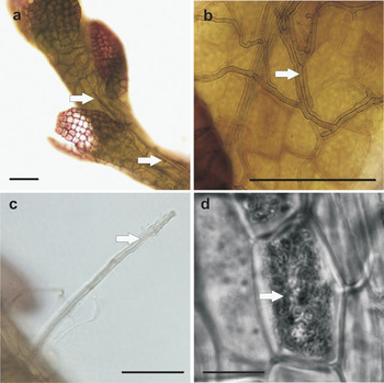
Further studies revealed that R. ericae is abundant in the tissues of C. varians along a 1875 km transect from Bird Island in the sub-Antarctic, through to Coronation, Lynch and Signy islands in the South Orkneys, King George and Livingston Islands in the South Shetlands, and Adelaide and Alexander islands, off the south-western Antarctic Peninsula (Upson et al. Reference Upson, Read and Newsham2007). Analyses of ITS1-5.8S-ITS2 and 28S large subunit sequences indicated that isolates are 98–99% similar to those of R. ericae from North America, Australia and Europe (Upson et al. Reference Upson, Read and Newsham2007). Fungal hyphae ramify through the tissues of the plant almost to the apical meristem, forming strands of hyphae on the shoot surface, and also frequently colonize rhizoids (Fig. 9a–c). Hyphal coils, similar to those observed by Williams et al. (Reference Williams, Roser and Seppelt1994) in plants from Bailey Peninsula, are also formed by R. ericae in the axial cells of axenically-grown plants (Fig. 9d).
Micrographs of a. fungal hyphae in Cephaloziella varians shoot (arrows), b. hyphae forming strands on shoot surface (arrow), c. hypha colonizing a rhizoid (arrow), and d. hyphal coil in axial cell (arrow). Bars in a–d are 80, 50, 25 and 10 μm, respectively. Reproduced, with permission from Wiley-Blackwell, from Upson et al. (Reference Upson, Read and Newsham2007).

Although R. ericae is widespread in the rhizoids of C. varians in the maritime and sub-Antarctic, other apparently non-pathogenic fungi also colonize the tissues of the plant. Sequencing of fungal 18S small subunit genes amplified directly from C. varians from Adelaide Island reveals a diverse fungal community, including sequences conspecific with a large group of fungi nested within the Chaetothyriales, and those similar to Rhizoscyphus spp. and Phialocephala fortinii Wang & Wilcox (Jumpponen et al. Reference Jumpponen, Newsham and Neises2003). Mats of C. varians from the Terra Firma Islands in southern Marguerite Bay, off the south-western Antarctic Peninsula, yield nematode-trapping ring fungi (Duddington et al. Reference Duddington, Wyborn and Smith1973). Geothermally-heated soils under C. varians on Mount Melbourne also yield Mucor, Penicillium and Cryptococcus spp. (Broady et al. Reference Broady, Given, Greenfield and Thompson1987).
Algae, cyanobacteria and protozoa
The microalga Stichococcus bacillaris Nägeli is frequently isolated from C. varians mats at Rothera Point and Signy Island, and the foliose alga Prasiola crispa (Lightf.) Kütz. is also commonly found growing amongst mats of C. varians at these locations. Broady et al. (Reference Broady, Given, Greenfield and Thompson1987) report the alga Chlorella cf. reniformis Watan., the testate amoeba Corythion dubium Taranek., and the cyanobacteria Gloeocapsa magma (Bréb.) Holl., Tolypothrix bouteillei (Bréb. & Desm.) Lemm. and Stigonema ocellatum Thuret, to be present with C. varians in geothermally-heated soils on Mount Melbourne.
Disease
No specific reports of any pathogens on C. varians exist in the literature. However, rings formed by Thyronectria antarctica occur in mats of C. varians in the northern maritime Antarctic (R.I.L. Smith, personal communication 2009). A species of Pythium, which bears phylogenetic affinities to snow moulds and causes dieback symptoms in Deschampsia antarctica (Bridge et al. Reference Bridge, Newsham and Denton2008), is present in mats of C. varians at Adelaide Island, as is Phoma herbarum Westend (Hughes et al. Reference Hughes, Lawley and Newsham2003), a globally-distributed weak fungal pathogen.
Adaptations of C. varians to the Antarctic natural environment
In their review of the phytogeography of Antarctic hepatics, Bednarek-Ochyra et al. (Reference Bednarek-Ochyra, Váňa, Ochyra and Smith2000) note that (the) distribution pattern of liverworts in the maritime Antarctic implies that particular species differ markedly in their tolerance of severe environmental conditions, and there are only a very few which have adapted physiologically to very low temperatures and long periods of continual daylight. Cephaloziella varians is clearly one such species: from the literature reviewed above, it is evident that it tolerates a range of stresses in the Antarctic natural environment. The foremost of these are the wide diurnal (20–30°C) and annual (50–60°C) fluctuations in temperature to which the species is exposed, the absence of solar radiation for several months each year, and exposure to high irradiances of solar radiation, including damaging shortwave UV-B radiation, during the brief growing season.
What adaptations does C. varians possess in order for it to survive these hostile conditions? Although the photosynthesis of the species displays rapid responses to snowmelt, enabling it to maximize carbon acquisition during the short growing season (Snell et al. Reference Snell, Convey and Newsham2007), it seems unlikely that the ability of the species to fix carbon is substantially greater than that of other bryophytes in Antarctica. Assuming a photosynthetic quotient of 1 for Antarctic bryophytes (Longton Reference Longton1974), the estimated carbon fixation rates of the species (up to 87 and 57 μmol CO2 g-1 dry weight h-1 at 10°C for the sun-exposed and shade forms; Snell et al. Reference Snell, Kokubun, Griffiths, Convey, Hodgson and Newsham2009) is similar to that of other hydric species of Antarctic mosses and liverworts (33–83 μmol CO2 g-1 dry weight h-1 at 10°C; Davey & Rothery Reference Davey and Rothery1997). The concentrations of chlorophylls in its tissues (0.2–0.4 mg g-1 fresh weight) are also similar to those of other Antarctic mosses (0.3–0.9 mg g-1 fresh weight; Rastorfer Reference Rastorfer1972, Roser et al. Reference Roser, Melick, Ling and Seppelt1992).
Although the concentrations of photosynthetic pigments in the tissues of C. varians apparently do not differ from those of other Antarctic bryophytes, it is probable that the flavonoid pigments synthesized by the species play a significant role in its survival in the natural environment. Of note is the presence of riccionidin A in sun-exposed apical leaves: this anthocyanidin enables efficient heat absorption by C. varians tissues, increasing their temperature by several degrees relative to associated moss species. The shading of chloroplasts by this flavonoid most probably also reduces photoinhibition at high irradiances, with the oxygen evolution of shade-adapted plants, which lack the pigment, saturating at 200–400 μmol photons m-2 s-1, but that of sun-exposed plants, which possess the pigment, not becoming saturated at irradiances of < 1090 μmol photons m-2 s-1 (Post & Vesk Reference Post and Vesk1992, Snell et al. Reference Snell, Kokubun, Griffiths, Convey, Hodgson and Newsham2009). Two other Antarctic hepatics, Lophozia excisa and Barbilophozia hatcheri, which extend to c. 68°S on the south-western Antarctic Peninsula, also synthesize dark pigments in apical leaves (Bednarek-Ochyra et al. Reference Bednarek-Ochyra, Váňa, Ochyra and Smith2000), which might similarly contribute to tolerance of high solar radiation exposure in the southern maritime Antarctic. Tolerance of high irradiance, although not ascribed to dark pigmentation, has been put forward as a reason for the success of Deschampsia antarctica and Colobanthus quitensis in the Antarctic natural environment (Smith Reference Smith2003). Riccionidin A and other colourless flavonoids, which most probably scavenge free radicals during periods of high solar radiation exposure, are aglycones, and can thus be synthesized at a low metabolic cost to C. varians (Snell et al. Reference Snell, Kokubun, Griffiths, Convey, Hodgson and Newsham2009). Furthermore, the colourless flavonoids synthesized by the species appear to protect photosystem II from rapid changes in UV-B exposure during periods of ozone depletion in the stratosphere over the Antarctic (Newsham et al. Reference Newsham, Hodgson, Murray, Peat and Smith2002).
Another key adaptation of C. varians appears to be its association with the mycorrhizal symbiont Rhizoscyphus ericae. This fungus, which facilitates the uptake of nitrogen by the roots of ericaceous plants in acidic, nutrient-poor soils at lower latitudes (Smith & Read Reference Smith and Read2008), has a widespread and consistent association with C. varians throughout the maritime Antarctic (Chambers et al. Reference Chambers, Williams, Seppelt and Cairney1999, Upson et al. Reference Upson, Read and Newsham2007). At present, the nature of any possible benefit to C. varians from R. ericae is unclear, but the formation in the cells of axenically-grown plants of hyphal coils, which maximise the surface area of contact between symbionts in ericoid mycorrhizas (Smith & Read Reference Smith and Read2008), is suggestive of active nutrient exchange between the liverwort and the fungus (Upson et al. Reference Upson, Read and Newsham2007). However, with the exception of nitrate at some locations, inorganic nutrient availability in soils on which C. varians occurs in the Antarctic is within the range of that considered to be optimal for plant growth at lower latitudes (Holdgate et al. Reference Holdgate, Allen and Chambers1967, Allen Reference Allen1989). Nutritional benefits of R. ericae colonization to C. varians might hence not be expected under such conditions of optimal inorganic nutrient supply. Resynthesis experiments under axenic conditions are hence required to resolve the question of whether R. ericae enhances the flow of nitrogen into the tissues of C. varians or not, and whether the liverwort benefits from its association with the fungus in this way in the natural environment.
A further adaptation enabling the survival of C. varians in the Antarctic terrestrial biome is the presence of cryoprotectants, notably sucrose, mannitol and trehalose, in its tissues. The last is an effective cryoprotectant that is considered to confer desiccation tolerance and osmotic protection on a range of Antarctic plants and animals by stabilizing cell membranes during dehydration (Montiel Reference Montiel2000). The presence of cryoprotectants in the tissues of C. varians might explain the survival of the liverwort for several years at temperatures below freezing point (Longton & Holdgate Reference Longton and Holdgate1967, Bednarek-Ochyra et al. Reference Bednarek-Ochyra, Váňa, Ochyra and Smith2000) and the ability of the species to remain frozen in a dehydrated state during winter in the natural environment. Previous studies suggest that polyols are synthesized by leafy liverworts, and not by their associated fungi (Christie et al. Reference Christie, Pocock, Lewis and Duckett1985). However, the presence in C. varians of mannitol, a sugar alcohol frequently used as a fungal storage compound, at 60 times greater concentrations than in those of associated mosses, and at similar concentrations to those in lichens, suggests that fungi present in C. varians may benefit the liverwort by synthesizing polyols in its tissues (Roser et al. Reference Roser, Melick, Ling and Seppelt1992).
Further comparative studies between C. varians and other Antarctic hepatics are required to determine the precise reasons for the success of the species in the high maritime and continental Antarctic. At present, however, it seems likely that the presence in C. varians of riccionidin A, colourless flavonoids, cryoprotectants and fungal symbionts account, at least in part, for the remarkable geographical range of the hepatic and its tolerance of environmental stresses in the Antarctic terrestrial biome.
Acknowledgements
Ron Lewis Smith and the late David Wynn-Williams initiated my interest in C. varians. The former, along with two anonymous referees, supplied extensive and helpful comments on the manuscript. Peter Fretwell and Peter Bucktrout kindly assisted with the preparation of the figures and Tetsuo Kokubun, Rolf Maslen, Pete Convey, Rebecca Upson, Katherine Snell, Helen Peat and Richard Hall provided useful data, comments and discussions. The Analytical Chemistry Service at CEH Lancaster provided the chemical data shown in Table IV. Funding from the UK Natural Environment Research Council, in part through the British Antarctic Survey’s Long Term Monitoring and Survey programme, is gratefully acknowledged.