Scimitar syndrome is a rare congenital anomaly. In its complete form, the right hypoplasic lung drains totally into the systemic veins, whereas its lower lobe is supplied by a systemic arterial collateral branch arising from the thoracic or abdominal aorta. Reference Neill, Ferencz, Sabiston and Sheldon1 Hypoplasia of the pulmonary arteries and abnormal bronchial branching accompany this syndrome. Reference Neill, Ferencz, Sabiston and Sheldon1
In this brief report, we describe the repair of a Scimitar syndrome associated with dextrocardia using an autologous vascularised pericardial flap tunneling technique.
Materials and methods
Our patient is a 15-year-old Algerian female with a late diagnosis of Scimitar syndrome associated with dextrocardia.
On evaluation, her chest X-ray was suggestive of right-sided Scimitar vein. Cardiac echocardiography demonstrated moderate right ventricular dilatation with preserved function and the absence of atrial septal defect. The left pulmonary veins were normally located, the right Scimitar vein drained in the inferior caval vein with a mild restrictive flow (mean gradient 4mmHg).
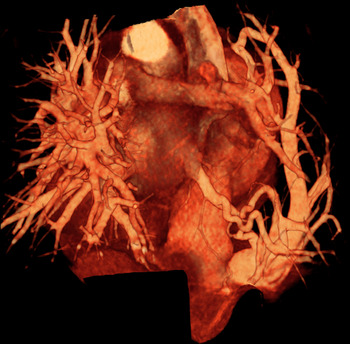
At angio-computed tomography scan, the Scimitar vein connected to the inferior caval vein underneath the diaphragm, the right lung was hypoplasic. Its lower lobe was supplied by a systemic arterial branch arising from the thoracic aorta (Fig 1).
3D computed tomography scan reconstruction (posterior view) illustrating the Scimitar vein draining into the distal inferior caval vein (supra-hepatic veins not shown)

Preoperative catheterisation confirmed the absence of pulmonary hypertension. The systemic arterial branch was coil-occluded during the same procedure.
At surgery, as a first step, a 5 cm length muscle-sparing right lateral thoracotomy was performed, allowing the dissection of the triangular ligament, the diaphragm, and the mobilisation of the Scimitar vein. Surgical repair was performed through a sternotomy, under normothermic cardiopulmonary bypass, and femoral venous cannulation for the inferior caval vein drainage.
To allow appropriate surgical exposure in the presence of dextrocardia, two stay sutures were placed on the apex of the left ventricle and on the right atrial wall to allow clockwise heart rotation.
On a beating heart, as originally described by Shumacker, Reference Shumacker2 a pericardial flap was created between the superior and inferior atrio-caval junctions to create a bowl-like pericardial baffle. The right phrenic nerve pedicle was mobilised from its pericardial attachment using bipolar coagulation.
Once the pericardial tunnel was partially created, cardioplegic arrest was performed.
Through a short right atriotomy, a trans-septal approach was used to delineate the precise location of the left atrium within the pericardial baffle. As in any Senning-type repair, the widest incision was made on the left atrial wall near the Sondegaard’s groove.
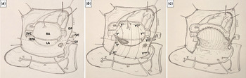
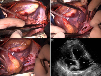
The Scimitar vein was divided at its insertion site on the inferior caval vein. The distal stump was oversawn. The proximal end was rotated toward the pericardial “well” to identify the site of anastomosis. A 2 cm elliptic portion of the pericardium was excised, and a termino-lateral anastomosis was performed The repair was completed by trimming the pedicled pericardial flap and suturing it to the epicardial surface of the right atria (Figs 2 and 3a–c).
Illustration of the technical aspects to create a bowl-like pedicled autologous pericardial baffle to which the Scimital vein can be connected with a tension-free anastomosis (DP: diaphragm, IV: inferior caval vein, LA: left atrium, RA: right atrium, RPA: right pulmonary artery, SV: Scimitar vein, SVC: superior caval vein)

( a ) The proximal end of the Scimitar vein was rotated toward the pericardial “well” to identify the site of anastomosis. ( b ) A 2 cm elliptic portion of the pericardium was excised. ( c ) a termino-lateral anastomosis of the Scimitar Vein was performed. ( d ) Post-operative transthoracic echocardiography demonstrating a wide channel towards the left atrium

Post-operative echocardiography demonstrated unrestricted and laminar flow from the Scimitar vein to the left atrium (Fig 3d). The patient was extubated on the first post-operative day. Her intensive care unit stay was 2 days.
Her final cardiac evaluation confirmed unobstructed pulmonary venous pathways.
Discussion
First reported by Neill et al in 1960, Scimitar syndrome is a rare association of congenital cardio-pulmonary anomalies. Reference Neill, Ferencz, Sabiston and Sheldon1 Its hallmark is an anomalous right pulmonary vein that drains all or part of the right lung into the inferior caval vein, with this vessel producing the Scimitar sign on the chest radiograph.
Several techniques have been described to redirect the Scimitar vein into the left atrium, including intra-atrial (right) baffling, Reference Zubiate and Kay3 direct reimplantation, Reference Tornvall, Jackson, Alvayay, Vargas, Koch and Zarate4 and more recently prosthesis interposition Reference Lam, Reemtsen, Starnes and Wells5 or pericardial flap tunneling. Reference Lugones and Garcia6
Though short-term results are excellent for both intra-atrial baffling and direct reimplantation techniques, long-term studies have demonstrated residual pulmonary vein or systemic venous pathway stenosis ranging from 17 to 50% requiring re-interventions (dilatation or stenting) or reoperations. Reference Vida, Padalino and Boccuzzo7,Reference Dusenbery, Geva and Seale8
The autologous pericardium has been successfully used in total cavo pulmonary connection, Reference Hvass, Pansard, Bohm, Depoix, Enguerrand and Worms9 as well as in simple Senning-type repair of late-presenting transposition of great arteries-intact interventricular septum, Reference Rubay, de Halleux and Jaumin10 and in double-switch procedures Reference Mee11 with excellent functional results and low incidence of tunnel stenosis.
We modified the technique described by Lugones et al in 2014, by using a termino-lateral anastomosis which requires only a 45° anterior rotation of the Scimitar vein on its long-axis.
The suboptimal long-term results of well-established techniques of Scimitar vein rerouting justify the search of alternative technique of repair to improve outcome and reduce the rate of re-intervention or reoperations.
Though short-term results appear to be very good with this new technique, additional patients and longer follow-up is required to validate its value as part of the armentarium to the cardiac congenital surgeon.
Acknowledgements
The authors thanks Dr Jérome Duisit for his drawing skills to generate the brilliant illustration of the technical aspect of the repair.
Financial support
This research received no specific grant from any funding agency, or from commercial or not-for-profit sectors.
Conflict of interest statement
None.
Ethical standards
All patient identifiers were removed from this case report.
Informed consent
Written informed consent was obtained from the patient’s parents.